
Project Gutenberg's An Introduction to Nature-study, by Ernest Stenhouse

This eBook is for the use of anyone anywhere in the United States and most
other parts of the world at no cost and with almost no restrictions
whatsoever.  You may copy it, give it away or re-use it under the terms of
the Project Gutenberg License included with this eBook or online at
www.gutenberg.org.  If you are not located in the United States, you'll have
to check the laws of the country where you are located before using this ebook.

Title: An Introduction to Nature-study

Author: Ernest Stenhouse

Release Date: January 30, 2020 [EBook #61273]

Language: English

Character set encoding: UTF-8

*** START OF THIS PROJECT GUTENBERG EBOOK AN INTRODUCTION TO NATURE-STUDY ***


Produced by Chris Curnow, Paul Marshall and the Online
Distributed Proofreading Team at http://www.pgdp.net


MACMILLAN AND CO., Limited
LONDON · BOMBAY · CALCUTTA
MELBOURNE
THE MACMILLAN COMPANY
NEW YORK · BOSTON · CHICAGO
ATLANTA · SAN FRANCISCO
THE MACMILLAN CO. OF CANADA, Ltd.
TORONTO
AN INTRODUCTION
TO
NATURE-STUDY
BY
ERNEST STENHOUSE, B.Sc. (Lond.)
ASSOCIATE OF THE ROYAL COLLEGE OF SCIENCE, LONDON; JOINT-AUTHOR
WITH A. T. SIMMONS, B.SC. OF “SCIENCE OF COMMON LIFE”
MACMILLAN AND CO., LIMITED
ST. MARTIN’S STREET, LONDON
1910
First Edition 1903.
Reprinted 1904 (twice), 1905, 1906, (with additions) 1908, 1910.
GLASGOW: PRINTED AT THE UNIVERSITY PRESS
BY ROBERT MACLEHOSE AND CO. LTD.
PREFACE.
One of the most encouraging of recent educational movements is the increasing importance attached, both in this country and abroad, to what is called Nature-Study. It is evident that the instruction contemplated differs as widely, on the one hand, from the traditional object-lessons on polar bears and ironclads, as it differs from formal Biology on the other. This difference is abundantly shown, not only by the circulars and syllabuses issued by our own Board of Education, but by the publications of the leading educational authorities of Europe and America. The aim of Nature-Study, as thus laid down, is not primarily the acquisition of the facts of natural history: it is rather a training in methods of open-eyed, close, and accurate observation, especially of familiar animals and plants, which shall teach the student to see what he looks at, and to think about what he sees.
It is in a spirit of entire agreement with these views that this book has been written. No previous knowledge of Biology on the part of the reader is assumed, and technical terms have as far as possible been dispensed with. In drawing up the course, I have had in mind throughout the attitude of an intelligent youth of sixteen, and the work will be found to be well within the powers of such a student. Teachers will, [Pg vi] however, find no difficulty in adapting the exercises to the needs of younger pupils.
Care has been taken to select, as types for study, animals and plants which are at the same time representative and easily obtainable,[1] and I have been further guided in the selection by the Board of Education Syllabuses of the King’s Scholarship Examination and Section I. of the Elementary Stage of General Biology, the subjects of which are included in the volume. The book has, however, a considerably wider scope than is indicated in these syllabuses, and will therefore, I hope, be found useful not only in schools and training-colleges, and to examination candidates, but also to members of field clubs and to students of natural history generally. It has been necessary to arrange the chapters with some attempt at logical sequence, but it is not supposed that this order will be adhered to in practice; by the aid of the monthly nature-calendar, together with numerous cross-references, it will be found easy to take up the work at any point.
The chapters are divided into sections, each of which consists of two parts: First, precise instructions for practical observations and experiments, designed to exercise the reasoning faculties of the students; and, second, a descriptive portion, in which the meaning and relation of the results obtained are discussed. At the end of each chapter is a number of additional exercises, either original or taken from past examination papers. Of the latter class, questions to which dates are affixed have been set by the Board of Education, while those [Pg vii] marked “N.F.U.” are selected from National Froebel Union tests. In many cases, the exercises provide subjects for further observation and experiment, as well as for written description.
Much trouble has been taken in the selection of the illustrations, many of which have been expressly drawn or photographed for this book. Through the kindness of the publishers I have been able to include illustrations from Strasburger’s Text-Book of Botany, Parker and Haswell’s Text-Book of Zoology, The Cambridge Natural History, and other books; and Mr. Ernest Evans has courteously consented to the use of a number of figures from his Botany for Beginners. The following illustrations have been prepared from photographs supplied by Mr. J. C. Shenstone, F.L.S., Vice-President of the Essex Field Club: Figs. 27, 57, 65, 67 to 71, 74, 75, 80, 81, 83, 84, 85, 87, 89, 92, 94, 95, 102 to 110, 120, 136, 145, 149, 152 and 153; while Figs. 180, 196, 200, 201, 203, 205, and 211 are reproduced, by permission, from Pike’s Woodland, Field, and Shore (Religious Tract Society).
Finally, I must acknowledge gratefully the continuous help which, at every stage in the preparation of the book, I have received from Professor R. A. Gregory and Mr. A. T. Simmons, B.Sc.—help as valuable as it was generous.
The issue of a new edition has provided the opportunity of adding a section on School Journeys, originally contributed by me as an article to The School World, and reprinted here by kind permission of the Editors of that journal. For the illustrative sketch-map (Fig. 237), and for Figs. 19, 21 and 138, I am indebted to my friend Mr. T. D. Tuton Hall.
CONTENTS.
| PART I. PLANT LIFE. | ||
| chapter | page | |
| I. | Seeds and their Early Stages of Growth, | 1 |
| II. | How a Green Plant Feeds, | 26 |
| III. | The Forms and Duties of Leaves, | 37 |
| IV. | Buds. The History of a Twig, | 55 |
| V. | How Stems do their Work, | 67 |
| VI. | Some Common Flowers, | 88 |
| VII. | Grasses, | 125 |
| VIII. | Common Forest Trees, | 140 |
| IX. | Fruits: How Seeds are Scattered, | 165 |
| X. | Ferns and Horsetails, | 183 |
| XI. | Mosses, Mushrooms, and Moulds, | 199 |
PART II. ANIMAL LIFE. |
||
| XII. | The Rabbit: A Typical Mammal, | 211 |
| XIII. | How a Rabbit Lives, | 222 |
| XIV. | Some other Mammals, | 246 [Pg x] |
| XV. | The Pigeon: A Typical Bird, | 265 |
| XVI. | The Development and Education of the Chick, | 282 |
| XVII. | Some Familiar British Birds, | 301 |
| XVIII. | Frogs and Tadpoles, | 332 |
| XIX. | The Habits and Life-Histories of Common Insects, | 349 |
| XX. | Some Crustaceans, Molluscs, and Worms, | 372 |
| XXI. | Field-Work. The School Journey, | 388 |
| Monthly Nature Calendar, | 400 | |
1. Preparation of the seeds.—Obtain several seeds of the broad bean, pea, mustard, yellow lupine, vegetable marrow, and sycamore; soak them in cold or slightly warm water until they are soft enough to be cut through easily with a sharp knife. The time necessary will vary with different seeds according to the size of the seeds, and with the temperature of the water. The beans should be left in the water for a few days. When the seeds are soft enough, examine one or two of each, and in the meantime put about six of each (except the mustard) in damp sawdust in a warm place. Put the mustard seeds on damp flannel in a saucer.
2. The outside of a broad bean.—Notice the flattened oval shape, with an indentation at one place (Fig. 1). What is the colour of the skin (seed-coat) of the bean seed? Is all the skin of this colour? A black scar extends along the edge from the indentation for about ¾ in. What is this scar? If beans in the pod can be obtained, see that the scar is the place of attachment of the seed stalk. Make drawings to scale, showing side and edge-views of the seed. Wipe the [Pg 2] bean dry and then squeeze it gently. Notice that a drop of water comes out at a point at one end of the stalk scar. There is evidently a little hole here. This little hole is called the micropyle. Mark its position by a dot on the drawing.
3. The inside of a broad bean.—With a sharp knife cut the seed-coat open, beginning at the side of the seed furthest from the micropyle, and carefully remove the seed-coat. Notice that near the micropyle the seed-coat forms a funnel-shaped depression, and that the point of the funnel is at the micropyle. Does anything fit into the funnel? A little cone may be seen to fill the funnel; this conical body is called the radicle. Make a drawing of the seed after the removal of the seed-coat. Look at the edge opposite the radicle and notice that a crack divides the body of the seed into halves. Put the point of your knife blade into the crack, and gently force the halves apart. They come apart without tearing, showing that they are naturally separate, although they fit so closely together.
These two swollen bodies are called the cotyledons. Separate them and see, at the point where they join the radicle, a little curved rod, evidently a continuation of the radicle, lying between them. This rod is the plumule. Take off one cotyledon, and make a drawing of the inner face of the other cotyledon, with the adhering plumule and radicle (Fig. 2).
4. Starch present in the cotyledons of the bean.—Scrape the inner surface of a cotyledon and then pour on it a drop of iodine solution.[2] Is there any change? Pour also a drop of iodine solution on a piece of laundry-starch. Is a similar blue colour formed? What substance is probably present in the cotyledons of the bean?
5. The pea.—Examine a pea in a similar manner. Make drawings showing the stalk-scar, the micropyle, and the plumule and radicle with their manner of connection with the cotyledons. Does the end of the radicle point towards the micropyle? How many cotyledons has this seed? What shape and colour are they? Do they contain starch? [Pg 3]
6. The seed of the yellow lupine.—Compare this with the bean and the pea, and find out how many cotyledons it has, and whether they contain starch. Can you find the plumule? It is very small, but occupies a position similar to that of the plumule of the bean. Does the end of the radicle point to the micropyle?
7. The vegetable marrow seed.—Notice the peculiar shape (somewhat like a pocket-flask) of the seed, and the thickened margin which runs round it. Carefully cut the seed-coat away so as not to injure the part inside. How many cotyledons are present? What is their colour? Do they contain starch? Can you see the plumule and radicle clearly? If not, do not decide that they are absent, but leave the question to be settled later, when you watch a vegetable marrow seed “come up.”
8. The mustard seed.—Notice how much smaller this seed is than the others. With a balance, find how many mustard seeds are equal in weight to one bean seed. Observe the stickiness of the seed-coat of the soaked seed, and then remove it carefully with needles, exposing two thin plates, each one folded on itself, and one tucked inside the other, like two sheets of note-paper. These are the cotyledons; it seems that the smallness of the seed may be mainly due to the small size of the cotyledons. What is their colour? Remember these characters and try, when you watch the young plants come up later, to find an explanation of them.
9. The sycamore fruit.—The seed of the sycamore is enclosed in a case which has a wing attached to it. The wing, the case, and the enclosed seed together constitute the fruit of the sycamore. The fruits occur in pairs (Fig. 137). Notice that a cord runs out to each fruit from the stalk on which the pair of fruits is borne. Make a drawing of a pair of fruits, then separate the fruits.
10. The sycamore seed.—Cut open a fruit. Can you see anything between the seed and the fruit-case? Would the hairy covering of the seed tend to keep it warm during the winter? Why? Why do you prefer to wear flannel in winter and linen in summer? Flannel is more fluffy than linen. [Pg 4]
Remove the seed-coat carefully. Running down one side you will see a little curved rod. This is the radicle. Gently raise it with the point of your knife. Notice that the rest of the seed seems to consist of a green part, which is curled up. Uncoil the curls carefully. You find that they are two green leaves, fixed at the top of the radicle. These are the cotyledons. In the seed each cotyledon is first folded in two across the middle and then coiled up. Make a sketch showing the coils (Fig. 4). Can you see the plumule? It is just at the top of the radicle, where the cotyledons are fixed on.
Plants are living things.—One of our foremost naturalists[3] tells us that when he goes out into the woods, or into one of those fairy forests which we call fields, he finds himself welcomed by a glad company of friends, everyone with something interesting to tell. Such a feeling would be quite impossible to one who did not vividly recognise the fact that plants are alive; for it is precisely this recognition or its absence which makes the observation of the forms and habits of plants fascinating or the reverse. Let the Nature-Student, then, at the outset of his work, keep the idea of life inseparably bound up with his every thought about plants. It may at first require a little effort, but before long it will enable him to understand how the friendship of the more silent half of animate nature may form one of the great pleasures of life.
The study of seeds.—The manifestation of life is so striking, and the changes in form and size take place so rapidly, in the germination of seeds, that the study of plants cannot better be commenced than with this stage of their growth. The method has also the logical virtue of beginning at the beginning, or nearly so.
These early changes can be well observed by taking various common seeds, soaking them in water until they are soft, and then allowing [Pg 5] them to germinate in damp sawdust, taking a few out at intervals and noting their progress. The growth of the seeds takes place more rapidly if they are kept in a warm room, but in any case some days will probably elapse before much change is noticeable in them.
During the interval of waiting, some of the seeds themselves should be carefully examined, and drawings of all the parts should be made. The drawing ought on no account to be omitted. It compels the student’s attention to details which would otherwise pass unnoticed; and a careful sketch is a much better record of an observation than any amount of description alone could be. The drawing need not be elaborate; an outline pencil-sketch to scale will usually be sufficient.
The seed of the broad bean.—The seed of the broad bean (Fig. 1) is large, having a diameter of perhaps an inch and a half, and a thickness of half an inch. In shape it is oval, but at one region the edge is indented, and a black scar (st. sc.) runs from the indentation along the edge for a distance of about three-quarters of an inch. This scar is the place of attachment of the stalk which formerly carried the seed in the bean-fruit (pod). It may be called the stalk-scar. If a soaked bean is wiped dry and then gently squeezed, a small drop of water escapes from the end of the stalk-scar [Pg 6] nearest the indentation. The hole out of which the water comes is very small and difficult to see, but its position is thus made clear. This hole (m) is called the micropyle,—a word meaning the “little gate.”
The bean seed is covered by a tough brown skin, the seed-coat (Fig. 2, s.c.), a funnel-shaped depression in which leads to the micropyle (m). The depression is occupied by a part of the seed which is shaped like a conical peg and called the radicle (R); the point of the radicle is directed toward the micropyle. The great body of the seed is composed of two fleshy, cream-coloured lobes, easily wedged apart by inserting a knife-blade between them; these fleshy lobes are the cotyledons (Cot.). Between them, and continuous with the radicle, is a small yellow body, the plumule (pl.). The relations of the radicle, plumule and cotyledons are best seen by removing one cotyledon (Fig. 2).
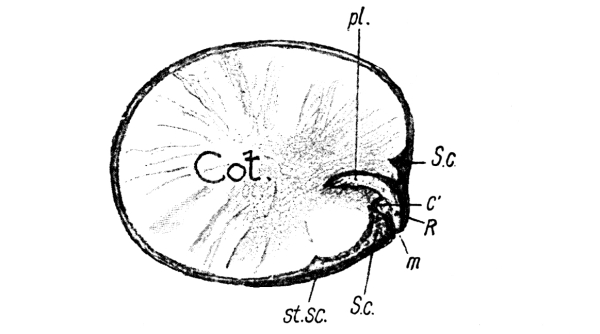
Fig. 2.—Broad-Bean seed, seen from the inside, after the removal of half the seed-coat and one cotyledon. Cot., the inner face of remaining cotyledon; C′, area of attachment of other cotyledon; m, micropyle; pl, plumule; R, radicle; S.c., seed-coat; st. sc., stalk-scar. (× 1.)
A scraped cotyledon at once turns blue when a drop of dilute iodine solution is poured on it, thus showing the presence of starch. We shall see in Chapter II. what use the growing seedling makes of the starchy food which is stored in its cotyledons. [Pg 7]
The seed of the pea.—Except in size and shape the seed of the pea is very similar to the bean seed. Its form is spherical, and the scar left by the stalk which formerly attached it to the wall of the pea-pod (Fig. 3) is plainly to be seen. Pointing towards the micropyle is the peg-like radicle; the plumule lies between the hemispherical cotyledons. As before, the cotyledons can be proved to contain starch, by the blue colour which is formed when a drop of iodine solution is poured on the scraped surface.
The seed of the yellow lupine.—The seed of the yellow lupine is about as large as a pea, but it is slightly flattened in shape. The seed-coat is prettily mottled; when it is removed, the greater part of the seed is found to consist of two cotyledons. They are somewhat swollen, but the stored food is not starch. The plumule and radicle occupy positions similar to those of the bean and pea.
The vegetable marrow seed.—This seed has a rather curious shape, and somewhat resembles a pocket-flask. It is flattened, and the border of the seed-coat is thickened and of silky appearance, the rest of the “skin” having some resemblance to kid. The two cotyledons, which compose the greater part of the seed, are white and only slightly fleshy. The plumule and radicle are at the pointed end of the seed, and are difficult to see.
The mustard seed.—In comparing the mustard seed with those [Pg 8] already described, one is struck with the great difference in size. An average broad-bean seed weighs about 600 times as much as the mustard seed. While the two fleshy cotyledons make up the bulk of the seed of the bean, pea, lupine and vegetable marrow, the cotyledons of the mustard seed are thin and leaf-like. They are folded on themselves, one inside the other (as at g, Fig. 61), and enclose the radicle. The characters of the cotyledons account very largely for the small size of the mustard seed. It will be seen, when the growth of the young plants is watched, that the difference is associated with the special duties which the cotyledons perform in the various cases.
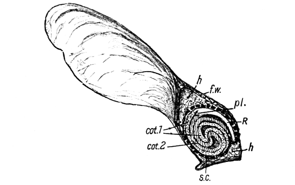
Fig. 4.—Sycamore Fruit, cut through in the plane of the wing. s.c., seed-coat (indicated by a thick, broken line); f.w., fruit-wall; h, layer of fine hairs; R, radicle; pl., plumule; cot. 1, cot. 2, cotyledons (diagrammatically shaded). (× 2.)
The sycamore seed.—What is generally called the seed of the sycamore is really a fruit. The fruits are in pairs (Figs. 33 and 137), and each half consists of a flat wing and a rounded case in which the seed itself is enclosed. The round seed-cases of the two [Pg 9] fruits are connected together. When they come apart, a scar marks the place where they were formerly in contact, and a little cord runs out to each fruit from the stalk on which the pair of fruits is borne.
Between the sycamore seed and the wall of its case is a layer of fine hair (h, Fig. 4), which forms a warm nest for the seed in winter. The seed is surrounded by a thin, brown seed-coat, and consists mainly of two cotyledons, but these are very different from any yet described. Each is a green leaf, measuring, when unfolded, about an inch in length. It is first folded across the middle of its length, and then rolled up into a close coil with its fellow. The coils are very plainly to be seen when the seed coat is removed, or when the whole seed is cut through, by a sharp knife, in the plane of the wing. Running down one side of the seed is a green rod, the radicle (R, Fig. 4). The two cotyledons (cot. 1 and cot. 2) spring from its upper end, and between them is the tiny plumule (pl.)
The sycamore seed bears more resemblance to the mustard seed than to the others, but it is on a much larger scale. In each of these two seeds the cotyledons are plainly leaves, while in the others their nature is disguised by the great accumulation of stored food in them.
If the seeds which were sown in damp sawdust and on flannel are kept warm they will soon be ready for study. You should remember that at present your object is not so much to rear the plants as to find out how they grow. As soon, therefore, as any sign of growth is to be seen when you take a seed out, you should begin to examine them at regular intervals, taking one or two out every day and leaving the rest to continue their development. Keep the sawdust damp, but not wet.
1. The pea and bean.—(a) General development.—Very soon [Pg 10] the seed-coat splits at the micropyle-end of the stalk-scar, and the end of the radicle protrudes. Does the radicle grow upwards or downwards? Observe that even if the seed was so planted that the micropyle was at the top, the radicle turns over and grows straight down. Turn over a seedling and see if you can persuade the radicle to grow upwards. Open a seed when the radicle is about an inch long, and see what the plumule is doing. It is still enclosed in the seed-coat, and lies between the cotyledons, but is larger than at first. As the growth proceeds the cotyledons begin to separate near the top of the radicle, and you can get a glimpse of the plumule.
(b) The growth of the radicle.—Day by day the radicle becomes longer. Is it all growing longer, or does the increase in length take place more at one part than another? To answer this question, take five or six inches of cotton thread and moisten the middle part with Indian ink. Lay the seed on a flat ruler, so that the radicle lies over an inch divided into—say—tenths. Hold the thread tight, and press the inked part gently on the radicle, making about five marks at equal intervals from the point upwards. The ink will dry almost immediately. Then carefully replant the seed, taking care not to injure the radicle. After a few days take it out again, lay it once more on the ruler, and measure the distance between the marks.
The radicle is evidently the young root.
(c) The root-cap.—Hold up the radicle to the light, and examine its tip with a lens. Try to see that the tip is covered by a little cap, somewhat like a very small thimble. This is called the root-cap.
(d) The root-hairs.—Hold a seed, with the radicle about an inch long, against a dark surface. Is the surface of the radicle smooth, or can you see any fluffiness on it? Is all the radicle fluffy, or only a part? Which part? As you examine older and older seedlings notice how much of the radicle is fluffy, and where the fluffy part is. The fluffy appearance is caused by fine, closely-set hairs, called root-hairs.
(e) The plumule.—How soon after planting does the plumule become free? Does it grow upwards or downwards? The plumule is evidently the young stem.
[Pg 11] As soon as the young stem is old enough mark it with Indian ink as you marked the young root, and replant it to find if there is any difference in the rates of growth of its different parts.
(f) The fate of the cotyledons.—From time to time examine the cotyledons and notice that as the seedling grows larger they become more and more shrunken. Something is evidently being taken from them, perhaps to feed the young plant. We shall inquire into this by further experiment (Chapter II.).
Do the cotyledons remain in their original position, or are they carried upwards with the growing stem or downwards with the growing root?
2. The yellow lupine.—In the same way observe how this seed grows. Do the cotyledons shift their position or change in colour? Do they become leaf-like? How do they differ from later-formed leaves? What becomes of them at last? What becomes of the seed-coat?
3. The mustard seed.—Notice that, soon after the radicle has come out of the seed-coat, a sort of hump forms at its upper end, and at length the cotyledons are pulled out of the seed-coat and turn up towards the light. What is their colour? Observe that the two cotyledons are soon raised on the end of a little stalk. Like the cotyledons of the yellow lupine they are plainly leaves. Notice their shape. Are they of equal size? Why not? When they are about three inches above the seed-coat gently separate them and notice the little bud between them. Draw the seedling. How large can you get a mustard seedling to grow on damp flannel? Plant a few mustard seeds on earth, and notice the difference between the shape of the cotyledons or seed-leaves and that of the leaves which appear later. What becomes of the cotyledons?
4. The vegetable marrow seed.—Make similar observations upon the vegetable marrow seeds, noticing particularly whether the cotyledons remain in their original positions and shrink up as the plant increases in size, or whether they are pulled out of the seed-coat by the elongating stem, and become green and leafy. How does the plant hold down its seed-coat whilst it pulls out its cotyledons?
5. The sycamore seed.—From what you have seen of the cotyledons [Pg 12] of the sycamore seed, will you expect them to behave like those of the mustard seed, or like those of the pea and bean? Even in the seed they are green, and plainly leaves. How do they escape from the seed-coat? What is their shape? Do they come out before or after the radicle? Do they get any larger as the stem grows? How large can you get a sycamore seedling to grow in damp sawdust? As large as a seedling of pea or bean? Plant some sycamore seeds on earth and compare the shape of the cotyledons with that of the next-formed leaves. How soon do the “true” leaves appear after the cotyledons have escaped from the seed? Do any “true” leaves grow on the plants in sawdust? What becomes of the cotyledons at last?
The embryo.—The plumule, radicle, and cotyledons, which have now been seen in the seed, form the embryo of the plant. The adult plant will be wholly formed by the growth and development of these parts, and we must now follow carefully the changes which take place when the seed germinates, and try to find out what becomes of each part. It is better to put the seeds at first in damp sawdust rather than in earth, as the young roots can then be more readily cleaned and observed. With small seeds the early stages of growth are better seen if damp flannel is used.
Germination.—Under the influence of moisture and warmth the embryo in the seed begins to swell and unfold its parts. The radicle makes its appearance first (Fig. 5), breaking through the seed-coat at the micropyle; it is the young root. The radicle always grows downwards, that is, toward the centre of the earth. If the seed lies in such a position that the micropyle is directed upwards, the point of the radicle turns over and grows downwards as soon as it escapes from the seed-coat. As the young root becomes longer and thicker (Fig. 6) the seed-coat opens more and more, showing the cotyledons beneath, and these, too, are gradually forced apart. [Pg 13]
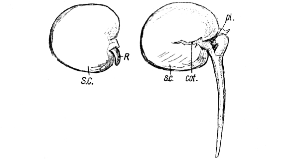
Fig. 5.—An early stage in the germination of a Broad-Bean seed. R, radicle; s.c., seed-coat. (× ⅔.) |
Fig. 6.—A slightly later stage in the germination of a Broad-Bean seed. cot., cotyledon; pl, plumule; R, radicle; s.c., seed-coat. (× ⅔.) |
The cotyledons.—During the germination of various seeds, a very marked difference in the behaviour of the cotyledons is to be seen. In the case of the broad bean and pea the cotyledons remain in their original positions, partially enclosed by the split seed-coat. Presently a hump (Figs. 6 and 11) forms at the upper end of the radicle, as if the plant were making an effort to pull its plumule out of the seed. It soon succeeds (Fig. 12), and the plumule turns up to the light. It is the young stem. At its end is a little bud, formed by a number of small, overlapping, green leaves which surround the growing point. Henceforth the stem grows upwards, that is in a direction precisely opposite to that of the root’s growth. Both stem and root are attached to the cotyledons, which gradually shrivel up as the stem and root become larger and larger.
When, however, the seed of the mustard, or sycamore, germinates the cotyledons behave very differently (Figs. 7 and 8). Soon [Pg 14] after the root has become well established the cotyledons come quite out of the seed-coat and unfold themselves. Instead of remaining on or under the surface of the ground they are carried upwards at the end of a stalk toward the light, and for some time the little plant appears to consist of root, stalk, and cotyledons only. If, however, the cotyledons are gently pressed apart, a tiny bud is seen between them. This evidently corresponds to the bud at the end of the stem of the bean or pea.
In the case of the lupine (Fig. 9) or vegetable marrow (Fig. 10) the cotyledons appear to combine these two conditions. They are swollen and contain stored food; yet they come out of the seed-coat early, become green, and open out to the light. They are evidently leaves, though their shape differs from that of the later leaves.
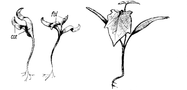
Fig. 8.—Three stages in the growth of a Sycamore Seedling. cot., cotyledons; fol., first pair of foliage leaves. (Slightly reduced.)
[Pg 15] The germinating vegetable-marrow seed possesses a curious contrivance for pulling its cotyledons out of the seed-coat. This is a peg (p, Fig. 10) which develops at the top of the radicle, and holds down the lower half of the seed-coat whilst the other half is forced upwards to allow the cotyledons to be withdrawn.
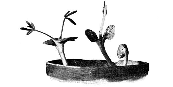
Fig. 9.—Three stages in the growth of the Yellow Lupine. On the right the cotyledons are still enclosed in the mottled seed-coat. In the middle plant the cotyledons are spreading out; the first foliage leaves have not yet unfolded. On the left, the first two foliage leaves are unfolding, and the cotyledons have spread out flat. (Slightly reduced.)
After a little thought a possible explanation of these differences in the cotyledons suggests itself. It may be that, in the case of the mustard and sycamore, leaves are required as early as possible, while the bean and pea have no immediate need for leaves because their cotyledons contain so much stored food. The cotyledons of these plants shrivel up as the seedling grows, and this seems to indicate that [Pg 16] during its early stages the plant lives upon this food. In Chapter II. we shall make experiments to see if this explanation is the true one. If so, the lupine and vegetable-marrow seeds evidently rely partly upon their stored food and partly upon setting the cotyledons to work as leaves, whilst the plant is still very young.
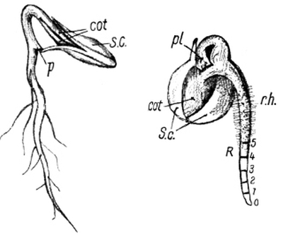
Fig. 10.—Germinating Vegetable Marrow seed. p, the peg by which the seed-coat (s.c.) is held down to allow the cotyledons (cot.) to be withdrawn. (× 1.) (After Bailey.) |
Fig. 11.—A germinating Pea; cot, cotyledon; pl, plumule; R, radicle; r.h., root-hairs; S.c., seed-coat. The radicle has been marked with Indian ink at intervals of 1/10”. |
The true leaves.—The cotyledons are really makeshift leaves, which are already formed in the seeds. Even when they expand and become green they do not live long, but as soon as the next few leaves are well established, shrivel up and wither. The true or “foliage” leaves first make their appearance as a bud which surrounds the growing point of the stem. As this part of the stem increases in length, the foliage leaves become separated from each other and spread out to the light and air.
The lengthening of the stem and root.—Unless an experiment to test the truth of the matter is really made, it might be supposed that the different parts of the stem and root of the seedling grow in length at the same rate. This can be tested by marking the stem and root with lines of Indian ink at equal distances. In one experiment with a pea seedling five lines were marked upon the young root at regular intervals of one-tenth of an inch, beginning at the tip (Fig. 11). The seedling was carefully replanted and examined again a few days later. Between the tip and the first mark there was then (Fig. 12) a distance of seven-tenths of an inch; that is, this part had grown to seven times its former length. The second interval was four times as long as [Pg 17] before, the third was one and a half times as long, while the fourth and fifth intervals had not increased in length at all. Such experiments prove that the root grows in length either at or just behind the tip. When a young stem is treated in the same way the lengthening is found to take place more evenly.
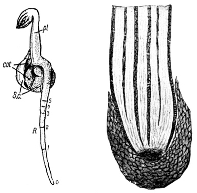
Fig. 12.—The Pea seedling of Fig. 11, a few days later. cot, cotyledons; pl, plumule; R, radicle; S.c., seed-coat. (× 1.) |
Fig. 13.—The tip of a root, showing the root-cap. (Magnified.) |
Rootlets.—After a time the radicle begins to put out branches called rootlets. These come off the main root in rows. In some cases rootlets make their appearance whilst the radicle is still very short, as in the vegetable marrow of Fig. 10, but in others the radicle may be a few inches long before it produces rootlets.
The root cap.—The tip of the root, and of each of its branches, is covered by a little cap, shaped somewhat like a thimble (Fig. 13). This protects the tender growing point from the friction of particles of soil, and is continually renewed by growth from within as its outer layers are worn away.
Root hairs.—When a young root is held against a dark background it appears fluffy. This appearance is caused by a large number of very fine hairs upon its surface. The hairs are not found all over the root and its branches, but only for a short distance a little way behind the [Pg 18] tips (Figs. 7 and 11). These root hairs are of very great importance to the plant, as will be seen in Chapter II.
1. Preparation of the seeds.—Soak grains of maize (Indian corn) and wheat in water until they are soft. The grains of maize will need soaking for several days. Plant about a dozen of each in damp sawdust, and in the meantime examine others.
2. The maize grain.—A grain of maize is really a fruit, as a pea-pod is. A pea-pod contains several seeds; a maize fruit contains only one seed, which fills it. Notice the shape of the grain—flattened, rounded along one edge, and bluntly pointed at the opposite edge (Fig. 14). Notice a whitish patch on one of the flattened sides; a ridge (E) down the middle of this marks the position of the embryo. Cut through the grain lengthwise, so as to divide the embryo into two equal parts, and examine the cut surface (Fig. 15). Identify:
(a) The embryo, lying somewhat obliquely, and to one side. The radicle (rad.) is directed towards the pointed end of the grain, and the plumule (pl.) towards the rounded end.
(b) The endosperm (end): a mass of material outside the embryo, and forming at least half of the grain.
(c) The scutellum (scm): a plate lying between the endosperm and the embryo.
(d) The coats of the seed and fruit, surrounding the whole.
Draw. Add a drop of iodine solution to the cut surface of the half-grain. The endosperm turns blue. What does this indicate?
3. The wheat grain.—A grain of wheat is also a one-seeded fruit. Notice the groove along one side, and—on the opposite side near one end—the white patch marking the position of the embryo. At the other end is a tuft of very fine hairs. Cut the grain lengthwise, so as to divide the white patch into two equal parts, and make out the embryo, endosperm, and scutellum (Fig. 16). Draw. Test the endosperm with iodine solution. Does it contain starch? [Pg 19]
Grains of maize and wheat.—A grain of maize or wheat is really a one-seeded fruit. In other words, the grain consists not only of the seed with its seed-coat, but also of the seed-case. In this respect it resembles the fruit of the sycamore (p. 8). When, however, a grain of maize or wheat is carefully examined, it is found to differ greatly from all the seeds hitherto mentioned. A maize grain is somewhat flattened, and rather pointed along one edge (Fig. 14). On one flat side, near the pointed end, may be seen a whitish patch, and, along the middle line of this, a ridge which marks the position of the embryo.
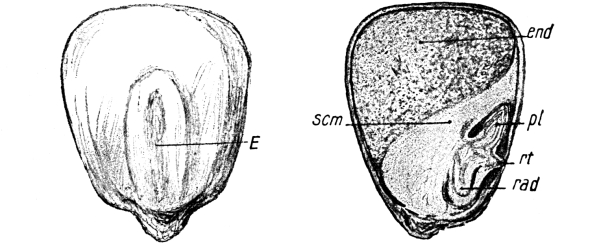
Fig. 14.—Maize grain, showing the position of the embryo (E). (× 4.) |
Fig. 15.—A longitudinal section of a Maize grain, through the middle line of the embryo. end, endosperm; pl, plumule; rad, radicle; rt, origin of a root; scm, scutellum. (× 4.) |
A wheat grain has a different shape. It is oval, with a deep groove running down one side. One end is clothed with a tuft of very fine hairs; near the other end, on the side opposite to the groove, is a white patch beneath which is the embryo.
These differences in shape are of small importance, for the two grains are really of very similar structure, as may be seen when they are cut through lengthwise with a sharp knife, in a direction which divides the embryo along its middle line. It is then plain that each grain consists [Pg 20] of two principal parts: the embryo and the endosperm (Figs. 15 and 16). The embryo lies to one side and in the lower half of the seed. At its upper end the young stem and at its lower end the young root—each still enclosed in protecting sheaths—are easily seen. The greater part of the seed is quite outside the embryo; it is a mass of food called the endosperm, which has been stored up for the use of the young plant during its earliest stages of germination. This food mass at once turns blue when a drop of iodine solution is placed upon it, showing that it contains a considerable amount of starch. It is the endosperm which is made into flour when corn is ground.
Lying between the embryo and endosperm is a flat plate called the scutellum. Covering embryo, scutellum, and endosperm is the seed-coat proper, and outside that come the various layers of the fruit case.
In the seeds previously examined, the embryo—consisting of plumule, radicle, and cotyledons—was seen to fill the seed-coat completely. In some cases the cotyledons were found to be more or less swollen with stored food-material. In the maize and wheat, however, the embryo forms only a comparatively small proportion of the seed, the bulk of which consists of stored food called endosperm. This is a difference of some importance. Still more important, however, is the fact that the two cotyledons, which were so conspicuous a feature of the other seeds, cannot apparently be seen at all in these seeds. Are there then no cotyledons in the seeds of the maize and wheat? If there are, how and when do they appear, and what is their number? [Pg 21]
1. The roots.—Watch for the appearance of the roots. Is there, as in the seedlings previously studied, one principal root, or are there soon several, all apparently of equal or nearly equal importance? Do the roots grow straight down as before, or do they spread horizontally?
2. The stem and the cotyledon.—Notice that a rod, somewhat thicker than a root, grows out near the origin of the roots, and curves upwards towards the light. When this is about an inch long on a maize seedling, slit it open carefully, and observe that it consists of a pale outer sheath and a green core. The sheath is the single cotyledon; the green core is the young stem enclosed in a young foliage leaf. Cut open the grain and notice how the endosperm has shrivelled. As the seedlings become larger watch the young stem growing out at the end of its sheathing cotyledon. What is the length of the cotyledon when the stem first appears (a) in the maize, (b) in the wheat?
3. The foliage leaves.—As soon as a foliage leaf unfolds make a drawing of its shape. Contrast it with the young foliage leaves of the other seedlings. Hold up the leaves to the light and compare the arrangement of their veins.
How maize and wheat seeds grow.—When the maize and wheat fruits have been kept in damp sawdust for a few days, the seeds—one in each fruit—begin to germinate. As a rule the wheat plants have grown to a height of some inches before the roots and stems of the maize plant have emerged from the seeds.
The roots.—As might be expected, the first signs of life make their appearance at the white scar which indicates the position of the embryo or young plant. Instead of one main root growing out, several little roots make their appearance almost at the same time. They do not grow as directly downwards as the radicle of a pea or bean (Fig. 6), [Pg 22] but tend to spread in a horizontal direction (Fig. 17). It is clear that in this way the roots are more independent of each other than if they grew directly downward side by side.
The cotyledon and the stem.—Growing out from the seed close to the roots is another rod (Fig. 17, C), rather thicker than the roots, which at once curves upwards to the light. It is pale green in colour. This is the cotyledon or first leaf. When it is carefully slit open, it is found to be a hollow sheath, enclosing a bright green core. In the seedlings which are left undisturbed, the core at last breaks through the tip of the cotyledon. It consists of the young stem and its surrounding foliage leaves. As the growth of the plant continues, these sheathing leaves unfold themselves into the long narrow blades characteristic of grass leaves (Fig. 98). The bottom of each leaf is tubular and forms a sheath round the stem.
The endosperm.—The endosperm, which at first made up more than half the seed, gradually shrivels up as the little plant continues its growth. The food material which it contains is absorbed by the scutellum and is passed on to afford the plant the necessary nourishment for those early stages when it is too young to feed itself. By the time the first few foliage leaves are well developed, all that remains of the grain is an empty husk.
Comparisons and contrasts.—The examination of these seeds and [Pg 23] seedlings will enable the student to see that differences, which at the first glance appear great, are often of only minor importance; while apparently small variations may prove, on closer inspection, to be caused by deeply-seated differences of structure and habits of life. He should always set himself the questions, “In what ways do such and such objects resemble each other; and in what ways do they differ from each other? Which of the differences and resemblances are of most importance?” He should also notice that a mere difference of size is often of very small consequence.
Above all, the student should get into the habit of asking the reasons for the differences and resemblances which he notices in his nature-study. To learn what these reasons are he must observe closely, think carefully, and then make experiments to test the accuracy of his conclusions. “Be sure you are right; then look again”[4] should be his motto.
It is at once plain that the seedlings fall into two classes, according to the number of cotyledons or seed-leaves which they possess. The wheat and maize have only one such seed-leaf, while the mustard, bean, pea, sycamore, and vegetable marrow have two each. We shall see later on that one-seed-leaved plants differ from those with two seed-leaves not only in the number of their cotyledons, but also in the characters of their leaves and flowers and in their method of growth. These differences are so constant and so important that botanists have agreed to call all plants of the first class (such as maize and wheat) Monocotyledons, and plants of the second class Dicotyledons.
One of these differences is that the main roots of dicotyledons are formed directly by the growth of their radicles; while in [Pg 24] monocotyledons there is, after a short time, no such main root to be found, but several roots of almost equal size spring from the base of the stem and spread outwards in all directions (Fig. 17).
Both maize and wheat seeds contain—outside the embryo—a large store of food called endosperm (Figs. 15 and 16), which is not seen in any of the dicotyledonous seeds described in this chapter. This is not a very important difference, for, if we examined a very large number of dicotyledon seeds, we should find that most of them possessed endosperm. On the other hand, many monocotyledonous seeds are destitute of endosperm. Only after observing a very large number of facts is it safe to make general statements.
Confining our attention to dicotyledons, we are impressed by the great variation in size of the cotyledons. Those of the bean and pea are swollen with food material and form a large proportion of the bulk of the seed. As a consequence, the seedling has enough food to enable it to grow into quite a sturdy little plant before it needs any foliage leaves. The cotyledons of the mustard and sycamore, however, are thin, and they unfold almost immediately into green leaves, and set to work to help to maintain the plant until the first foliage leaves can be formed. The cotyledons of the lupine (Fig. 9) and vegetable marrow (Fig. 10) serve a double purpose. They not only contain a store of food ready to hand, but they also set to work early to make new food, until the new leaves are sufficiently advanced to take up their duties. It should be remembered that cotyledons are makeshift leaves.
EXERCISES ON CHAPTER I.
1. Make a collection of the seeds of various trees; try to find, in each seed, the cotyledons, radicle, and plumule. Which of the seeds contain stored starch? [Pg 25]
2. Soak pine and larch seeds in water for several days and then sow them, with a covering of half an inch of soil. Make notes of the number, shape, size, and behaviour of the cotyledons. How large are the seedlings at the end of the first season?
3. Make similar observations on the growth of sycamore, ash, and beech. Cover the seeds with an inch of soil.
4. Plant seeds of oak and chestnut two inches deep, and make drawings and notes of the stages of growth.
5. Investigate the structure and method of germination of a barley seed, and find out whether barley is a dicotyledon or a monocotyledon.
6. Make experiments to discover the effects, upon the germination of various seeds, of differences of temperature, moisture, and light, and write full accounts of the results obtained.
7. Draw from memory a young seedling of maize, and notice its chief peculiarities. (1898)
8. Draw the seedling of the sycamore in two or more stages, and add short notes. (1898)
9. Draw the root of any seedling that you have studied, giving its name. Mark the exact position of the root-hairs. (1898)
10. Open the nut provided. Draw what is to be found in it in one or two positions. Name the parts and give short explanations. (1901)
11. Explain, with drawings, how certain seedlings withdraw their seed-leaves from the seed-coat. (1901)
12. Describe and explain as far as you can the principal changes to be observed during the germination of a bean or pea. (1901)
13. Describe the germination of a bean, and compare it with that of a grain of wheat. (1898)
14. Describe the structure of a grain of wheat, and contrast it with that of an acorn. (1896)
15. Plant seeds in wet, sticky soil (so that the air cannot easily get to them), and compare their growth with that of similar seeds in a light, open soil.
16. Two acorns are allowed to germinate, one in the neck of a bottle full of water, and the other in an ordinary flower pot. What differences will be noted in the two plants as they grow? (Certificate, 1904)
1. A plant cannot grow permanently in damp sawdust or clean sand.—Notice that the seedlings which were grown in damp sawdust presently wither and die, while those which were grown in soil flourish, and, with proper care, come to maturity. Obtain some clean sand, and, to be sure that there is nothing in it which water can dissolve, wash the sand in several changes of clean water. Germinate some seeds in the sand, keeping it damp. The resulting plants in this case also wither and die. Evidently soil contains some plant-food which the plant cannot obtain from sawdust or clean sand. What is this food?
2. The amount of water and mineral matter in plants.—Take a healthy plant, say a bean plant, and weigh it. Then dry it thoroughly in the oven and weigh it again. It will be found very much lighter; the difference in weight represents the water which has been driven off. [Pg 27] Burn the dried plant. When the flame goes out notice the black charcoal which is obtained. Continue the heating and observe that at last nothing is left but a little grey ash. This experiment can be performed over an ordinary fire by using an old shovel or a tile, but if you can use a porcelain crucible (without lid) and a Bunsen burner (Fig. 18) you will get better results. If a chemical balance is available, weigh the ash and compare it with the weight of ash obtained from an ordinary bean seed, such as that which gave rise to the plant you have used.
The ash from the plant is much greater than that got from the seed. This extra ash must have been taken from the soil during the plant’s growth.
3. A nutritive solution.—Make, or ask a dispensing chemist to make, the following solution:
| Potassium nitrate | ||
| (consists of potassium, nitrogen, and oxygen), | 1 | gram. |
| Sodium chloride | ||
| (consists of sodium and chlorine), | ½ | ” |
| Calcium sulphate | ||
| (consists of calcium, sulphur, and oxygen), | ½ | ” |
| Magnesium sulphate | ||
| (consists of magnesium, sulphur, and oxygen), | ½ | ” |
| Calcium phosphate | ||
| (consists of calcium, phosphorus, and oxygen), | ½ | ” |
| Water, | 1 | litre. |
(A few drops of a dilute solution of sulphate, or chloride, of iron should be added.)
Water, with this solution, a plant growing in wet sand, and when it is well grown, dry and burn it. As much ash is obtained as from a plant of the same size grown in soil. Notice the difference between such a plant and one which has had water only supplied to it.
4. Water culture.—Fix two similar young plants in corks as shown in Fig. 19, and put the corks into two bottles, the first of which contains pure water and the second the nutritive solution, and let the roots of the seedlings dip into the liquids. Cover the outsides of the bottles with rolls of paper to keep out the light. Notice that [Pg 28] the plant living in the nutritive solution thrives, while the other presently withers. Dry and burn the former, and observe that it yields more ash than does a seed such as that from which it sprang.
5. Plants obtain their mineral food from the soil by their roots.—As the roots are the only parts of the plant which are in contact with the nutritive solution, or which (under ordinary conditions) are in the soil, the mineral matter must be taken in by the roots.
6. The root-hairs.—Take up a seedling which has been growing in damp sand, and observe the small particles of sand adhering to the root-hairs (p. 17). The hairs of a plant’s root and rootlets apply themselves very closely to particles of soil (Fig. 20), and the mineral food (dissolved in water) passes into the hairs and so gets into the root and thence to the other parts of the plant.
7. Roots as storehouses of food.—Examine, before the plants flower, the roots of a turnip, a carrot, and a radish, and notice how greatly they are swollen. You know that these roots are valued as foods; of what use do you think the stored food is to the plants themselves?
The food of a young seedling.—When such a seed as that of a bean is germinated in damp sawdust or wet clean sand, and kept in a warm and light place, it puts out a radicle, which grows downwards and becomes the main root, and a stem which grows upwards and bears green leaves. After a time the main root branches, giving off side roots, which spread in all directions through the sawdust or sand. The main root and the rootlets bear very fine fluffy hairs for a short length, which is situated just behind their points (Fig. 11), and these root hairs come into very close contact with particles of damp sawdust or sand (Fig. 20), the moisture passing into them and thus reaching the main root, from which it is distributed to the various parts of the plant. The stem likewise flourishes, growing in length and thickness, and putting out new leaves.
[Pg 29] All this time the young bean plant is living on the food material stored up in its cotyledons (p. 6); and if the sand or sawdust is kept moist, with even pure water, this seed food is at first quite sufficient. When at last the seed food is all used up, however, and all that remains of the cotyledons is a shrivelled skin, the plant begins to droop and wither from lack of food.
Plants obtain food from the soil.—Contrast this with the condition of a seedling which has been grown in soil. It still flourishes, even when the seed food is used up, for it is drawing up food from the soil—food which could not be obtained from the damp sawdust or clean sand.
That the plant really has taken up some solid matter from the soil can be proved by a few simple experiments. A plant which has been growing in soil for some time after its seed food is used up is dried and burnt, and the ashes are weighed. The weight of ash or mineral matter thus obtained is found to be considerably greater than that of the ash obtained from an ungerminated seed, or from a seedling grown in damp sawdust or sand which has only been supplied with pure water.
The mineral food of plants.—The composition of the ash obtained from various plants has been carefully determined by chemists, and in this manner they have been able to find out what substances must be present in soil in order that the plant may obtain all the mineral food it requires. A mixture of potassium nitrate (nitre), sodium chloride (common salt), calcium sulphate (plaster of Paris), magnesium sulphate (Epsom salts), calcium phosphate, and chloride (or sulphate) of iron—dissolved in water in the proportions specified on p. 27—has been found to supply the necessary elements of the mineral food in a form which the plant can readily use. That such a mixture is capable of [Pg 30] supporting the plant, while water alone is incapable of doing so, may be seen by growing a plant—in the manner shown in Fig. 19—in this solution. If, in addition, the plant is supplied with light and fresh air, it will grow in a perfectly healthy and normal manner. If any of the constituents (except the common salt) are omitted, the plant will suffer. On the other hand, a plant which is growing in pure water will presently die, from the lack of the necessary mineral food.
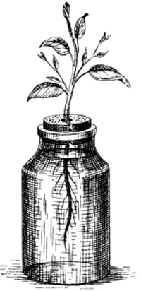
Fig. 19.—Plant growing in a nutritive solution
of salts. The bottle should be covered with a
roll of paper to keep out the light.
The work of the roots.—These experiments show that water—of which a large proportion of a plant consists—and the mineral constituents of its food (dissolved in the soil-water) are taken up from the soil by the roots. In ordinary soil the rootlets spread out on all sides, dividing and subdividing, seeking for this very weak solution of mineral salts. Even when soil appears practically dry, a very thin film of moisture covers each little particle of earth, and the root hairs become closely applied to these little particles (Fig. 20), so that the water passes through their walls and gradually makes its way to the main root, the stem, and the leaves.
Roots sometimes perform other duties in addition to those of fixing the [Pg 31] plant in the soil and providing it with water and mineral food. It is usual, for example, for biennial plants—which produce flowers and seeds in their second year, and then die—to take in much more food during their first season than they require at the time, and to store up the surplus in readiness for the great effort of the second year. These reserve materials are often stored in the roots, which then become swollen and fleshy, like those of the turnip, radish, and carrot.
1. Plants contain much carbon.—Char a stick, and notice the black charcoal which is formed. Charcoal is an impure form of carbon.
2. Carbon dioxide gas is formed when wood burns.—Fasten a shaving or a splinter of wood on a piece of wire, light it, and lower it into a clean glass jar. When the wood has burned for a few seconds take it out, and pour a little clear lime-water into the jar. The lime-water turns milky. Similarly, pour a little lime-water into a jar in which nothing has been burning, and notice that it remains clear. There is evidently a difference in the nature of the air of the two jars. The difference is caused by the burning of the wood, during which some of the carbon unites with the oxygen of the air in the jar, forming an invisible gas, called carbon dioxide. Carbon dioxide can always be detected by the milkiness it causes in clear lime-water.
3. Carbon dioxide present in ordinary air.—Pour some clear lime-water into a blue saucer and let it stand exposed to the air for half an hour, then examine it. A white scum has formed on the surface of the lime-water. Stir with a glass rod; the solution becomes milky. The scum and the milkiness are produced by the union of the lime with carbon dioxide from the air. Carbon (in the form of carbon dioxide) is therefore present in the air.
4. No carbon in the food solution.—Examine again the list of [Pg 32] elements (p. 27), which compose the mineral salts which have been found to replace satisfactorily the food which a plant obtains from the soil. There is no carbon in it. A plant evidently does not depend on the soil for the carbonaceous part of its food. From what other source can a plant obtain its carbon? Carbon has just been proved to be present in the air. Does the plant obtain its carbon from the air?
5. Green leaves contain starch after exposure to sunlight.—Take a green leaf from a plant which has been exposed to the sunlight, boil the leaf in water for a minute or two to kill it. Then put it in methylated spirit until the leaf-green is dissolved out. When the leaf is bleached rinse it in water, then put it into a dilute solution of iodine (p. 2) and notice that it becomes blue or purplish brown. The formation of this colour proves the presence of starch in the leaf.
6. Starch contains carbon.—Char a piece of laundry starch and observe the charcoal formed.
7. Green leaves do not contain starch after being left in the dark for 24 hours.—Keep a leafy plant in the dark for 24 hours and then test a leaf as in the previous experiment. No starch can be detected. Put the plant in the sunlight for an hour or two and test another leaf. It contains starch. Plainly, starch is only formed in leaves if they are exposed to light, and any starch previously present disappears when the plant is kept in the dark.
8. A green plant kept in air from which the carbon dioxide has been removed will not form starch in its leaves.—Obtain a large glass bottle, such as those used by confectioners, and fit it with a cork or india-rubber stopper through which passes a glass tube bent[5] as in Fig. 21. Care should be taken to make all the joints tight, and it may be necessary to soak the cork in melted paraffin to ensure this. Pack the bend of the tube loosely with pieces of soda lime, and in the bottle place a small jar containing lumps or a strong solution of caustic soda. When the apparatus is ready, place in the bottle a small plant or a leafy twig in water (fuchsia answers very well), which has been kept in the dark for 24 hours. The caustic soda in the jar very [Pg 33] soon absorbs all the carbon dioxide which is present, and the soda lime in the bend of the tube prevents any carbon dioxide from getting into the bottle from the outside air. Place the jar in bright sunlight for a few hours and then test a leaf for starch. None can be detected. It is plain that one of the carbonaceous food stuffs—starch—is not formed in the leaves of plants unless the plant is grown (in the light) in air containing carbon dioxide.
9. Seedlings are at first independent of light.—Germinate pea or bean seeds in wet sand or sawdust in the dark. Notice that for some time the seedlings grow almost as well as when in the light. Soon, however, the stem becomes long, weak and straggling, and the leaves are pale in colour, even if the plant is supplied with mineral food.
10. The formation of sugar in a germinating pea-seed.—Take up a pea-seedling when the stem is one or two inches long, and chew the partly shrunken cotyledons. Notice the slightly sweet taste. Contrast this with the taste of an ungerminated seed.
A plant contains much carbon.—When a piece of wood or other part of a plant is strongly heated it first blackens or chars, showing the presence of a large proportion of charcoal or impure carbon. On continued heating, this carbon “burns away.” In the process of burning it unites with some of the oxygen of the air, and forms a colourless, invisible gas known as carbon dioxide. Though this substance cannot be seen, its presence can be easily detected by means of clear lime-water, which, when exposed to the gas, absorbs it and becomes milky owing to the formation of a white precipitate of chalk. If, for example, a splinter of wood is burnt in a glass jar, and a little clear lime-water is immediately afterwards poured into the jar and shaken up, a milkiness at once proclaims the presence of carbon dioxide.
The air contains carbon.—If lime-water is poured into a blue [Pg 34] saucer and left exposed to the air for half an hour, a white scum of chalk is seen to have formed on its surface, showing that carbon dioxide is present in the atmosphere. It is important that the student should realise this presence of carbon—as invisible carbon dioxide gas—in the air. Although the proportion is very small, amounting to only 3 parts of carbon dioxide in 10,000 parts of fresh country air, it is of incalculable importance to plants, and indirectly to ourselves and all other animals.
A green plant obtains its carbon from the air.—Since the parts of a plant contain much carbon, and the food which a plant obtains from the soil need not contain any carbon; while the air, on the other hand, does contain carbon, it seems likely that a plant obtains its carbonaceous food from the air. This surmise is confirmed by experiments. One of the most easily recognisable of plant products containing carbon is starch, for it yields a very characteristic blue, or purplish-brown, colour when treated with iodine solution. By means of this test starch can easily be proved to be present in the green leaves of a plant which has been exposed to the air and sunlight. The leaf is first killed by being boiled in water for a minute or two, and then its green colouring matter is dissolved out by immersion in alcohol (methylated spirit). The bleached leaf is rinsed in water and then put in iodine solution, and the blue or purplish-brown colour which is formed shows the presence of starch. There is a marked difference when a leaf, which has been kept in the dark for twenty-four hours, is similarly tested. In this case no starch can be detected.
One compound of carbon—i.e. starch—may thus be recognised easily; and if we found that a leaf made no starch when supplied only with air from which the carbon dioxide had been removed, this fact would be strong evidence in favour of the conclusion that a green plant obtains [Pg 35] its carbon from the carbon dioxide of the air. To test this, a large bottle is fitted, by means of a tightly fitting cork or stopper, with a tube containing lumps of soda lime, a substance which eagerly absorbs carbon dioxide from air. A small jar of caustic soda is placed inside the bottle (Fig. 21). A green plant or a leafy twig, which has been kept for twenty-four hours in the dark to free it from starch, is then put in the bottle, and the whole exposed to sunlight for a few hours. At the end of this time it is found, on testing the leaves, that no starch has been formed. By this, and other experiments, botanists have proved that green plants obtain all their carbon from the carbon dioxide of the air, and that sunlight is indispensable for the process. We shall examine this question more fully when we study leaves (Chapter III.).
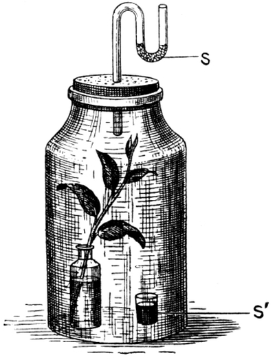
Fig. 21.—Experiment to prove that leaves do not make starch
unless the air with which they are supplied contains carbon
dioxide. S, tube containing lumps of soda lime;
S′, jar containing a solution of caustic soda.
The carbonaceous food of a young seedling.—Just as a bean or pea seedling is for a time independent of an outside supply of mineral food—its roots needing only to be supplied with water—so there is enough carbonaceous food also stored up in the seed to satisfy for some time the needs of the growing plant—the stored starch being gradually changed into sugar and absorbed. For this reason a young seedling will live healthily in the dark. When, however, the seed food is exhausted, or nearly so, the plant draws upon the store of carbon, which is present as carbon dioxide in the air, for a renewal of the starch and allied substances which are necessary to it. As it cannot make use of [Pg 36] this atmospheric carbon in the dark, it must henceforth be supplied with sunlight or it will not thrive. Plants kept in the dark after their seed-food is exhausted are pale in colour and unhealthy. Their stems grow long and straggling (Fig. 22), but are usually too weak to stand upright.
EXERCISES ON CHAPTER II.
1. Make experiments to discover the effects, upon seedlings growing in a nutritive solution (p. 27), of each of the following modifications in the composition of the solution: (a) omit the sodium chloride; (b) omit the potassium nitrate; (c) omit the compound of iron; (d) omit the magnesium sulphate; (e) substitute sodium nitrate for potassium nitrate.
2. Explain how it is that a green plant cannot carry on its nutrition in darkness.(1892)
3. What part of its food does a green plant obtain from the air? In what form, and under what conditions, is it taken in? (1889)
4. Describe the method of water cultures, and give the general results of a set of experiments. (1898)
5. Give experimental proof that green plants require to be fed with combined nitrogen. (1897)
6. What are the necessary conditions for the formation of starch in a plant? Mention experiments which support your statements. (1896)
7. Explain the influence of light on a growing plant. Illustrate your answer by reference to the changes in a ripening and germinating bean. (King’s Schol. 1903)
8. How can it be proved experimentally that a green plant draws some, but not the whole, of its nourishment from the air? (1904)
1. The shapes.—Make a collection of the leaves of a large number of different plants, for example, elm, beech, lime, oak, birch, ash, blackberry, pine, yew, horse chestnut, rose, holly, woodsorrel, grass. Lay each in turn flat in your notebook and trace the shape of the leaf blade by passing the point of your pencil round the edge. Measure the length and greatest width; write down these dimensions. Is the greatest width at, above, or below, the middle of the leaf blade?
Most of the leaves are flattened green plates. Those of the pine and yew are long and needle-shaped. Do you know any other leaves like these?
2. The veins.—What enables the leaf to keep stretched out? Turn it over and notice the “veins” on the lower side. Do they act like the ribs of an umbrella? Fill in the positions of the main veins in your drawings. Are the veins parallel to each other in any of your leaves? Write a list of as many leaves as you can find which have parallel veins.
3. Skeleton leaves.—Put some leaves in a saucer with a little soft water, and allow them to rot. Clean away the soft stuff from time to time by gently brushing the leaves with an old tooth-brush. Notice the “skeleton” which remains. Skeleton leaves may be made much more quickly by soaking the leaves for some time in a weak solution of bleaching powder. Wash them well before drying. [Pg 38]
4. The colour.—Is the green deeper on the upper or the lower surface of a leaf? Which surface usually receives more light?
5. The apex.—In how many of your leaves is the apex (a) pointed, (b) blunt, (c) rounded?
6. The margin.—Examine the edge or margin of each leaf. In how many is it (a) quite plain, (b) hairy, (c) wavy, (d) saw-edged, (e) doubly saw-edged, (f) spiny? Do you find spines on holly leaves which are so high on the tree as to be out of the reach of cattle? What is the use of the spines?
7. Blackberry leaves.—Gather several leaves from a blackberry bush. Notice that in addition to the “saw cuts,” the margins of some are cut into slightly, while others are divided quite to the midrib, the leaf being thus cut into two or more leaflets. Select specimens which form a gradual series between the “simple” leaf and the “compound” leaf (consisting of three or five leaflets) and draw them.
8. The horse chestnut leaf.—Draw the compound leaf of the horse chestnut, and draw an even curved line joining the points of the leaflets. You can imagine that the compound leaf may have been formed by a leaf of this shape being cut into until it was divided into seven complete leaflets.
9. The rose leaf.—Draw an imaginary simple leaf such as may have been the original form from which the compound leaf of the rose was derived. Notice the difference in the arrangement of the main veins of the leaves of the horse chestnut and the rose. Does this account for the leaflets coming off at the sides of the midrib in the rose leaf, and springing from one point, like fingers from the palm of the hand, in the case of the horse chestnut?
10. Sycamore and ivy leaves.—If the large indentations in these leaves were continued to the midrib, would the compound leaves thus formed be of the type of the rose leaf or of that of the horse chestnut leaf?
11. The leaf-stalk.—What attaches the leaf-blade to the stem or branch of the plant? Can you see signs of the main veins joining the top of the leaf-stalk? Do you know any plant with the blades of the leaves fixed directly on the stem, i.e. without any leaf-stalks? [Pg 39]
12. Stipules.—Examine the rose leaf again and notice the two leaf-like outgrowths at the bottom of the stalk. These are called stipules. Make a list of as many leaves as you can find which have stipules. How many leaves can you find with a sheath at the bottom of the stalk?
13. The leaf of the sweet pea.—Notice the large stipules of this compound leaf (Fig. 28). What are the tendrils? Do you think they may be mainly the larger veins of the upper leaflets? Is the leaf of the type of the rose or of the horse chestnut leaf?
14. Other compound leaves.—Compare and contrast ash, lupine, woodsorrel, strawberry, and other compound leaves with those of the rose and horse chestnut.
A leaf.—The leaves of different plants vary much in size and shape, but in general a leaf is a thin, broad, and more or less oval blade of green colour, attached by a leaf stalk to the stem or branch. In some cases, however, the leaf stalk is absent and the blade is attached directly to the stem or branch.
The veins.—The leaf is kept taut by a number of branching ribs, somewhat as the silk of an opened umbrella is stretched tightly by the ribs. The ribs or “veins” of the leaf run beneath the skin, but are generally nearer the lower surface than the upper, and are easily seen when the leaf is turned over. If a leaf is allowed to rot in a little soft water, the skin and the soft green stuff of the interior decay and leave these veins as a white “skeleton” (Fig. 23). The process may be assisted by gently brushing the leaf from time to time. A skeleton leaf may be obtained still more quickly by putting the leaf in a weak solution of bleaching powder until the skin and interior are soft enough to be brushed away. Care should be taken to use a weak solution, or the veins also will be rotted. The skeleton should be well washed in water before drying.
The arrangement of the veins in a leaf varies widely, but it falls [Pg 40] broadly into two classes, according as the main veins run parallel or nearly parallel to each other (Fig. 24), or form a less regular network (Fig. 23). The venation of a leaf is curiously associated with the number of cotyledons possessed by the seedling; for nearly all dicotyledons (p. 23) have net-veined leaves, while the leaves of monocotyledons are almost invariably parallel-veined. Careful drawings of several typical leaves should be made, and the principal veins indicated on them.
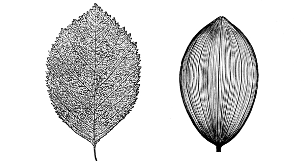
Fig. 23.—Net-veined leaf of |
Fig. 24.—Parallel venation of |
The shapes of leaves.—Although the blade of a leaf is most commonly flattened, and roughly oval in outline, there are several exceptions. The leaves of pine, spruce, larch, and yew are needle-shaped; those of grasses (Figs. 102-110) are very long in proportion to their width; while the leaves of many moorland plants are rolled up into hollow cylinders. There is some reason—could we find it—for every such variation, and the significance of some of these shapes will be referred to later (p. 47). When the dimensions of leaves are carefully measured, the proportion of the length to the width [Pg 41] will be found to vary much in the leaves of different plants, but will be found to be pretty constant for the same sort of plant. This holds good, too, for the position of the greatest width (e.g. at, above, or below, the middle of the blade), the form of the apex of the blade (blunt, pointed, spiny, or rounded), the nature of the margin (smooth and “entire,” hairy, saw-edged, doubly saw-edged, lobed, etc.), and the extent and positions of the larger indentations. Thus, while any particular elm leaf (Fig. 124) is probably slightly different from every other elm leaf in the world, it resembles every other elm leaf more than it does any leaf from any other plant than an elm. No leaf of this shape ever grows on an oak tree or a sycamore. Thus, in spite of minor variations there is a wonderful conformity to type, and the student will find that by carefully examining the shape, venation, margin, apex, etc., of all his leaves, and above all by drawing them, he will soon be able to recognise them at sight. It is by doing this and noticing in each case the methods of folding and arrangement of young leaves in the bud that it may be possible in the future to explain some variations which are at present not understood. It has already been seen that the peculiar forms of the leaves known as cotyledons are associated to some extent with the shape and size of the seeds containing them, and with the amount, if any, of the food stored in them.
Simple and compound leaves.—Blackberry leaves (Fig. 76) well repay close examination. Some of the leaves on the bush will be found to be simple—having one blade only on the leaf stalk. Here and there, however, a leaf may be discovered which is so deeply cut into along one side, that it is almost completely divided into two leaflets; and other leaves will easily be found which consist of three or five leaflets, much resembling the leaves of the rose (Fig. 25), a near relative of the blackberry. Here, then, we have a plant which produces simple or [Pg 42] compound leaves according to its needs. It seems as if the blackberry were still trying, as an experiment, a device which the rose tree has found so advantageous as to have adopted for good. Some other plants, the ash, for example, have compound leaves broadly similar to the rose leaf—the leaflets springing in pairs from the sides of the midrib.
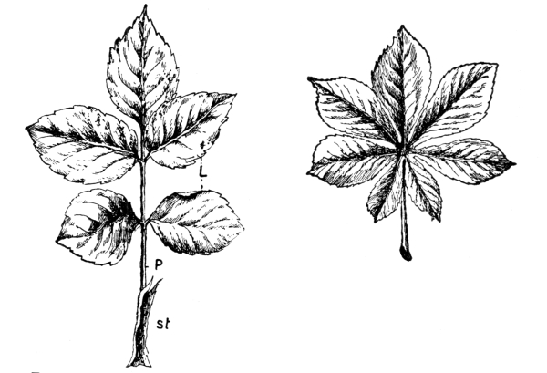
Fig. 25.—Compound leaf of the Rose. |
Fig. 26.—Compound leaf of the Horse Chestnut. (× ⅙) |
The compound leaf of the horse chestnut (Fig. 26) is of a different type, for the seven leaflets all arise from one point; and the leaves of the lupine (Fig. 9) are arranged on the same plan. When the venation is examined, the reason for this becomes plain. In these cases the main veins all diverge from the top of the leaf stalk; whereas in the rose and ash the midrib gives rise to side ribs in pairs. The leaflets are naturally arranged so that one of the larger veins shall support each. The next question arising is, “What causes the differences in the methods of branching of the midrib?” At present this is a mystery. Compound leaves consisting of three leaflets are found in woodsorrel, strawberry (Fig. 50), clover, etc. [Pg 43]
Intermediate leaves.—The ivy (Fig. 27) and sycamore (Fig. 33) have leaves which seem intermediate between truly simple and truly compound leaves. From the arrangement of the veins it is seen that they approach the horse chestnut type more than that of the rose. On the other hand, if the deep indentations of the oak leaf (Fig. 113) were carried to the midrib, the simple leaf would be divided into leaflets arranged, somewhat like those of the rose or ash leaf, along the sides of the midrib.
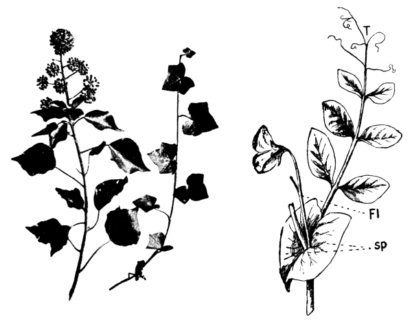
Fig. 27.—Ivy. (× ¹⁄₁₀) |
Fig. 28.—Compound leaf of Pea. |
Stipules.—At the bases of many leaf stalks, close to the stem, are leaf-like outgrowths called stipules. They are well seen in the rose (Fig. 25) and pea (Fig. 28). Some leaves have a sheath at the bottom of the stalk, partially enclosing the stem.
Tendrils.—The pea also affords an interesting case of leaflets [Pg 44] being modified to do special work. Here the upper leaflets seem to have remained undeveloped except for their main veins, and these have acquired a remarkable power of twining round suitable objects and so supporting the stem. Many other plants have tendrils, but these are not always modified leaflets.
1. Opposite leaves.—Examine a deadnettle plant (Fig. 92). Do the leaves come off the stem haphazard? How many come off at each level? Are both leaves on the same side of the stem, or opposite each other? Are the two leaves at the next level above just over these, or do the directions cross? Do the leaves get as much light or more than they would if each pair were just over the pair next below? How many other plants do you know which have leaves arranged in this manner? Examine leafy twigs of sycamore, horse chestnut (Fig. 40), and ash. Are the stalks of the lower leaves of these twigs of the same length as those of the upper ones? Is it any advantage for the lower leaves to have longer leaf stalks?
2. Box leaves.—What is the arrangement in the box? Examine particularly the young leaves near the end of the twig. Are the lower ones twisted? Can you suggest a reason for this twisting? Can you find any twigs in which no twisting has taken place? Are these untwisted twigs so placed that they are equally exposed to the light on all sides?
3. Alternate leaves.—Examine the arrangement of the leaves on a wallflower stem. They come off alternately, each springing from a rib on the stem. How many ribs are there? Look at the bottom of the stem, where the leaves have fallen off, and notice that each has left a scar. Mark one of the scars with your pencil and then count how many scars you pass before coming to another on the same rib. How many times do you wind round the stem in doing this? You pass five leaves and wind spirally round the stem twice. This is always the case in the wallflower.
Examine leafy twigs of oak and pear trees. Here, too, the leaves are alternate, and every sixth leaf is above the first, and a line [Pg 45] joining all the leaf-bases or scars between the first and sixth leaves would wind spirally round the stem twice.
What is the arrangement in the elm and lime?
4. Leaves which form a rosette.—Examine plants of primrose (Fig. 81) and daisy. The leaves in these cases spring from close to the ground and form a rosette. Notice that the bottom of the leaf blade is much narrower than the upper part. Is any saving of material obtained by this arrangement?
5. The position of branches and buds.—Look in the upper angle between a leaf and the stem in all your specimens. This angle is called the axil of the leaf. Can you see a bud in the axil of the leaf? Can you find that a bud or a side branch ever arises in any other position? (The former positions of fallen leaves are marked by scars.)
To the beginner in nature-study leaves seem in the majority of cases to be arranged on the stem of a plant in a haphazard and confusing manner, and it is only after very careful observation that a definite order and regularity is seen to be always maintained.
Nodes and internodes.—The level at which a leaf springs from the stem is called a node (Lat. nodus, a knot), and the length between two consecutive nodes is called an internode (Lat. inter, between).
Opposite leaves.—It is best to begin the study of leaf-arrangement by examining some such plant as the deadnettle (Fig. 92). The leaves come off in pairs: two at the same level, set opposite to each other. The next pair above or below springs from the stem in a direction at right angles to the first—a device which allows the leaves to get a more equal share of light than if each pair were placed directly over the next below.
A similar arrangement is adopted by various other plants, including the horse chestnut (Fig. 40), sycamore (Fig. 34), box, privet, etc., but in some instances it is disguised. Box [Pg 46] twigs afford an interesting example of this. Those twigs which are equally, or almost equally, illuminated on all sides, have their leaves arranged in pairs at right angles to each other like those of the deadnettle. Some twigs, however, receive the light from one direction only, and in these cases the leaves turn themselves until they face the light; so that at a casual glance the pairs of leaves seem to lie all in the same plane. One only needs to examine the end of the twig, where the leaves are just unfolding, to see that the arrangement is really in pairs alternately at right angles. In the case of the privet the efforts of the leaves to face the light often cause the stem itself to be twisted between the leaf-levels.
The alternate, or spiral, arrangement.—Perhaps the commonest leaf-arrangement is one in which only one leaf is given off at any particular node, the next leaf being a little further round the stem, and so on. As a result, an ink line or a piece of thread joining the leaf-bases would wind spirally round the stem. In the case of the wallflower, oak, pear, and many others, such a line would wind spirally twice round the stem before coming to a leaf vertically above the first, and in so doing it would pass five leaves. This may be shortly described as the ²/₅ arrangement. A less common one is ⅜, where in winding spirally round the stem 3 times, 8 leaves would be passed.
Efforts of leaves to obtain light.—It would be difficult to imagine any order of insertion which would secure a more equal distribution of light to each leaf than the spiral arrangement; but here also cases of leaves twisting in order to face the light are not uncommon. Lime leaves very often turn for this reason, so as to lie in almost the same plane—adopting a device similar to that of the box and privet described above. Further, the lime leaves arrange themselves at such angles that there is very little overlapping. Elm [Pg 47] twigs also often exhibit similar instances of a mutual accommodation of leaves to each other’s light supply.
The lower leaves on a horse chestnut twig have longer stalks than those nearer the end (Fig. 40). This enables the leaves to stand well out to the light and escape the overshadowing of those above.
The positions of branches.—A branch of the stem of a flowering plant always arises as a bud in the upper angle between a leaf and the stem. This position is called by botanists the axil of the leaf, from the Latin word axilla, the arm-pit. Clearly, then, the arrangement of the branches is primarily dependent upon that of the leaves, and we shall, for example, never find “opposite” branches on a tree which bears its leaves on the “alternate” system. It is easy to notice, however, that not all the buds develop into branches. In other words there are many buds which remain dormant, and the final arrangement of the branches is often somewhat irregular on this account. But wherever an ordinary bud or a branch occurs, we may be perfectly sure that there was once a leaf immediately below, even if the leaf-scar can no longer be seen.
Economy of leaf surface.—All these things seem to indicate that a good supply of light is of the greatest importance to leaves, and this conclusion is supported by the fact that leaves are usually either narrow or actually cut away in places where the light cannot reach them. The leaves of the daisy and of the primrose (Fig. 81), for example, all spring from nearly the same point, and form a rosette. Evidently there would be a certain amount of overlapping at the leaf-bases, unless the blades there were very narrow, as they are. Again, the greatly-indented leaves of the ivy are often arranged so that a point of one leaf fits over an indentation of another—a beautiful example of plant economy. [Pg 48]
1. In sunlight leaves make starch.—Expts. 5, 7, and 8 (Sec. 6) have already proved (a) that leaves of a plant growing in ordinary air and exposed to the sunlight make starch; (b) that in the dark this starch somehow disappears; (c) that in air destitute of carbon dioxide leaves are unable to make starch even in sunlight.
2. The parts of a leaf which are not exposed to light do not make starch.—Keep a plant, say of tropæolum—or, if not convenient, a single leaf (Fig. 31)—in the dark for 24 hours to free the leaves from starch. Split a small cork and pin the halves on opposite sides of a leaf, and then expose the plant to bright sunlight for an hour or two. (If a single leaf is used let the end of the stalk dip into water.) Take off the cork, kill the leaf with boiling water, dissolve out the green colouring matter with methylated spirit, rinse, and test with iodine solution. The part from which the light was excluded remains bleached, and therefore contains no starch; while the rest of the leaf becomes blue or purplish brown owing to the presence of starch.
3. Parts of a leaf which are not green do not form starch in sunlight.—Take a variegated leaf from a plant (e.g. the variegated geranium or maple) which has been in bright sunlight for some hours. Apply the usual test for starch. The parts which were originally green contain starch; the originally white parts remain bleached.
4. Leaves supplied with carbon dioxide, and exposed to sunlight, give off oxygen gas.—(a) Take a bunch of fresh watercress or any green water-weed and put it in a beaker or glass jar. Cover the plant with an inverted funnel which is shorter than the beaker. Now fill the beaker with ordinary tap water or river water (not distilled water), so that the end of the neck of the funnel is covered. Completely fill a narrow test tube with water, close it with the thumb, and invert it over the neck of the funnel. If this has been done carefully the test tube will still be full of water. Expose the arrangement (Fig. 29) to bright sunlight, and notice the bubbles of gas which are given off from [Pg 49] the plant and collect at the top of the tube. When a few inches of gas have collected, raise the test tube, close it with the thumb whilst still under water, and hold it mouth upwards. In the meantime, light a splinter of wood with the other hand. When it is well burning, blow out the light, remove the thumb from the test tube, and plunge the glowing splinter into the gas. It bursts into flame again, showing that the gas is oxygen.
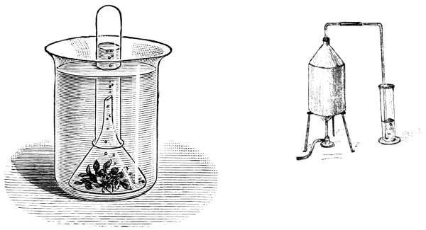
Fig. 29.—Experiment to prove that green leaves supplied with |
Fig. 30.—Experiment to prove that tap-water, or |
(b) Repeat the experiment, (i) placing the apparatus in the dark, (ii) without using any plant. No gas collects in the test tube.
(c) To show that the water used contains carbon dioxide in solution, completely fill a gallon can or a large flask with similar water, and attach a cork and a delivery tube which has also been filled with water—dipping the end of the tube into a little clear lime-water (Fig. 30). Put the same quantity of lime-water into another vessel for comparison, and then heat the can. Gas is given off, and as it bubbles through the lime-water the liquid is gradually turned milky.
5. Leaves wither in sunlight unless supplied with water.—(a) Cut off a leafy twig and leave it exposed to sunlight for an hour or two; notice the change in the appearance of the leaves.
(b) Put a similar twig in the dark for the same length of time; again notice the leaves. Is the difference due to a difference in light or to [Pg 50] one of heat? (c) To test this, keep, if possible, a similar twig in the dark in a warm place. Do the leaves wither as much as in (a)?
(d) Smear with vaseline the under surface of some of the leaves of such a twig and again expose to sunlight. Do the smeared leaves remain fresh longer than the others?
(e) Cut off the end of a twig with a sharp knife whilst it is under water, and leave it exposed to sunlight, dipping in water. The leaves remain fresh. How do you explain these differences?
6. In sunlight, leaves give off water.—Take a piece of cardboard about 4 in. square and make a small hole in the middle. Pass the end of a leafy twig through the hole and make up with wax any chinks between the twig and the card. Put the card on a tumbler containing water, so that the end of the twig dips under water; and invert on the card—covering the leafy end of the twig—a second tumbler which is clean and dry. Put the apparatus in the sunshine and notice the mistiness (or even visible drops of water) forming on the inside of the upper tumbler. Where does this moisture come from?
7. The skin of a leaf is perforated by little pores.—Dip a fresh laurel leaf into boiling water in a beaker or tumbler. Can you see bubbles of air escaping from the leaf? Are they to be seen on both surfaces of the leaf, or only on one? Which?
Examine both surfaces of box leaves with a strong lens, and try to see the little dots (pores) on the lower surface.
The student who has performed the experiments described in this section, and who has thought about the results obtained, cannot but have gained some insight into the main duties of leaves. The meaning of these results must now be discussed.
The formation of starch in leaves.—When the green leaves of a plant are exposed to sunlight in ordinary air—that is in air containing a certain proportion of carbon dioxide—the leaf forms starch in its interior, and the starch can be detected by applying the iodine test (p. 34). When part of a leaf is protected from the light, [Pg 51] as by pinning the halves of a split cork on opposite sides of it (Fig. 31), no starch is formed in the shaded parts, but only in the regions which are exposed to the light. Further, if a variegated leaf is treated in the same way, starch can be detected only in those parts of the leaf which were originally green; the parts which were white are free from starch. It is plain that it is the green colouring matter which puts the energy of the sunlight at the disposal of the leaf and enables it to manufacture starch.
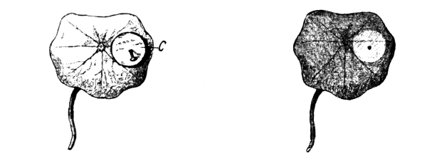
A B
Fig. 31.—A, Tropæolum leaf, on which have been pinned the halves of a split cork (C). (× ½.)
B, the same leaf tested for starch with
iodine solution, after exposure to sunlight for an hour.
The part shielded
from the light remains bleached; the rest of the leaf has turned blue.
At least three conditions are therefore necessary for the formation of starch in leaves: (1) the green colouring matter; (2) sunlight; (3) carbon dioxide.
Oxygen is liberated when leaves form starch.—Carbon dioxide gas, which has been seen to be indispensable for the manufacture of starch in leaves, consists of carbon, or charcoal, chemically united with the gas oxygen. The green-stuff of the interior of the leaf makes the starch by causing this carbon to combine with water which has come up from the roots, but it returns to the air the unnecessary oxygen. Water plants, the leaves of which are not directly exposed to the air, use carbon dioxide which the water has dissolved from the air. They [Pg 52] also give off the surplus oxygen, after fixing the carbon. This is the explanation of the bubbles of gas which, in sunlight, are often seen rising from the plants in an aquarium. By such an arrangement as is described in Expt. 9, 4, this evolved gas can be collected, and proved to be oxygen.
The use of water to the leaves.—It is common knowledge that if a twig is allowed to become dry its leaves hang limply and wither; but if the twig is allowed to dip into water the leaves will keep fresh and crisp for a considerable time. This necessity for supplying the twig with water seems to indicate that leaves give off water, and that this is so may be proved by a few simple experiments. Two tumblers may be arranged as in Fig. 32: separated by a card through which passes the end of a leafy twig. The end of the twig dips into water in the lower tumbler. In order to prevent water vapour from passing from the lower tumbler to the upper, the chinks between the twig and the card are sealed with paraffin wax.
When this arrangement is placed in the sunlight, a dew soon collects on the inside of the inverted upper tumbler. This water must have been given off in the form of vapour from the leaves. That the loss of water from leaves is due rather to the light than to the heat of the sunshine may be shown by keeping leafy twigs in the dark. The leaves keep fresh much longer than when placed in the light, even if they are kept in as warm a place.
The pores of the leaf-surface.—An ordinary leaf remains fresh much longer if its lower surface is smeared with vaseline. The [Pg 53] explanation of this lies in the fact that the waterproof skin of a leaf is perforated by a multitude of little pores, especially on the lower surface. In most leaves, indeed, the pores are confined to the lower surface. Smearing the surface with vaseline blocks up these pores and thus prevents the escape of water vapour from the interior of the leaf.
These little mouths (known as stomata)[6] open in the light and close in the dark. During the daytime, therefore, the air (containing its small proportion of carbon dioxide) has free access to the interior of the leaf through the stomata, and, on the other hand, any water which the leaf does not require can escape in the form of vapour. A leaf requires water not only because all its mineral food (p. 29) is brought to it dissolved in water, but also because water as well as carbon dioxide is required for the manufacture of the starch and other plant-foods.
How plant-food is distributed.—The water which comes up from the roots is distributed to the various parts of the leaf through the veins. These are therefore not only supports, which stretch out the soft leaf-stuff to the light and air, but they also form a very complete network of irrigating channels or water pipes. Further, the starch and other foods which a leaf makes are drained off into the stem through other minute channels which are bound up with the water pipes. The starch, for example, is changed into a kind of sugar which dissolves in water and drains away. From the stem, the food solutions are distributed to all the parts where growth is taking place. [Pg 54]
EXERCISES ON CHAPTER III.
1. Make drawings of as many cases as possible of economy of leaf surface.
2. Grow various plants, e.g. Tropæolum, Geranium, Fuchsia, Mustard, etc., in the window, and notice the effect which the direction of the light has upon the positions of the leaves.
3. Smear with vaseline the lower surfaces of various growing leaves, and on the following day test the leaves for starch, comparing each with an unsmeared leaf from the same plant.
4. How is the transpiration of water from a green leaf effected and controlled? Discuss the uses of transpiration. (1897)
5. Put the same quantity of water into each of two similar test-tubes, and let the end of a leafy twig dip into one. Weigh the tubes, place them together in the sun for an hour, weigh again, and estimate roughly the weight of water lost by one square inch of leaf surface per hour. Compare various plants in this respect. Repeat the experiments, (a) in a moderate light, (b) in the dark.
6. Make a list of plants in which the leaves are so arranged as (a) to conduct rain-water towards the base of the main stem, (b) to cause rain-water to fall to the ground from the outside of the foliage. Try to discover whether the difference has any relation to the arrangement of the roots.
7. Under what conditions can plants use carbon dioxide as a source of food? Mention experimental and other proofs of the principal statements made. (1905)
8. What part of its food does a green plant obtain from the air? In what form and under what conditions is it taken in? (King’s Scholarship, 1905)
1. A typical bud.—Split a cabbage, or a lettuce “heart,” down the middle, and observe how the leaves are arranged round the conical end of the stalk. The leaves which are fixed lowest on the stalk are the largest, and they cover the outside of the bud. The leaves are smaller and smaller as they are fixed nearer and nearer the end of the stalk, until round the tip they are almost too small to be recognised as leaves. A bud is the tip or “growing point” of a stem or branch, together with the young leaves which crowd round it. Draw the section.
2. A sycamore twig.—Look in the axils (p. 47) of the leaves of a sycamore twig in summer or autumn, and notice the buds. Can you see any buds on the lower parts of the twig—which bore leaves twelve months or longer ago? Did these also arise in the axils of leaves? How can you be sure they did? Tie a piece of string, or tape, round this twig and look at it often, making a note of every change which you see in it.
3. The fall of the leaf.—On what date did you first notice that the leaves of the sycamore were falling? Did the first leaves fall from the twigs at, or near, the ends of the branches, or lower down near the trunk? Examine the scar left by the first leaf which falls from your twig. What is its shape? What is the meaning of the dark dots on the scar? Gently take off the leaf nearest to the scar. Does it come off easily? Are there any dark dots on this scar? Can you see anything on [Pg 56] the end of the leaf stalk corresponding to the dots? Do you think the dots are the ends of the food pipes referred to on p. 53?
Make a drawing of a fallen leaf and write down on the drawing at the proper place, the colour of that part of the leaf, or better still, colour the drawing with water-colour paints. Collect good specimens of the autumn leaves of as many other trees as possible, and make coloured drawings of them.
On what date did you first notice that all the leaves were shed? Make similar notes for other trees.
4. The structure of a sycamore bud.—In winter take off a sycamore twig which has a big bud at the end, and examine it. The bud is clothed with overlapping scales of a pale green colour. Make a drawing of the bud, double the size, and then take off the scales one by one and count them. Look with a lens at the upper end of one of the largest scales. Can you see any trace of a rudimentary leaf blade? When the last scale is removed there remains on the end of the twig a tiny tuft of delicate green leaves surrounded by a little down. Count them, and notice how each is folded up fanwise. In some buds you may find a bunch of green flowers in the middle. These buds are larger than those containing leaves only.
Cut a bud across with a sharp knife, and examine the cut surface with a lens to see more clearly how the scales and young foliage leaves are arranged. Draw what you see.
5. How the bud bursts.—In spring watch your twig very carefully to be sure you do not miss any stage of the opening of the buds. Make drawings every day or two of the end bud, when it has begun to unfold. Notice how the scales fall off—those on the outside first—and how the foliage leaves push open the upper scales and come out. Watch the way in which each leaf opens its folds and spreads itself out flat.
The buds may be made to open earlier by cutting off a twig about two or three feet long and keeping it in water in a warm room.
6. Starch is stored up in the twigs.—Cut off a twig and pour a drop of iodine solution on the cut end. What does the blue colour indicate? [Pg 57]
7. The growth of the bud.—Watch your bud growing, and notice that the tip of the twig—which was surrounded by the young leaves—elongates so that each pair of leaves is soon separated from the next pair. Notice the rings of scars which the scales left when they fell off.
8. A year’s growth.—Look down the twig until you find the rings or scars which were left last spring, when the scales fell off the expanding terminal bud. A year ago the end of the twig was at this point, so that the length between one such set of rings and the next marks a year’s growth.
9. Side branches.—The buds on the length formed last summer but one may have grown out into side twigs. Try to find the leaf scars below each of these side twigs.
10. A horse chestnut twig and its buds.—Examine in the same way a horse chestnut twig and trace its history. The buds are larger than those of the sycamore, and each is covered with a shining, sticky layer of resin. What do you think is the use of the resin? Put a bud in water, and when you take it out notice how the water runs off and leaves the bud dry.
Pull one of the terminal buds to pieces. The scales will come apart more easily if the bud is soaked for some time in methylated spirit, to dissolve out the resin. Notice the thick layer of down inside the scales. What do you think is its use? Scrape the down gently, and carefully clean it away from the little foliage leaves in the middle. See how the leaflets of each leaf are folded. In some terminal buds you may also find a little pink flower-spray.
Tie a piece of string round a growing twig, so that you can recognise it, and watch all the stages of the expansion of the terminal bud, the unfolding of the leaves, and the elongation of the tip (Figs. 37, 39 and 40). Cut off other twigs two or three feet long, in February or early March, and keep them in water in a warm room.
11. Other buds.—Examine the buds of the beech, lilac, violet, dock, fern, etc., and make drawings showing (1) how the leaves are arranged with regard to each other, and (2) how each leaf is folded or rolled. As a rule these points can be easily made out by examining with a lens the cut surface of a bud which has been cut across with a sharp [Pg 58] knife; but the buds should also be examined at frequent intervals when they are unfolding.
A typical bud.—An excellent idea of the structure of a typical bud can be obtained by splitting down the middle an ordinary cabbage, or a lettuce “heart,” and examining the manner in which the leaves crowd round and cover the conical end of the stalk. Around the tip or “growing point” the leaves are extremely small and tender. They are overlapped by slightly larger leaves, which spring from the stalk a little lower down. These in their turn are covered by still larger leaves, inserted at a yet lower level, and so on—the largest and oldest leaves folding over the smaller and more recently formed. The growing point of a stem, or of a branch of a stem, surrounded by a cluster of immature leaves, is called a bud.
The history of a sycamore twig.—The student who would know the meaning of the various marks and scars on the surface of a twig, should select one and follow carefully for a year the fate of the buds which it bears. It is convenient to begin by studying a twig on a sycamore tree. It may be marked for ease of recognition by tying a piece of tape on it. If several students are working, each should write his name or number on a luggage-label and fix this to his twig.
[Pg 59] The positions of the buds.—In summer the younger twigs of the sycamore tree are covered with large five-pointed leaves (Fig. 33). The leaves come off in pairs, each pair being at right angles to the pair next above or below. Every leaf is engaged throughout the day in building up—by means of the green stuff in its interior—starch and other foods (p. 50), and in giving off excess water, in the form of invisible vapour, through its stomata (p. 53). In the axil (p. 47) of each leaf is a little bud, called from its position an axillary bud (Fig. 41, B), and at the very tip of the twig is a larger terminal bud.
Autumn colours and the fall of the leaf.—As the summer wanes, the soil becomes colder, and the chilled roots lose much of their power of absorbing moisture. It is plain that if the leaves continued giving off water when no fresh supplies were forthcoming the tree would suffer. How is the danger to be met? Starch and other foods have already been stored up in quantity sufficient to supply the needs of the winter and the early spring. The leaves have finished their work, and one by one they fall off. But this does not take place until careful preparation has been made. Their green colouring matter breaks up; the part which may still be useful to the plant drains into the stem, leaving little heaps of yellow grains in the leaves. Or a special colouring matter may be formed, which, united in various ways with the materials of the dying leaf, gives the warm shades of red, orange, and purple which make the woods so beautiful in autumn. When all is ready, a layer of cork (Fig. 41, C) forms at the junction of the leaf stalk and the twig so that no raw wound may be left; the leaf-base splits across, just above the cork layer, and the leaf flutters to the ground, there to rot and make rich leaf mould.
The leaf scars.—The former position of each leaf is now marked [Pg 60] by a curved scar (l.s. Fig. 34), and a row of brown dots (v.b.) in the scar still shows where the food-pipes bent outwards from the twig into the leaf. A line (a) stretches across and joins the two scars of each pair.
The buds.—Just above every scar is the bud which arose in the axil of the fallen leaf. It is covered with overlapping scales of light green colour. At the end of the twig is a single terminal bud, similar in appearance to the axillary buds, but of larger size. It is instructive to take the terminal bud of a twig to pieces. The outer scales are tough and green, while the inner ones are thinner and have a beautiful silvery appearance. Usually there are fourteen scales. Each is long and narrow and bears at its upper end the rudiment of a leaf blade, which cannot usually be seen well without a lens. The scale is really a leaf which has been arrested in its development.
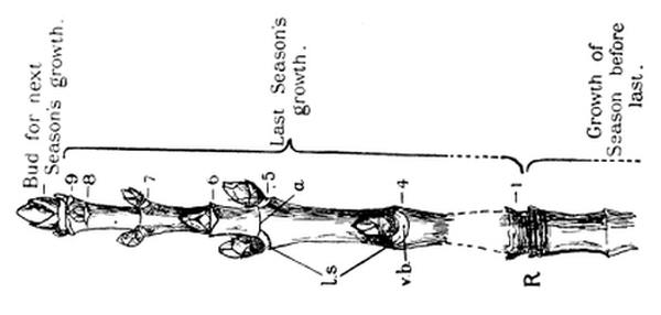
Fig. 34.—Sycamore twig in winter. l.s.,
leaf-scars; v.b., ends of food-pipes;
R, rings of scars left by
scales of last winter’s terminal bud.
(Slightly reduced.)
When all the scales have been removed, there remains a tiny tuft of delicate green foliage leaves surrounded by a little down. Each leaf is folded fanwise, both for convenience of packing, and to protect its tender tissues from the cold and the damp when the bud is expanding. The complicated folding of these leaves is well shown in Fig. 35, [Pg 61] which is a magnified sketch of a cross section through a sycamore bud. The large sheaths surrounding the young leaves are four of the overlapping scales. In a large terminal bud a bunch of green flowers may often be found.
The bursting of the buds.—About the middle of April the tree wakens to new life. The stored food makes its way to the terminal buds; invigorated by the rich sugary sap the young leaves swell and push forward, burst apart the scales, and open out their folds to the light and air, as if eager to get to work at the earliest possible moment. The scales fall off to the ground, leaving close-set rings of scars; the growing point elongates, and new leaves—which were indistinguishable in the bud—grow out and expand. During the summer, food is plentiful, and a little bud appears in the axil of every leaf. Only with the autumn does the activity of the tree slow down. Except for some two pairs at the tip, the newest leaves now remain stunted. They form scales and close round the tender growing point protectingly, in readiness for the winter.
If necessary, the axillary buds could have behaved as the terminal bud did, in which case they would have grown out into side twigs. They usually remain small, however, until next year (Fig. 36), for the leaves are so busy making food, and the terminal bud is so busy growing [Pg 62] in length, that no energy can be spared for their further development. If any accident had befallen the terminal growing point, one or more of the axillary buds would have grown out into side twigs. Gardeners take advantage of such reserve buds when they clip off the ends of twigs to make a plant grow “bushy.”
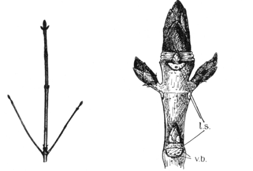
Fig. 37.—Horse Chestnut twig in winter. |
Fig. 38.—The terminal part of a Horse Chestnut twig in winter. |
A year’s growth.—When the bud scales drop off they leave, as we have seen, a series of closely-set rings of scars. The distance between one set of rings and the next (as at R., Fig. 34), therefore represents a year’s growth. The student should get a twig two or three feet long and find out for himself, by examining the marks on it, what the twig did last year, two years ago, and three years ago. With care he will be able to say definitely in which year any fairly recent side-twig began to grow out from its bud.
A horse chestnut twig.—A winter twig of horse chestnut (Fig. 37) [Pg 63] is very similar in its general features to what we have seen in the sycamore. The buds are in pairs at right angles to each other, and below each bud is a large corky leaf scar (l.s. Fig. 38), with the positions of the former food-pipes marked by black dots (v.b.). These buds, however, are larger than those of the sycamore, and each is covered with a shining layer of resin, to keep out insects and the rain.
We may slip the end of a penknife under the bud scales and remove them one by one. The first few are thin and papery, and soaked in resin. Those next inside are woody and much thicker. Next comes a layer of papery scales, inside that a coat of cottony down, then another soft papery layer, and lastly a thick pad of down. When we carefully scrape away this down, we find—warm and cosy in its midst—a tuft of little objects with a most quaint resemblance to hands clad in woollen gloves. We remove one of these, and on scraping it gently with a knife we see that the “hand” has seven fingers; and it presently becomes clear that each finger is a tiny green leaflet folded on itself, and that the hand is a young leaf. If we take off these gloved leaves in turn one by one, we find as we proceed that they become smaller, until they are almost too small to be distinguished in their fluffy nest. And when all the down and the baby leaves are scraped away, the tender growing point of the twig is left alone at the summit of a little cone, with steps showing where the leaves were.
Had the twig been left undisturbed on the tree, the bud would have awakened in spring and begun to grow (Fig. 39).[7] The scales and the down would have been shed, leaving only the rings of scars as a memento of the winter sleep; the growing point would have pushed on and on, lengthening perhaps a foot or more in three weeks; [Pg 64] the leaves would have opened their bright green fingers to the spring air, and begun their work (Fig. 40), only to cease when in the autumn they too dropped off, and the new buds tucked themselves up in their beds to go to sleep. The leaves of the horse chestnut fall off, as do those of the sycamore, owing to a “separation-layer” arising at the base of the leaf stalk (Fig. 41), and in this case each leaflet also becomes separately detached in the same way.
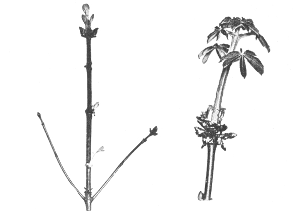
Fig. 39.—Later stage of the Horse Chestnut |
Fig. 40.—The later development of the terminal |
Other forms of buds.—Surrounding the young silky-fringed leaves of the beech bud are several crimson membranous scales which are really the stipules (p. 43) of undeveloped leaves. The thin, soft parts of the leaf blade are sharply pleated (Fig. 42) between the side veins which spring from the midrib. [Pg 65]
The way in which the branching of a tree depends on the buds is well shown in a lilac. The terminal bud does not usually develop, so that each of the two lateral buds just below grows out into a branch, producing the characteristic “forking.” In the lilac bud every gradation between scales and ordinary foliage leaves may be seen.
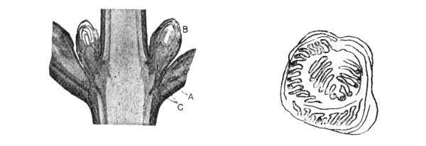
Fig. 41.—Longitudinal section of twig of Sycamore. |
Fig. 42.—Cross section through leaf-bud |
In the violet bud the two margins of the leaf are rolled inwards towards the midrib; while in the dock they are rolled backwards.
Young fern leaves (Fig. 146) are not folded from side to side like the examples referred to above, but are rolled into a tight coil from apex to base. It is the upper surface of the leaf (frond) which is to the inside. As the leaf grows, the coil straightens out.
EXERCISES ON CHAPTER IV.
1. Where and when are the buds of common English trees formed? (1901)
2. Show, by describing and drawing one example, that the branch of a tree may preserve a record of past seasons in the bark. (1901)
3. Draw an unopened bud of sycamore. Of what parts is it composed, and how are the parts arranged? (1901)
4. What can be seen inside a large bud of horse chestnut? On what part of the branch are the largest buds to be found? (1898) [Pg 66]
5. Upon what does the method of branching of a tree depend? Give examples.
6. Examine pollarded trees; what is the cause of their peculiarities?
7. Examine buds of various trees in spring, and try to discover in which cases the bud scales are (a) modifications of entire leaves, (b) modified leaf stalks, and (c) modified stipules.
8. Examine and describe the method of protection, during the summer, of the axillary buds of the plane.
9. Take a poplar shoot during late summer, and examine the corky separation-layer forming at the base of each leaf. Write an account of its use, and make drawings of its appearance.
10. The “heart” of a cabbage or lettuce is of lighter colour, sweeter taste, and more tender texture than the external leaves. How do you explain these differences? (King’s Schol., 1902)
11. Describe and explain the effect of clipping a privet hedge.
12. Give an account of the changes in appearance in any common leaf during the whole period of its growth. Explain briefly what part the leaf plays in the life of the plant. (Certificate, 1904)
13. Explain precisely how you would decide whether a given specimen consisted of (a) one compound leaf, or (b) a twig bearing several simple leaves.
1. The shapes of stems.—Cut across a deadnettle stem and a wallflower stem and examine the shape of the sections. The former is square, the latter is five-ribbed. Is there any relation between the form of the stem and the arrangement of the leaves?
2. The “bleeding” of stems.—Cut through the lower part of a scarlet-runner plant in spring. Can you see any water escaping from that part of the stem still in the ground? Similarly, cut back a sunflower stem when it is from ½ to 1 inch thick. Does the water exude from all parts of the cut surface equally, or does it come from definite channels? To see this better, dry the cut end with blotting paper and examine the surface with a lens. “Bleeding” is best seen in vine stems; if a young vine is available, cut it back in spring and observe the large escape of water.
3. The water-current travels along definite channels.—Colour some water with red ink and put in it the stalks of cut white flowers such as snowdrops or narcissi. The stalks and flowers soon become veined with red. Cut the stalks across and see that the strands in the interior are also coloured. The coloured water has evidently travelled along definite channels.
4. The food-channels in a soft stem.—(a) Take a stout piece of the stem of a deadnettle, including three nodes (p. 45), and slit it [Pg 68] down in a line between the end pairs of leaves. Then boil the stem in water until the internal tissue is soft. Take the stem out and carefully scrape away all the soft material until the woody strands can be well seen. These are the food-channels. Notice their arrangement in the stem, and the manner in which they run out to the leaves.
(b) Similarly, examine the strands in a piece of sunflower stem, and in an old cabbage stalk.
5. The path of the water-current in a woody stem.—Take a leafy twig of elder or laurel and, from the part of the shoot below the leaves, remove a ring of about an inch of the bark and the soft tissues which lie beneath it, so as to expose the wood. Put the end of the twig (below the ring) in water. The leaves remain fresh and crisp, showing that the water travels either along the wood or the pith, or along both.
6. The water travels along the wood.—Put a similar leafy twig of elder dipping in water which has been coloured by red ink. Expose to sunlight, and after an hour or two cut open the twig and observe which parts are coloured. The bark and the soft tissues between it and the wood are not coloured. The wood is stained red. The pith is not coloured.
7. The path of the leaf-made food.—(a) Take a leafy branch of a tree—e.g. willow—and near the bottom remove a ring of the bark and the soft tissues lying between bark and wood. Put the twig in water, so that the ringed part and a few inches above shall be immersed. After a time roots are produced above the cut. If any arise from the stripped part they are few in number and much shorter than those above.
(b) Place a similar but uninjured twig in water, and notice that the new roots are produced at the end. What is the reason for the difference?
The duties of stems.—No one can study leaves without being impressed by the great importance to the plant of the work which they do. Even the casual observation that year after year thousands of fresh leaves make their appearance would indicate this. And when it is learned that throughout the day every leaf is busily engaged in [Pg 69] decomposing carbon dioxide, and joining up the carbon with the water and mineral compounds which have come up from the root—thus forming sugars, starch and a host of other valuable substances—some general idea is obtained that the trunk and spreading boughs of a tree may, after all, be of minor importance, and may exist mainly to help the leaves to perform their duties as perfectly as possible.
This is the true view to take of the stem and its branches; their duties are (1) to bear the leaves and spread them out, so that these will receive as much sunshine and fresh air as possible; (2) to supply the leaves with the water and mineral substances which they require for their work; and (3) to receive from the leaves and distribute to the rest of the plant the food materials which the leaves have prepared.
A stem, together with the leaves and branches which it bears, is called a shoot.
It will be found that almost all the variations in the structure and habits of stems are connected with the arrangements of the leaves. For example, the weight of the mass of leaves borne by a forest tree is very great, and they offer a great resistance to the wind; the trunk and its branches, therefore, are correspondingly strong and stout. Again, when a stem is ribbed or ridged in any particular manner it will generally be found that the ridges have a definite relation to the leaves and to their points of insertion on the stem. This has been seen (p. 44) to be the case in the stem of the wallflower.
The tip of a stem or branch is occupied by a bud. As the tip elongates (Fig. 40), the outer leaves of the bud become separated by internodes, and new leaves continually arise around the growing point.
The path of the water in the stem.—It may easily be shown that water travels up the stem to the leaves. It is common knowledge that [Pg 70] slightly withered leaves or flowers become fresh and crisp again if the twig or stalk is put in water. If the stalks of white flowers—say snowdrops or narcissi—are put into water coloured with red ink, the flowers become veined with red, and, if the stalks are cut into, red strands may be seen. This experiment not only shows that water has travelled up the stalk, but it also shows that the water has passed along definite channels. When a vine stem is cut back in the spring, the water wells out rapidly from the end of that part of the stalk which is still in the ground. This bleeding, as it is called, may also be seen, though to a smaller extent, in stems of sunflower, scarlet-runner, and other plants. By drying the cut surface with blotting paper, and then examining it with a lens, it may be seen that the water escapes from the ends of certain little tubes; and it is possible in a boiled or rotting stem (Expt. 11. 4) to follow the distribution of the strands containing these tubes. The strands run along the length of the stem, outside the pith. Cross strands connect the main ones, chiefly at the leaf-levels (nodes); and other strands run out into the leaves to form the veins.
The tubes which convey the water current are in that part of the strand which is nearest the pith, and they become woody. Thus, if the current year’s growth of, say, a horse chestnut or an elder twig is cut across, a thin ring of wood is seen surrounding the soft pith. Outside the wood are the softer tissues, surrounded by the bark. A ring of the bark and soft tissues beneath it may be entirely removed from a growing twig, leaving the wood exposed, but the leaves above remain crisp and fresh, showing that this treatment has not interfered with their water supply. The water, therefore, travels along either the wood or the pith. That the wood and not the pith conducts the water may be shown by putting a leafy twig in water coloured with red ink. Only the wood is stained. [Pg 71]
The path of the food made by the leaves.—The plant food which the leaves make is drained off into the stem, and distributed to the parts where growth is taking place. This food travels in the soft tissue—called the bast—which lies below the bark. This fact can be shown indirectly by removing a ring of bast from the lower part of a branch, say of willow, and putting the branch into water. When at length the cutting puts out roots, these spring from the top of the ring. If any spring from the stripped part they are markedly smaller, and fewer in number. In an uninjured cutting, which is of course supplied with prepared food along all its length by the bast vessels, such roots spring from the cut end. The supply of leaf-made food can also be cut off by ligaturing a twig below the leaves, as by twisting a wire or cord tightly round it. In such a case growth usually ceases in the part of the twig below the strangled part, while the upper part of the twig, to which the leaf-made food is now restricted, grows much more luxuriantly than before. Gardeners often produce unusually fine fruits by ligaturing the lower parts of the twigs on which the fruits are ripening.
1. The formation of wood.—(i.) In summer take a horse chestnut twig of three or four years’ growth. Cut through it with a sharp knife at the following places, and trim the cut ends flat:
Make a drawing of what you see in each case:—In (a) the twig is covered on the outside by a green skin. In the middle is the soft pith. Between the two is a ring of separate strands. [Pg 72] In (b) and (c) the strands have joined up, and a distinct, though thin, layer of wood surrounds the pith. Bast and other soft tissues lie between the wood and the bark. (d) has two layers of wood. (e) has three layers of wood.
(ii.) Split each length longitudinally. Why is it easier to do this than to cut the twig across? In which direction does the grain of the wood run? Make out in each piece the pith, strands, or layers of wood, bast, etc., and skin or bark. You can tear off the bast in ribbon-like shreds. See how the strands run out into the young leaves. Cut lengthwise through the junction between the main twig and any side twigs, and notice that corresponding parts are continuous.
2. The strength of a grass stem.—Notice the relatively enormous strength of a straw and other grass stems.
Burn a straw and observe the tube of mineral matter which is left behind. Examine a piece of bamboo; is it hollow or solid?
Woody stems.—To enable them to bear the weight of the leaves and branches, and to withstand the force of the wind, the stems of plants are strengthened in various ways. Most commonly this is effected by the formation of wood in the walls of the water-vessels.
Even in succulent stems, such as that of the sunflower, the strands of vessels are stiffened by the long and narrow wood pipes which run along them; and when the strands join up to form a complete cylinder a very strong column is the result. Engineers make use of the same device, knowing that the same amount of material will bear a far greater stress when made into a hollow cylinder than it will in any other form.
The thickening of woody stems.—In dicotyledons (p. 23) and gymnosperms (p. 163) the cylinder, which is formed by the joining-up of the conducting strands, consists of three layers. The innermost of these is the wood, and the outermost is bast. Between them is a very delicate layer called cambium, which is [Pg 73] continually dividing and forming more wood on its inner side and more bast on its outer side. The wood is hard and resists pressure, while the bast is soft and is squeezed against the inside of the bark by the expanding wood (Fig. 43).
The formation of new wood and bast takes place vigorously during the summer, at the expense of the food which is manufactured by the leaves and travels along the bast to the active cambium. As autumn comes, the activity of the tree slows down, and the new wood is formed of closer texture. In winter the process stops altogether; but with the warmth and the plentiful food-supply of spring the formation of new, open-textured wood is resumed.
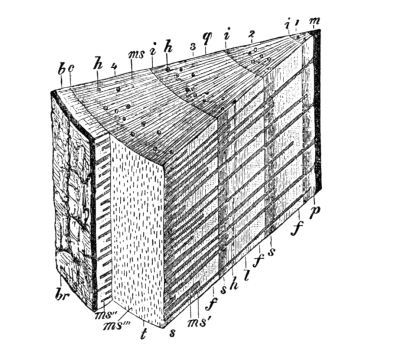
Fig. 43.—Portion of a four-year-old stem of the Pine, cut in winter. 1, 2, 3, 4, the four successive annual rings of the wood; b, bast; br, bark; c, cambium; f, spring wood; i, junction of wood of successive years; m, pith; ms, medullary rays; s, autumn wood. (× 4.)
This difference in texture between the autumn wood and the later spring wood is quite visible to the naked eye (Fig. 44), and gives rise to a series of annual rings, each of which represents a year’s growth. Thus, a cross cut through the four year old part of a branch (Fig. 43) shows four layers of wood, one of which was formed each year. The length formed last year has two layers of wood (if we look in summer), and the current year’s growth has one layer, all of which has been formed since spring.
The advantages of secondary thickening.—The formation of secondary wood and bast is very important. The new wood is required (1) to provide additional water-vessels to supply the demands of an [Pg 74] increasing number of leaves, and (2) to give the necessary increase of mechanical strength to the growing tree. Again, the increase in the quantity of food manufactured by the leaves makes a larger quantity of bast necessary for its distribution. Last of all, new tissues are required to take up the duties of those which are old and worn-out.
In forest trees, the central pith becomes almost obliterated and the old stem practically consists of wood, bast, and bark. The various annual rings, and the bast, of a woody stem are joined together by a number of radiating horizontal spokes called medullary rays (Fig. 43, ms, ms′, ms″, ms‴). These conduct water and food materials across the stem from layer to layer. The beautiful lines and patches called “silver grain,” which may be seen in oak furniture, consist of the medullary rays exposed in radial-longitudinal section.
Bark.—The very young stem is surrounded by a thin waterproof skin, perforated by stomata (p. 53) as the skin of a leaf is; and the part immediately below the skin possesses leaf-green, and can therefore decompose carbon dioxide as a leaf does (p. 51). An old stem, on the other hand, is covered by a layer of tough bark (br, Fig. 43), [Pg 75] which splits from time to time, owing to the stretching which is caused by the increasing thickness of the wood. The bark begins as a layer of cork, which forms on the outer side of the bast. The cork cuts off the food supply from all the external tissues, which die. Bark, therefore, consists of the cork and the dead layers outside it.
Grass stems.—The great strength of grass stems—so apparent when we try to bend a straw—is largely due to silica, the substance of which rock-crystal and ordinary sand are composed. When a straw is burnt, this remains as a hollow cylinder of mineral matter. The great strength of the cylindrical form has already been referred to. It is very well seen also in a bamboo stem, and anyone who has blunted the edge of his knife on a piece of bamboo will appreciate the additional hardness which is given by the presence of mineral matter.
1. Hooking stems.—Examine a bramble or a wild rose plant in a hedge. Why does it need to climb? How does it climb? Pull a branch and notice by what means it resists the pull. What is the shape of the prickles? Do they point upwards or downwards? On what parts of the plant are the prickles found? Notice how easily a prickle may be pushed off, sideways. Is it as easy to tear it off lengthways? Is the prickle a little branch, or merely an extension of the rind?
Contrast a prickle with the thorn of the hawthorn. The thorn does not come off easily, and it contains a woody core which is continuous with the wood of the branch. Cut lengthwise through the thorn and the branch which bears it to see this. The thorn is a short pointed branch; it arises in the axil of a leaf, and sometimes bears leaves itself.
2. The ivy.—Observe how the climbing stem of the ivy is attached to a wall or tree. It puts out a line of roots on its [Pg 76] shaded side. These roots give out a sticky fluid, which, on hardening, fixes them to the wall.
3. Twining stems.—Watch the growth of a convolvulus seedling. At first the young stem grows straight up, but soon the tip begins to move round and round. Try to find out how long it takes to describe one revolution. Put a long stick in the ground near the plant and notice how, when the revolving stem touches the stick, the spiral is henceforth described round the support and the stem consequently clings to it. Lay your watch face-upward, and notice whether the stem moves in the same direction as the hands (the “clockwise” direction), or in the opposite (“counter-clockwise”) direction.
Make similar observations on the hop, honeysuckle, and scarlet runner, and note the results.
4. Leaf climbers.—Examine a climbing tropœolum (often, though wrongly, called “nasturtium”). Which parts of the plant clasp the support? Watch a young plant coming up. Does the stem revolve before the leaf stalks come in contact with the support? Compare the clematis.
5. Tendril climbers.—Examine plants of sweet pea, bryony, vine, passion flower, and cucumber. Try to find out in each case which part has been modified to form the tendrils. Watch a plant day by day until a free tendril grasps a support, and notice how it becomes spirally coiled. Can you straighten the tendril by pulling, without leaving any kinks? Why? Is the spiral of the tendril continuous, or does it change its direction in the middle? Make a continuous spiral with wire, and notice the kinks formed when the wire is straightened by pulling the ends. Which is better for the plant in a gale of wind, a continuous or a reversed spiral? Why?
Notice the sucker-like tendrils of the Virginian creeper, and the way in which they fix the plant to the wall.
Plants whose stems are not strong enough to stand erect without support must adopt some special means of spreading out their leaves to the light and air. One of the commonest devices of such plants is that of climbing up other and stronger plants, walls, trellis-work, etc. [Pg 77]
Scramblers and climbers.—In the simplest cases the plant simply scrambles over other plants. Many brambles and roses are merely scramblers, but more often they are true climbers, weaving themselves among their neighbours by the help of hooked prickles (Fig. 76). The prickles point backwards, and therefore anchor the twigs firmly, as is very evident on trying to pull a branch out of the hedge. A prickle may easily be broken off by a side push, for it is merely an outgrowth of the rind, and does not contain any woody core. It is so attached, however, that it resists much greater force in the lengthwise direction—a manifest advantage to the plant.
[Pg 78] The differences between such a prickle and a thorn like that of the hawthorn should be carefully noticed. A thorn is really a little twig which has remained short and become pointed at the end. It has a core of wood which is continuous with the wood of the branch bearing it. That a thorn is really a little branch is shown by its origin in the axil of a leaf, and by its often giving rise to leaves and buds.
The stem of the ivy climbs by means of little roots, which it puts out on the side furthest from the light (Fig. 45). [Pg 79] These give out a sticky liquid which, on drying, cements them to the wall or tree.
Twining stems are much in advance of these. There seems to be something approaching intelligence in the manner in which the young stem of a hop, honeysuckle, or convolvulus, which at first grows straight up, begins to wander round and round, tracing a spiral path in the air until it touches a support. Then, however, as if the plant could feel, the movement below the point of contact stops; but the upper part of the stem still revolves and therefore twines round the support. The stems of the honeysuckle (Fig. 46) and the hop turn in the same direction as the hands of a clock. This is called the “clockwise” direction. On the other hand the convolvulus (Fig. 47) and most other twining stems are “counter-clockwise” climbers. The stem of the bittersweet revolves indifferently in either direction.
Sensitive clasping organs in their simplest form are seen in the twining leaf stalk of the clematis and tropœolum; the stem itself revolves as if to give its leaf stalks every opportunity of finding suitable supports. The leaf stalks seize these and twine round them.
Most wonderful of all climbing organs are the tendrils. They are well seen in Fig. 48. A part of the plant—sometimes a leaflet, as in the pea (Fig. 28); sometimes a branch, as in the passion flower; or a flower stalk, as in the vine—becomes modified into a thread, slight but strong. When the end of the thread touches and then twines round a support, the whole tendril forms itself into a spiral which, like a wire spring, draws the plant up to the support, and can yet lengthen and yield to the wind when necessary. In the middle of the tendril the direction of the spiral is reversed, so that the tendril can be straightened without being twisted. [Pg 80]
The tendrils of the Virginian creeper do not twine, but on meeting a wall they form round red suckers at the end, and attach the plant (Fig. 49).
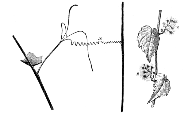
Fig. 48.—How a tendril grasps a support.
Fig. 49.—Virginian Creeper.
The spiral is reversed at x. R, R, stem tendrils. (× ½.)
1. A creeping stem.—Examine a plant of the ground ivy. Is the stem strong enough to stand upright? How does it spread out its leaves to the light and air? The stem grows along the ground, and at intervals it gives off a pair of leaves which grow upwards, and a tuft of roots which grow down to the ground.
2. A runner.—Is the “runner” of the strawberry of the same nature, i.e. is it a continuous stem like that of the ground ivy? When the plant is carefully examined, the creeping “stem” is seen to be a branch arising in the axil of a leaf. The branch runs along the ground for a little distance, and then roots itself and gives off a [Pg 81] number of leaves. In the axil of one of these another branch arises and runs on in the same direction. The same branch does not run on and on.
3. A stolon.—Follow carefully the underground part of the couch grass and make out its connection with the main shoot. It is a branch like the runner of the strawberry, which arises in the axil of a leaf, and extends only to the next shoot.
Compare the stolons of the cinquefoil.
4. A potato tuber.—Examine a potato tuber (the part which is eaten). Notice the “eyes.” These are buds, with scale leaves. Leaves never occur on roots, so that the potato must be an underground stem. Put a pin into every eye, and wind a thread round the tuber along the bases of the pins. It forms a spiral. Cut the potato into halves, and pour a drop of iodine solution on the cut surface. What is the meaning of the blue dots which at once make their appearance? Plant a potato in warm, moist earth, and when it has sprouted notice that each bud (eye) has given rise to a branch.
5. Bulbs.—Cut an onion or snowdrop bulb down the middle, and draw what you see, marking on your drawing the outer scale leaves, the swollen bases of last year’s leaves, the young leaves in the middle, the short, thickened stem, and the roots. Also cut other bulbs across and again draw. What is the similarity and what is the difference between these bulbs and such a bud as a cabbage?
Also examine hyacinth, tulip and daffodil bulbs. Put them in glasses with water touching their bases, and watch them grow. What do they live upon?
6. A crocus corm.—Obtain a few crocus “bulbs” in the early winter. Observe the tough outer tunic springing round the edge of a circular scar on the base. If there are any roots they come off from the scar. Take off the tunics from one “bulb” and observe the bud or buds at the top of the white mass inside. Other tunics cover the buds. Cut lengthwise through the mass of the “bulb” so as to bisect the largest bud. Separate the parts of the bud with a needle and notice (a) the thin outer leaves, (b) the young foliage leaves, (c) the flower-sheath and flower. Pour a drop of iodine solution on the cut white mass (the stem) below the bud. It turns blue. Why? [Pg 82]
What is the principal difference between a crocus “bulb” and the bulb of an onion, hyacinth, or tulip? A true bulb is mainly composed of swollen leaves or leaf bases; in the crocus the thick, rounded stem makes up most of the bulk. It is better, therefore, to speak of a crocus corm, to indicate the difference.
Plant the remaining corms and examine them at intervals for a year. Notice the formation of the roots, the lengthening of the buds, the formation of the flowers, the activity of the foliage leaves after flowering (why?), the withering of the roots and leaves in summer, and the growth of the enlarged base of the branch into next year’s corm.
Creeping stems.—Instead of climbing, many stems find that the best method of spreading out their leaves is to creep along or under the ground, and give off leaves and roots at intervals. Not only does this device prevent the leaves of one node from interfering with the light and air supply of those of the next, but the plant is continually coming in contact with a fresh lot of soil. The ground ivy is an instructive example of this method of growth. The stem creeps along the ground, and at every node it gives off a pair of leaves which grow upwards, and a tuft of roots which grow down into the ground.
The runner of the strawberry (Fig. 50) appears at the first glance to grow in a similar manner. As a matter of fact, however, the [Pg 83] apparent stem is a branch arising in the axil of one of the leaves of the last node. The branch runs along the ground and gives rise to a new shoot, and from this another branch, springing from the axil of a leaf, forms another runner. The same branch does not run on from shoot to shoot.
The stolon of the couch grass (Fig. 51) is somewhat similar. The erect stem of the plant is divided, as usual, into nodes or knots (from which the narrow, sheathing leaves arise) and internodes. Branches (stolons) spring in the axils of the lower leaves, turn downwards, and run on underneath the soil, taking root again at some distance from the parent plant.
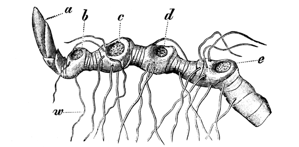
Fig. 52.—Creeping underground stem of
Solomon’s Seal. a, bud of next year’s aërial growth;
b, scar of
this year’s growth; c, d, e, scars of aërial growth of previous
years; w, roots. (× ¾.)
Underground stems.—Although the stem is usually that part of the axis of a plant which is above ground, there are many exceptions. The bracken fern, daisy, coltsfoot, Solomon’s seal (Fig. 52), and many [Pg 84] other plants have stems which are ordinarily buried in the ground, giving off leaves above and roots below. In some cases such underground stems become much swollen with stored food-material—manufactured by the leaves in excess of immediate requirements. In the potato, for example, certain underground branches of the stem store up starch to such an extent that their ends become fleshy, ovoid masses some inches in thickness (Fig. 53). The true nature of these tubers is revealed by the buds or “eyes” which spring upon them. The buds are arranged spirally—as may be seen by sticking a pin into each and joining up the pins with thread—and when the tubers are kept in a warm, moist place, each bud grows out into a new leafy shoot.
The structure of a bulb is easily made out in the onion, tulip (Fig. 54), hyacinth, or daffodil. When such a bulb is cut down the middle, it is seen to be mainly composed of leaves or leaf-bases, swollen with stored food. Inside these are the young leaves and the flower bud, which would have expanded next season; and on the outside are a few thin scale leaves. All these leaves spring from a fleshy button at the base, which gives off roots below. The button is the flattened stem. When a plant produces a bulb it will generally be found that it flowers either very early or very late in the season; that is, at a period which would not be very favourable for the work of the leaves. The flower (Fig. 55) is produced at the expense of the stored food in the bulb—made in excess of the requirements of the [Pg 85] previous season. After the plant has flowered, the new leaves work until they have made enough food for next season’s flower, and then they also die.
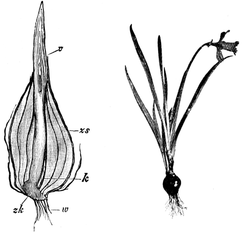
Fig. 54.—Longitudinal section of Tulip bulb.
Fig. 55.—Daffodil. (× ¹⁄₉.)
zk, modified stem; zs, scale leaves; v,
terminal bud; k, young bud; w, roots. (× 1.)
The so-called bulb of the crocus is technically known as a corm (Fig. 56). It differs from a true bulb in consisting mainly of a fleshy, rounded stem in which the surplus food made by last year’s leaves is stored up. The plant is thus able to flower early, without waiting for the new leaves to supply food. The swollen stem bears one or more buds, and the whole is surrounded by tough tunics of scales. When the corm begins to grow, roots are put out from the base, and the [Pg 86] flowers and—later—the leaves of the buds expand. The foliage leaves continue their work after flowering, and the food which they make accumulates in the base of the former bud, which becomes swollen to form the new corm for next year’s flower. The leaves then die down, their bases becoming the tunics of the new corm.
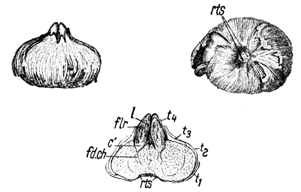
Fig. 56.—Crocus corm, seen from the side, from below, and in longitudinal section. c′, base of bud, which will grow into next year’s corm; fd.ch., food channel; flr, flower bud; l, young leaves; rts, roots; t₁, t₂, t₃, t₄, tunics. (× ½.)
EXERCISES ON CHAPTER V.
1. Mention experiments, which prove that organic substances are formed in the leaves, and distributed to other parts of a green flowering plant. By what channels are they distributed? (1895)
2. Draw a cross section through the stem of a flowering plant selected by yourself. Explain the uses of the chief things seen in the section. (1897)
3. Mention experiments or observations which show by what tissues water ascends to the leaves, and nutritive substance descends from the leaves. (1897)
4. By what tissues does water pass along the stem of a tree to the leaves? Give proofs of your statements. (1898)
5. Describe the effect of a tight ligature upon a growing hazel stem. (1898)
6. What proofs can be given that the stem of a tree draws nourishment from the leaves? (1898)
7. Show, by describing and drawing one example, that the branch of a tree may preserve a record of past seasons in its wood. (1901) [Pg 87]
8. Mention an experiment which shows that organic substance formed in the leaves travels down the stem outside the cambium. (1901)
9. Obtain thin sections (e.g. plane-shavings) of as many different kinds of wood as possible, and gum them into a book, writing under each section the name of the wood and the direction (transverse, radial-longitudinal, or tangential-longitudinal) of the section.
10. Make yourself familiar with the appearance and characters of the different kinds of timber used in carpentry and joinery.
11. Notice the light-brown spots on the bark of twigs of apple and horse chestnut. These are called lenticels; they are breathing-pores. In how many other trees can you find lenticels?
12. Observe whether an ivy stem puts out roots only where they can become fixed to a support, or indiscriminately.
13. Make a list of plants which you have observed to climb by twining stems, and note whether they are clockwise or counter-clockwise climbers.
14. Make careful drawings of all the tendrils you can find, and try to discover which part of the plant has been modified to form the tendril.
15. Gently stroke a tendril of the passion flower and write an account of any resulting movement.
16. Mention any three climbing plants which grow wild in this country, and explain in each case how the plant climbs. (1895)
17. What is the difference between (a) a thorn, (b) a leaf-spine, and (c) a prickle? Give examples. (1896)
18. Enumerate and briefly describe the principal varieties of tendrils, and explain how they act. (1897)
19. If a wire is fastened tightly about a growing branch of a common tree, and left for two or three years, what effect will be produced, and how can the effect be explained? (1904)
20. What processes of vegetable growth are accompanied by the presence of sugar? Give examples from plants within your own experience. (King’s Scholarship, 1904)
21. What are the chief uses of the vessels of a herbaceous stem? Mention observations and experiments in support of your statements. (1905)
22. How can you demonstrate experimentally that food substances, formed in the leaves of a tree, descend to the branches below? (1905)
I. The wallflower.—After noticing the general habit of growth of a wallflower plant (Fig. 57), and especially the shape and venation of the leaves, make out the following parts in one of its flowers. On the top of the flower-stalk (called the receptacle) are:
(a) Four small, narrow, purplish leaves, called sepals. The four sepals together constitute the calyx. Take off the sepals one by one. Notice that two opposite sepals are bulged out at their bases, forming pouches containing nectar. Try to get out a small drop of nectar on the point of a pencil and taste it.
(b) Four showy leaves arranged in the form of a Maltese cross, called petals. They are yellow, or red, or purplish in colour, are [Pg 89] delicately scented, and have beautiful velvety surfaces. The four petals together constitute the corolla. Take off the petals one by one.
(c) Six stamens, each consisting of a greenish stalk or filament surmounted by a yellow, boat-shaped body, called the anther. The anther is a four-chambered box containing an enormous number of tiny yellow grains called pollen grains. Two of the stamens are shorter, and are fixed at a lower level on the receptacle than the remaining four. Take off the stamens one by one.
(d) A central pistil, shaped somewhat like a slender bottle. At the top, where the cork would come in a real bottle, is the notched stigma, slightly sticky. The neck is called the style, and the part corresponding to the body of the bottle is the ovary. Tear open the ovary with a needle to see the ovules, which in an undisturbed flower would have become seeds.
Watch bees visiting flowers. Does each bee confine itself to one kind (species) of flower at each journey, or does it visit several kinds indiscriminately? Try to discover what the bees are doing. Avoid alarming them.
The work of flowers.—The roots, stem, and leaves of a plant do a great deal of work, but, as it is performed for the benefit of the plant itself, it is all, in a sense, selfish work. Plants, however, like animals, grow old in time, and at last die. If they are not to become extinct it is evident that they must devote part of their energies to producing new individuals, and to sending these forth into the world as well equipped as possible for the battle of life. This unselfish and self-sacrificing part of a plant’s life-work is called reproduction; in the higher plants it is carried out by flowers.
The structure of a wallflower blossom.—The flowers of different groups of plants vary greatly in structure, but a good general idea of the arrangement of the parts of a flower can be obtained by examining the blossoms of a wallflower plant (Fig. 58). Other flowers may afterwards be compared and contrasted. [Pg 90]
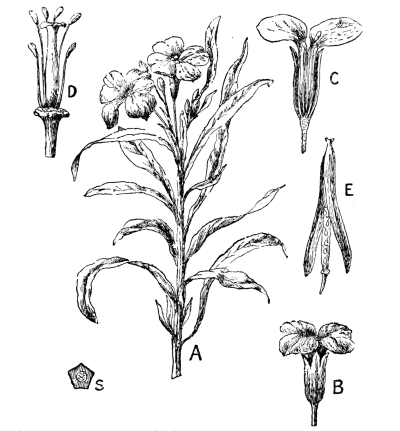
Fig. 58.—Wallflower. A, branch, bearing leaves and flowers; B, flower; C, longitudinal section of flower; D, stamens and pistil; E, fruit; S, transverse section of stem, (× ½.)
There are evidently at least eight leaves in the flower, but, unlike the green foliage leaves, these are not arranged spirally, but stand at nearly the same level on the end—called the receptacle—of the flower-stalk. The most external leaves are four in number, small, narrow, and purplish in colour. Each of these leaves is called a sepal, and the four sepals together constitute the calyx of the flower. Two opposite sepals are pouched at the base, forming pockets, in which a sugary fluid, called nectar, collects. Before the [Pg 91] bud opens, the calyx is the only part of the flower which is visible. It is probably developed in the wallflower solely for the protection of the more delicate structures within. Next, inside the sepals, placed alternately with them, and standing a little higher on the receptacle, are four showy leaves arranged in the form of a Maltese cross. These leaves are called petals, and the four petals together form the corolla. The petals are delicately scented, and their surfaces have a beautiful velvety sheen.
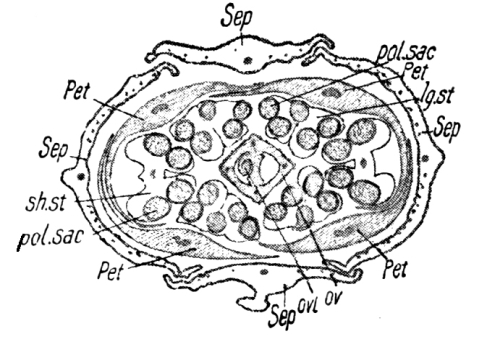
Fig. 59.—Cross section through a Wallflower bud. Sep, sepals; Pet, petals; lg.st., anther of a long stamen; sh. st., anther of a short stamen; pol.sac., pollen sacs; Ov, wall of ovary; Ovl, ovule. (× 8.)
When the sepals and petals are removed, there remain standing on the receptacle six stamens surrounding a centrally-placed pistil (Fig. 58, D). The stamens are the male part, and the pistil is the female part, of the flower. Each stamen consists of a greenish stalk or filament, surmounted by a yellow boat-shaped body called the anther. The anther is a box with four compartments (Fig. 59). When it is ripe, each compartment contains an enormous number of tiny, yellow grains called pollen grains; and when the anther bursts (as it does as soon as the flower opens) its inner face is covered by the yellow dust of the pollen.
The pistil bears a rough resemblance to a slender bottle, and consists of three distinct parts. The neck of the “bottle,” called the style, is short in the wallflower, and differs from an ordinary bottle-neck in being solid instead of tubular. At the top of the neck, where the cork would come in a real bottle, is a body called the stigma. The stigma of the wallflower pistil is hairy, notched, and slightly sticky, from the presence of a sugary solution which forms [Pg 92] upon it. The part of the pistil, corresponding to the body of the bottle, is the ovary. It contains four rows of little white ovules, which are destined to become seeds capable of growing up and forming new wallflower plants.
The relations of the parts of the flower are well seen in Fig. 59.
Fertilisation.—In order that an ovule may become a seed, its contents must mix with the contents of a pollen grain. The fusion of the two constitutes fertilisation. For fertilisation to take place, the pollen grain must first of all gain access to the stigma of the pistil. If this be prevented the flowers will wither without forming ripe seeds. (This may be proved easily by Expts. 18, 3 and 21, 2.) The sugary solution at the top of the stigma stimulates the pollen grains to growth, and each puts out a long tube which grows down the style. The living matter of the grains keeps near the tips of the tubes as these continue their journey down the style. At length the tubes enter the ovary and find the ovules. Each ovule has at one end a minute pore (the micropyle—p. 6), and a pollen tube finds this and enters it. The living matter of the pollen tube fuses with that of the ovule in the neighbourhood of the pore, and fertilisation is effected. It is now easy to understand that the comparatively insignificant stamens and pistil are the all-important parts of a flower.
How the wallflower advertises.—Botanists have proved that a flower produces more, and also better, seeds when it is fertilised by pollen from another flower of the same species. This is called cross fertilisation. The wallflower relies upon bees for the transference of the pollen from one flower to another; and it is solely to attract them that the petals are so delicately scented and brilliantly coloured, and that sweet nectar collects in the sepal-pouches. The gaily coloured petals are therefore advertisement placards which are [Pg 93] hung out to attract the attention of bees. A bee comes to a wallflower for the sake of both nectar and pollen—the “bee-bread.” As the bee thrusts its proboscis down between stamens and pistil in search of the sweet liquid in the pouches, its head is pretty certain to come in contact with, and to brush off, some of the pollen dust hanging loose on the inner faces of the anthers. When the bee flies off to another wallflower and continues its search for nectar, it almost invariably leaves some of the pollen, from the first flower, on the hairy and sticky stigma of the second.
In almost all cases when a flower is brightly coloured it depends upon the help of insects for cross fertilisation.
1. Shepherd’s purse.—Compare the shepherd’s purse (Fig. 60) with the wallflower. The flower is very much smaller, and white, but the parts have the same arrangement as in the wallflower, viz., four sepals, four petals arranged in the form of a cross, six stamens (two short and four long), and a central pistil, all arranged separately on the receptacle. Look down the plant, and notice that in the oldest (lowest) flowers, everything but the pistil has dropped off, and that this has become greatly enlarged to form a fruit. Cut some fruits open, both lengthwise and crosswise, and observe that each consists of two pocket-like chambers, separated by a thin partition on which the seeds are borne. Notice the manner in which the oldest fruits have opened naturally. [Pg 94]
2. Other relatives of the wallflower.—Compare also the flowers and fruits of the stock and candytuft, and also cress, mustard, radish (Fig. 61), cabbage, and turnip, which have been allowed to “run to seed.” Examine the roots of the turnip and radish.
Make a note of the earliest dates on which you see the above plants in flower.
The wallflower family.—The plants of the family to which the wallflower belongs are of very great importance to mankind; for while not one of them is poisonous, many are extremely valuable as food-crops. They are all dicotyledons; that is, their seeds contain two cotyledons or “makeshift leaves,” as has already (Chapter I.) been seen in the case of the mustard. The net-like venation of the leaves of the full-grown plant also indicates this. There is a great family likeness between the flowers of this group, and they are easily recognised (Fig. 61 a, c) by the cross-shaped corolla and the six stamens (two short and four long). All the parts of the flower—sepals, [Pg 95] petals, stamens, and pistil—are fixed separately on the top of the flower stalk or receptacle. The cross-arrangement of the petals has led to these plants being called Crucifers (cross-bearers). The shepherd’s purse (Fig. 60)—so-called from the shape of its fruit—is a common weed with small, white flowers. All stages of the flower may generally be found on the same plant. While at the top the buds may still be unopened, the flowers below have been fertilised (in this case generally self-fertilised); the sepals, petals, and stamens—having fulfilled their duties—have fallen off; the ovules have become seeds, and the ovule-box or ovary has become a seed-box or fruit, consisting of two bags separated by a partition which bears two rows of seeds on each side (Fig. 62).
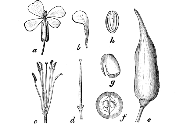
Fig. 61.—Wild Radish. a, flower (nat. size); b, petal; c, stamens and pistil (× 2); d, pistil (× 2); e, fruit (× 1); f, cross section of fruit; g and h, embryo (mag.)
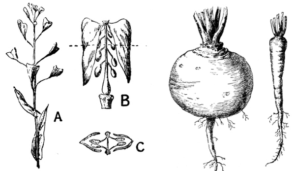
Fig. 62.—A,
Fruits of Shepherd’s Purse (× ½);Fig. 63.—Root of
Fig. 64.—Root
B, a single open fruit (mag.); C, cross section of B.
Turnip. (× ⅓.)of Radish. (× ½.)
Useful crucifers.—The turnip and the radish are largely cultivated for their roots (Figs. 63 and 64), and are then taken out of [Pg 96] the ground at the end of their first season. As these plants naturally flower in their second year of growth and then die, they are called biennials. The production of flowers and fruit is a great strain on a plant, and it is to prepare for the effort that the turnip and radish store so much food in their roots during the first year as to give them a globular and spindle shape respectively. A carrot is not a crucifer, but it also adopts this device.
The cabbage is grown for its leaves. Varieties of the cabbage are Brussels sprouts, broccoli, and cauliflower; it is the very small flower-buds of the last two which are eaten. Cress and white mustard are eaten in the seedling stage. The seeds of the black mustard are ground and eaten as a condiment.
1. The buttercup.—Notice the habit of growth, characters of the leaves, etc. Is the buttercup a dicotyledon? Make this observation with all flowering plants. (See, however, Chap. VIII., p. 163.)
In the flowers of a buttercup (Fig. 65) make out:
(a) The calyx of five green, separate sepals; they are the only parts to be seen in young, unopened buds. Take off the sepals of a fully-opened flower one by one.
(b) The corolla of five, golden-yellow, separate petals, alternate with the sepals. Notice the nectary—a little pocket—near the base of the upper surface of each petal. Take off the petals one by one and observe that they are fixed on the receptacle, a little higher than the sepals.
(c) The large number of separate stamens, inserted still higher on the receptacle.
(d) On the top of the receptacle the large number of separate, flask-shaped bodies, which together make up the pistil. Each of these is called a carpel.
Watch bees and other insects visiting buttercups and notice how they [Pg 97] stand on the flower to obtain the nectar from the nectaries. Does a bee on leaving go to another buttercup, or does it change to another kind of flower?
In a flower from which the sepals, petals, and stamens have fallen, notice the compound fruit (Fig. 66), consisting of ripened carpels. Open a carpel with a needle and pick out the single seed.
2. Other plants of the buttercup family.—Notice that in the anemone (Fig. 67) and marsh marigold (Fig. 68), the sepals appear to be absent (the three leaves under the anemone flower are not parts of the flower; they are called bracts). The apparent petals are really the sepals; it is the corolla which is absent. Observe the large number of stamens, and notice that the pistil consists of several separate carpels.
In what kind of ground have you seen these plants growing wild?
The buttercup family.—The buttercup (Fig. 65) and its relatives resemble the crucifers (1) in being dicotyledons (as is indicated (p. 40) by the venation of the leaves), and (2) in the fact that, of the parts which compose the flower, each group is arranged separately on the receptacle. For example, the stamens are not connected with either the calyx, the corolla or the pistil. Having noted these points of resemblance, however, we are met by some important differences. In the buttercup there are usually five sepals and five petals; there may be twenty or more stamens; and the pistil is not a single structure, but [Pg 98] consists of a number of separate parts, each of which is called a carpel, and contains a single ovule. The pistil of a crucifer consists of only two carpels; these are welded together into a single structure, and until the fruit ripens a slight notch in the stigma is the only external indication that the pistil is in two parts.
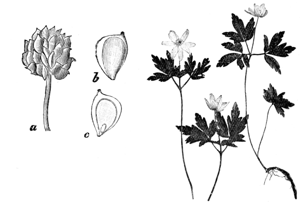
Fig. 66.—a, Compound fruit of Buttercup (× 2½);
Fig. 67.—Anemone. (× ⅓.)
b, a carpel (× 4); c,
carpel in longitudinal section (× 4).
Insects visit buttercups for the sake of nectar and pollen, and whilst creeping about the flower transfer pollen to the stigmas of the carpels. The insects may have brought some of this pollen from other flowers, and then cross-fertilisation is caused. If the pollen is derived from the same flower self-fertilisation is the result.
After fertilisation the ovules become seeds; the sepals, petals, and stamens drop off; and the carpels swell up, forming a dry compound fruit (Fig. 66), consisting of several nutlets.
Two other common plants of this family are the anemone (Fig. 67) and marsh marigold (Fig. 68). In neither of these cases has the [Pg 99] flower any petals, but the calyx has taken on the appearance of a corolla. In the marsh marigold it is large and yellow; in the anemone it is white or purple. The three green leaves immediately beneath the flower of the anemone are called bracts. They should not be mistaken for sepals.
The plants of the buttercup family are as generally poisonous as the crucifers are wholesome. Monkshood is especially poisonous, and its root has been mistaken, with fatal results, for that of horse-radish. It is often noticed that grazing cattle avoid the buttercups in a field. The bitter and disagreeable taste of the leaves is of course a valuable protection to the plant.
1. The garden pea.—Notice again the habit of the plant: its compound, net-veined leaves with large stipules, and its method of climbing by tendrils which are modified leaflets. Examine the flower and make out (a) the calyx of five united sepals; (b) the curiously shaped corolla. The large upper petal is called the [Pg 100] standard, the two at the side are the wings, and the lowest (really two locked together) is the keel; (c) the ten stamens. One (opposite the standard) is separate; the remaining nine have the lower parts of their filaments united to form a tube. Slit open the filament-tube and remove the stamens, noticing how they are attached to the other parts of the flower; (d) the pistil. Slit open the ovary and see the ovules in it. Watch the various stages of the formation of the fruit (pod).
Watch bees visiting the flowers. The insect alights on the wings, and its weight pulls them down and lowers the keel, bringing the stamens against the bee’s body.
Cut a complete flower down the middle with a sharp knife, and notice that calyx, corolla, and stamens seem not to be inserted separately on the receptacle, but to spring from a common base.
2. Other plants of the pea family.—Examine also bean, vetch [Pg 101] (Fig. 69), meadow vetchling (Fig. 70), clover (Fig. 71), laburnum (Fig. 73), and broom. Compare the habits of growth of the plants, and notice that they all have the same peculiar shape of flower. Dissect a flower of each. Notice that in laburnum and broom the ten stamens are all united. In clover the flowers are in heads. Notice how the leaflets of the clover plant close at sunset.
3. Fertilisation.—Dig up several red clover plants in early summer and pot them. Cover about half the plants with gauze, so fixed on wire frames that insects cannot get inside, and then put all the plants together where they will get plenty of sun. Water them regularly, and notice which plants ripen seed. How do you account for the differences?
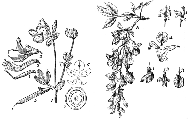
Fig. 72.—Bird’s-foot Trefoil. 1, flowering branch (× ⅔);
Fig. 73.—Flowering branch of Laburnum; st,
2, flower; 3, pistil and stamens; 4, pistil (× 1¹⁄₉); 5,
standard; w, wings; k, keel; 1, 2, 3, the flower
fruit (× ⅔); 6, corolla; a, standard; b, wings; c, keel; 7,
from different points of view. (× ½.)
diagram of flower.
The pea family.—Plants of the pea family are found in all quarters of the earth. They are of very diverse size and habit of growth; the laburnum, for example, is a tree; the gorse is a bush; the broad bean has a strong, erect, herbaceous stem; the pea is a weak-stemmed climbing plant; the clovers are small herbs with flowers forming “heads.” Most of the members of the family agree in having a “butterfly-shaped” corolla (Figs. 72 and 73), which consists of three well-marked parts, viz., a large standard, a pair of wings, and two closely-connected petals which form a boat-shaped keel. There are ten stamens, and the filaments of nine of these usually cohere to form a tube surrounding the ovary. In the laburnum, gorse, and a few others, all the ten stamens are united. When a bee visits the flower, in search of nectar, it alights on the “wings” of the flower, and its weight depresses these and pulls down the keel. The anthers of the stamens are so placed with respect to the keel that this results in a mass of pollen being scraped off the anthers and forced [Pg 103] out at the beak of the keel, or in the stamens being suddenly liberated and scattering pollen on the bee. The pollen sticks to the bee’s body, and some of it is almost certainly transferred to the stigma of the next flower visited. The quaint shape of the corolla is thus definitely adapted to the visits of insects; for the nectar is so placed that to obtain it the insect must carry off some of the pollen.
The calyx, corolla, and stamens are not obviously—as in the wallflower and buttercup—inserted separately on the receptacle, but seem to spring from a common base.
The fruit is a pod (Fig. 3), which opens when ripe along both margins and liberates the seeds. Laburnum seeds are poisonous, but the seeds of many other plants of the family (peas, beans, lentils, etc.) are valuable foods.
1. The wild rose (Fig. 75).—With a sharp knife cut vertically through the middle of a wild rose. Notice that the receptacle forms a deep cup, and that the carpels of the pistil are enclosed in the cup. From the edge of the cup spring the five sepals, five petals, and numerous stamens. What is the great difference between a rose and a buttercup?
Trace the formation of the succulent fruit or hip from the receptacle cup of the flower. Cut through a rose hip, and observe the ripened carpels in the interior.
2. The blackberry.—Similarly examine a blackberry flower (Fig. 76). This is still more like a buttercup, but—as in the rose and the pea—the calyx, corolla, and stamens seem to spring from a common base, and not to be inserted separately on the receptacle.
Trace the formation of the compound fruit, and notice that each part is like a little plum or cherry.
3. The cherry.—Similarly examine cherry blossom (Fig. 77). The pistil consists of one carpel, and is fixed at the bottom of the receptacle-cup, while the calyx, corolla, and stamens are fixed on the margin of the cup. Trace the origin of each part of the fruit.
Compare the plum and apricot. [Pg 104]
4. The apple and pear.—Cut vertically through an apple blossom (Fig. 74) or pear blossom (Fig. 78), and notice that the ovary is embedded in the receptacle, and that calyx, corolla, and stamens are fixed on the top of this. Cut across the ovary and see the five divisions (carpels).
The eatable part of the fruit is the swollen receptacle. Compare the hawthorn.
The wild rose.—The plants of the rose family are, most commonly, woody trees or shrubs. The leaves are provided with stipules in nearly all cases. The wild rose (Fig. 75) may be taken as a type of the group. It bears a superficial resemblance to a buttercup, but on dissection considerable difference in the arrangement of the parts is seen. In the rose, the receptacle is urn-shaped; from the margin of the urn spring the five sepals, five petals, and numerous [Pg 105] stamens; while inside the urn the separate carpels of the pistil are inserted. In the blackberry (Fig. 76), raspberry, and strawberry the receptacle is knob-shaped, and the carpels are arranged on the outside of the knob, somewhat as in the buttercup. Here again, however, the calyx, corolla, and stamens differ from those of the buttercup in seeming to arise from a common base.
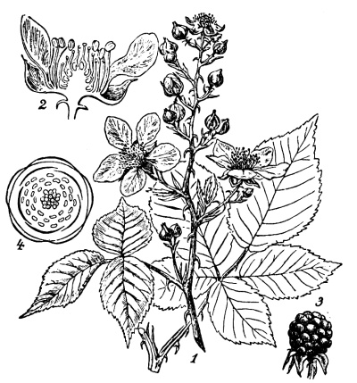
Fig. 76.—Blackberry. 1, flowering branch
(× ⅓);
2, longitudinal section of flower (× 1);
3, fruit (× ⅓); 4,
diagram of flower.
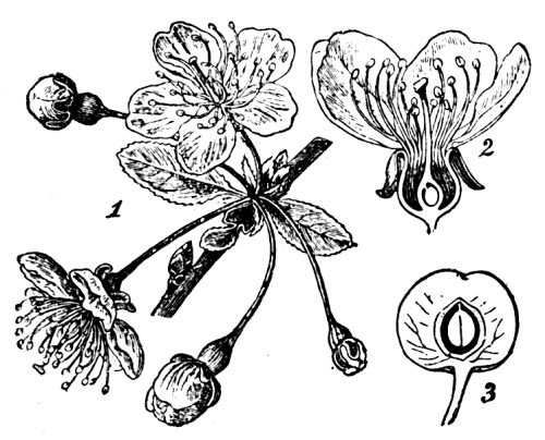
Fig. 77.—Cherry. 1, flowering branch (× ⅔);
2, longitudinal section of flower;
3, longitudinal section of fruit.
The differences between the rose, blackberry, raspberry, and strawberry are more marked when the pistil has become a fruit. The fleshy part of the rose hip is the urn-shaped receptacle which encloses the ripened carpels. In the case of the blackberry and raspberry the receptacle is dry, and is surrounded by the compound fruit (Fig. 76, 3) of the several bodies like little plums or cherries. The eatable part of the strawberry fruit (Fig. 144) is the swollen receptacle, on the outside [Pg 106] of which are the little yellow nutlets derived from the carpels of the flower.
In the cherry (Fig. 77), plum, and apricot the pistil consists of only one carpel, which is enclosed in the urn-like receptacle. After fertilisation, the greater part of the wall of the ovary becomes fleshy, and one of the two ovules contained in it becomes a seed. The “stone” is formed from the innermost part of the ovary wall.
The apple (Fig. 74) and pear (Fig. 78) have their five carpels embedded in the receptacle, and the rest of the flower stands on this part. The eatable portion of the fruit is the swollen receptacle. The hawthorn has usually only two carpels, and in the fruit the part derived from the receptacle becomes hard and horny. In other respects it is very similar to the apple.
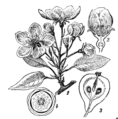
Fig. 78.—Pear. 1, flowering branch
(× ⅔);
2, longitudinal section of flower;
3, longitudinal section of
fruit;
4, diagram of flower.
The rose family is widely distributed, especially in temperate regions.
1. The poison hemlock.—Examine this plant (Fig. 79) very carefully, remembering that it is poisonous. Notice the general habit of growth; the characters of the sheathing, compound leaves; the hollow ribbed stem; and also the arrangement of the flowers, which is characteristic of the family. From the top of a main flower-stalk [Pg 107] several smaller stalks come off together, like the ribs of an umbrella. From the top of each of these spring the stalklets which bear the small white flowers. Notice the bracts at the points of origin of the stalks. Examine the flowers, and watch insects visiting them. Which insects are most commonly found on the flowers?
Compare the cow parsnip, the water hemlock, carrot, parsley, parsnip, and celery, carefully noting the points of resemblance and difference.
The parsley family.—The plants of this family may be recognised easily by the arrangement of the flowers. Several stalks spring together from the top of the main flower stalk and each of these again gives rise at its tip to a number of smaller stalks, at the ends of which the small flowers are borne (Figs. 79 and 80). The flowers are fertilised by the aid of insects, and as the nectar is on the surface [Pg 108] it is accessible to small insects such as flies, beetles, etc. The flowers are rendered more conspicuous by being placed close together. The stems are usually hollow, and the leaves are alternate, and generally compound, with sheathing bases. Many of the plants of this family are very poisonous, and such should be carefully distinguished and whenever possible exterminated.
The poison hemlock (Fig. 79) varies in height from two to seven feet. It has a hollow stem which is spotted with purple in the lower part, and when bruised the leaves give off a smell like that of mice. Cattle are often poisoned by eating the plant in hay, and children have been poisoned even by blowing whistles made from the stem. The water-hemlock is extremely poisonous. It grows along the sides of pools. The stem is hollow, and the leaflets of the compound leaves are finely toothed. The root is a cluster of fleshy swellings, and has unfortunately a rather pleasant taste. Other poisonous plants of the family are the water dropwort and the fool’s-parsley. Among the harmless and useful members of the group are celery (when cultivated), carrot, parsnip, and parsley.
1. The primrose.—Examine the habit (Fig. 81) of the plant, its underground stem, its spoon-shaped leaves—arranged in a rosette—and the manner in which the flowers spring from the stem. In the flower make out (a) the calyx, 5-pointed and with united sepals; (b) the corolla, consisting of 5 petals united below into a tube. Tear down the corolla-tube to see (c) the 5 stamens inserted on the corolla-tube. In some (“thrum-eyed”) flowers the anthers are at the top of the tube; in others (“pin-eyed”), they are halfway down; (d) the pistil, consisting of stigma, style, and ovary. In thrum-eyed flowers the style is short and the stigma is halfway down the corolla-tube; while in pin-eyed flowers the style is long and the stigma is at the [Pg 109] top of the tube. Do you find both pin-eyed and thrum-eyed flowers on the same plant, or does one plant bear only one kind?
2. Fertilisation.—Cover up a plant of each kind with gauze, to keep insects from the flowers, and notice whether the covered flowers ripen seeds like the others.
3. The cowslip.—Compare the cowslip (Fig. 83), and notice that the main stalk gives off from the same point several smaller stalks, each of which bears a flower. Observe that the cowslip also has both pin-eyed and thrum-eyed forms of flowers.
The primrose and cowslip.—In the primrose we have flowers of a type differing from all those previously considered in this chapter. Not only are the sepals joined together to form a five-toothed calyx-tube, but the five petals are also joined together to form a corolla-tube, and the stamens are fixed on the corolla-tube. There are two kinds of primroses, known to country children as pin-eyed and thrum-eyed flowers respectively (Fig. 82). The two forms grow on separate plants. In a pin-eyed primrose the style is long, and the stigma—looking somewhat like the head of a pin—is at the top of the corolla-tube; while the stamens are halfway down. The thrum-eyed primroses have their stamens at the top, while the stigma of the pistil is halfway down the tube, exactly opposite the place where, in the pin-eyed form, the stamens are inserted. This curious state of things [Pg 110] was a great puzzle to botanists until Darwin cleared up the mystery. A bee, thrusting its proboscis down a pin-eyed primrose in search of the nectar at the bottom, dusts it with pollen about halfway down—just in the place which will come in contact with the stigma when the animal visits a thrum-eyed flower. And the pollen from the thrum-eyed form adheres to the part of the bee which will presently touch the stigma of a long-styled flower. This beautiful and simple arrangement makes it practically certain that each primrose shall be fertilised by pollen from the other form. In the thrum-eyed form, however, it is possible for pollen to fall upon the stigma and produce self-fertilisation.
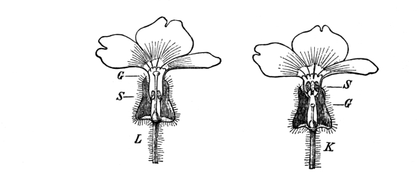
Fig. 82.—Chinese Primrose.
L, long-styled (“pin-eyed”) flower;
K, short-styled (“thrum-eyed”)
flower; G, stigma; S, anthers.
[Pg 111] It is obvious that the cowslip (Fig. 83) is closely related to the primrose. The difference lies chiefly in the character of the flower-stalk. In the cowslip this is long, and it bears at its top several stalklets, each of which ends in a flower. As in the primrose, cross-fertilisation is secured by some flowers being pin-eyed (long styled) and others thrum-eyed (short styled).
1. The daisy.—Take up several daisy plants (Fig. 84) entire, and wash away the soil from the roots. Notice how the stems—some of which are underground—are connected together. Draw a leaf. What advantage is it to the plant to have leaves of the shape noticed? Cut vertically through the “head,” and notice that what is usually called the “flower” really consists of a large number of small flowers.
The central or disc flowers are tubular. Which disc flowers open first, those near the middle or those nearer the edge of the disc? Pick off a flower and notice the 5-toothed corolla. Tear the corolla down with a needle, and observe the tiny stamens (5) fixed on the [Pg 112] corolla-tube. The anthers are joined together. Notice the divided stigma of the pistil. The white and pink ray flowers have strap-shaped corollas. They have no stamens, but each has a pistil like that of a disc flower. What do you think is the object of the ray flowers being so conspicuous? Why do they close over the disc at night?
Notice the large number of green bracts below the disc.
2. The dandelion.—Compare the dandelion (Fig. 85). Notice that all the flowers are strap-shaped, like the ray flowers of the daisy. Pull one out, and make out the strap-like corolla, the five stamens with joined anthers, and the double stigma (Fig. 86, 2). Notice the tuft of fine hairs below the corolla and above the knob-like ovary. The tuft of hairs is the top of the calyx-tube.
When the flowers have been fertilised, the yellow corollas wither, and each calyx-tube elongates until it is about an inch long, the tuft of fine hairs being still at the top (4). Blow a “clock,” and notice how easily the fruits are detached from the disc and how slowly they settle. What advantage is this to the plant?
3. The thistle.—Compare the thistle (Fig. 87). The bracts are very prickly. Is this an advantage? Are the flowers tubular or strap-shaped? Examine the fruits (“thistle down”) and compare with those of dandelion.
The daisy.—What is generally called the “flower” of the daisy (Fig. 84) really consists of a very large number of small separate [Pg 113] flowers set close together on a flattened disc or receptacle. The group of flowers is called a head. On the lower surface of the head are several green leaves, or bracts, which protect the bud before it opens. The flowers of a daisy-head are of two kinds. The white or pink straps set round the edge of the head are the corollas of the ray flowers. They have no stamens, but a pistil with a divided stigma is present in each. The disc flowers are yellow and tubular. The corolla consists of five united petals. On it are fixed five stamens, the anthers of which are joined together. The pistil is like that of a ray flower. No calyx is present in either the ray or disc flowers of the daisy.
The daisy is fertilised by the aid of insects which are attracted by the strap-like corollas of the ray flowers.
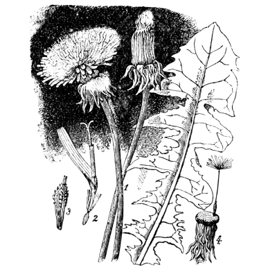
Fig. 86.—Dandelion.
1, two heads and a leaf (× ⅔);
2, a single flower (× 2); 3, fruit
(× ⁸⁄₃);
4, receptacle, with one fruit.
The dandelion “flower” (Fig. 85) is also really a head consisting of a great many separate flowers. There are often between two and three hundred of these little flowers present in one head. Below the head are several green bracts, and these protect the flowers both in the bud and at night (Fig. 86). The flowers of the dandelion are all of one type. The corolla is strap-shaped (Fig. 86, 2), and of a beautiful yellow colour to attract insects. On the end of the strap may be seen five small teeth which indicate that it really consists of five [Pg 114] united petals. At the base of the corolla is a tuft of fine hairs, which is the top of the calyx-tube, and at the bottom of the flower is a little white knob—the ovary. The five stamens are fixed on the inside of the corolla-tube. Their anthers are united to form a tube through which the upper part of the style, and the forked stigma protrude.
It will be noticed that a dandelion flower is practically like a ray flower of a daisy, with the addition of calyx and stamens.
The thistle (Fig. 87) is another common member of the family. Its bracts are prickly, and are a protection from the attacks of animals. The flowers (Fig. 88) are all tubular. The common thistle distributes its fruit by a plume of radiating fine hairs—the calyx. The fruit is commonly known as “thistle down.”
The Compositae, as plants of this family are called, are found in all parts of the world. The family is the largest in the vegetable kingdom, and many of the plants included in it are of considerable importance.
1. The foxglove.—Examine a flowering plant of foxglove (Fig. 89). Notice the general habit of growth. In the flower make out the five-lobed calyx, the irregular corolla with five petals joined to form a tube, the four stamens (two long and two short) fixed on the corolla tube (Fig. 90), and the form and attachment of the pistil. Watch bees [Pg 115] visiting the flower. 2. The speedwell.—Compare the speedwell (Fig. 91), and notice that the corolla is more nearly regular than is the case with the foxglove, that it consists of four combined petals, and that two stamens are fixed upon it.
3. The musk.—Compare the musk. Dissect a flower and notice the forms and positions of the parts. Especially examine the pistil with its two-lobed stigma. With a hair, carefully touch one of the lobes of the stigma of a growing flower and watch how the lobes close. Do the lobes open again? Put a little pollen on, and watch to see if this time the lobes open again after closing.
Watch insects visiting the flowers and try to make out how the pollination of the stigmas is brought about.
The foxglove family.—In the primrose and the disc-flowers of the daisy are seen examples of regular, dicotyledonous flowers with five petals fused to form a corolla tube, and with five stamens inserted on the corolla. In the strap-shaped flowers of the dandelion the corolla is irregular, but still consists of five fused petals and bears the five stamens.
The foxglove (Fig. 89) and its relatives have also irregular corollas of joined petals on which the stamens are fixed; but the stamens are usually only four in number, two being long and two short [Pg 116] as in Fig. 90, b. In the foxglove the stamens ripen and shed their pollen before the pistil of the same flower is mature. This prevents self-fertilisation, but bumble bees in passing from one flower to another convey the ripe pollen of the younger flowers to the stigmas of flowers which are ready for fertilisation.
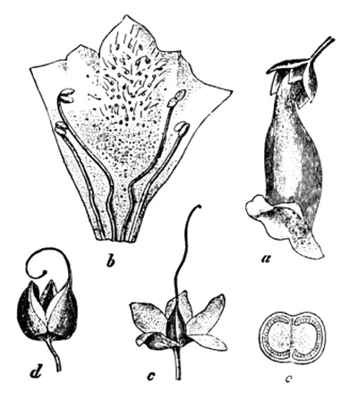
Fig. 90.—Foxglove.
a, flower; b, corolla cut open and spread out;
c, calyx and pistil; d, fruit; e, section of fruit. (× ⅔.)
The pretty blue speedwell (Fig. 91) is closely related to the foxglove, but its corolla has only four lobes instead of five, and some times these seem of almost equal size at the first glance. Generally, however, the corolla is very plainly irregular. The speedwell has only two stamens, while mullein has five.
Calceolaria, musk, gloxinia, and snapdragon—other members of the family—are often cultivated in gardens.
The flowers of the musk are especially interesting, because they show so well what is called irritability,—the power which all living things possess of acting in a definite manner in response to a definite [Pg 117] irritation or stimulus. We have already seen good examples of plant-irritability in the way a climbing stem winds itself round a support. The stigma of the musk flower has two flaps. If these are touched with a hair or bristle they quickly close together, but presently open again as if they had found out that they had been tricked. When, however, a little pollen is put on the flaps they close finally, for their whole object is accomplished.
Some of the plants in this family are poisonous, the foxglove being especially so in all its parts.
1. The deadnettle.—Examine a deadnettle plant (Fig. 92). Notice the habit of growth, and write down a description of the shape and appearance of the stem and leaves. What is the shape of the flower? How many sepals, petals, and stamens has it? Do the stamens ripen first, or does the pistil? What insects do you find visiting the flower? Try to find out how they pollinate the stigma.
2. Other labiates.—Compare the sage (especially in respect of its relation to bees), rosemary, thyme, marjoram, and mint, and distinguish between their various flowers, leaves, and scents.
The labiates.—The deadnettle is a type of an easily recognisable [Pg 118] family of plants. The stem is square in section, and the leaves are arranged upon it in opposite pairs at right angles to each other. The plants are hairy and have distinctive odours. The aroma of thyme, mint, marjoram, sage, etc., has led to the plants being used for flavouring food. None of the labiates is poisonous.
The shape of the flower is very characteristic, and is specially adapted to the visits of bees. The flowers are so modified that the lowest part of the corolla forms a platform on which the bee may conveniently alight, while the upper petals unite into an arched roof which protects the pistil and stamens.
The mechanism of cross-pollination is particularly well shown in the case of the sage (Fig. 93). The flower contains four stamens, but two of these have lost their use, and the others are modified in a strange manner. The whole stamen has somewhat the shape of a capital T, and at each end of the cross-piece is a pollen box. Usually the cross-piece (c, Fig. 93, 3) is not at right angles to the filament, but is swung up—the junction acts as a hinge—until it is nearly vertical (Fig. 93, 4). The pollen box s, which is at the lower end of the cross-piece c when this is vertical, contains hardly any pollen. [Pg 119] The entrance to the honey tube is thus guarded by two pillars, the filaments (f) of the stamens; and the lower pollen box (s) of the cross-piece of each stamen is directly in front of the bee’s head as it stands on the lower lip of the flower. When it pushes forward its head to reach the nectar it comes in contact with the lower pollen boxes, and the cross pieces swing round on their hinges, bringing the upper pollen boxes down with a smack on the bee’s back (Fig. 93, 1), and sprinkling it liberally with pollen dust. Having shed their pollen the stamens shrivel up, and the pistil comes to maturity. As the pistil ripens, the stigma arches over (Fig. 93, 2) so as to scrape along the back of any bee visiting the flower for the nectar, and thus to wipe off the pollen which has been brought from a younger flower.
1. The hyacinth.—Take up a plant (Fig. 94) entire and notice the underground bulb with roots springing from its lower surface, and the long narrow leaves. Is the venation of the leaves parallel or net-like? Is the hyacinth a monocotyledon or a dicotyledon? See the bract at the base of each flower-stalklet.
Examine the flower. Its leaves cannot be distinguished into calyx and corolla, but are quite similar to each other in size, shape, and colour. They are therefore called the perianth. The perianth leaves are united to form a tube. Tear down the tube to see the six stamens fixed on it. Are they all on the same level? Examine the pistil and cut the ovary across to see the ovules in the three joined carpels. Which is fixed at the higher level, the perianth or the base of the pistil?
2. Other plants of the lily family.—Examine also the white lily, tulip, star of Bethlehem, and lily of the valley, and notice that in spite of small differences they are all monocotyledons (how do you know this?) and all have the perianth fixed below the ovary. [Pg 120]
3. The snowdrop.—Compare and contrast the snowdrop (Fig. 95). Make out that it is a monocotyledon, but that its perianth is inserted above the ovary. This is the great point of difference from the lily family. Notice also how the stamens are fixed.
4. Other plants of the snowdrop family.—Examine the daffodil (Figs. 55 and 96) and narcissus. Arising from the short perianth tube of the daffodil is a longer one which is often mistaken for a corolla; it is called the corona. None of the flowers hitherto described contains anything corresponding to a corona. The corona in the narcissus is short. As in the snowdrop, the perianth is fixed above the ovary. Observe how the stamens are fixed, and notice the dry leaf beneath the flower.
The lily family.—Either the wild hyacinth (Fig. 94) or the cultivated single hyacinth may be taken as a good representative of [Pg 121] this family. The first point which strikes the student on examining the general “habit” of the plant is the character of the long sheathing leaves. Their veins do not form an obvious network, such as is seen in the leaves of dicotyledons, but run lengthwise and roughly parallel to each other in the manner characteristic of grasses and other monocotyledons (p. 40). The leaves are narrow, and are not divided into blade and stalk; they and the flower stalk spring from an underground bulb (p. 84) which consists chiefly of the swollen leaf-bases of a previous season. A separate calyx and corolla are not to be distinguished in the flower; the six leaves being all alike in size, shape, and colour. These six leaves hence receive a special name, and are called the perianth. The perianth leaves are united into a tube, on the inside of which the six stamens are arranged in two series of three each. In the middle of the flower, and fixed above the insertion of the perianth, is the pistil, which consists of three united carpels.
It will be noticed that the parts of the flower are in threes. There are six united perianth leaves (three inner and three outer), six stamens (also in two series), and three united carpels. This is very common—though by no means universal—in monocotyledons. [Pg 122]
All the plants of the lily family—including the tulips, the true lilies, lily of the valley, asparagus, onion, etc.—agree in being monocotyledons, and in their flowers having a conspicuous perianth (for attracting insects) and six stamens, and in the ovary being above the insertion of the perianth.
The snowdrop family.—Plants of this family are very similar to those of the lily family; in fact in only one respect can any sharp line of demarcation be drawn between the two groups; in the snowdrop and its relatives the other parts of the flower stand upon the ovary (Fig. 96). The flowers of some plants of the family, e.g. the daffodil, possess a tubular outgrowth of the perianth, which is called a corona. It is often mistaken for a corolla.
26. THE ARUM LILY.
1. The cuckoo-pint.—Examine the habit of the plant, and then cut open the “flower.” You will probably find a number of small flies inside. Examine the central rod and make out the stamens and pistils on it. Which ripen first?
The arum “lily,” with its humble relative the cuckoo-pint (Fig. 97), merits special mention; for, in the first place, it is not a lily at all, and secondly, it furnishes an extremely interesting example of pistils coming to maturity before anthers, which is rather rare.
What is generally called the “flower” of the cuckoo-pint, or “lords and ladies,” consists of a big curled leaf with a purple rod sticking up in the middle. Near the bottom of the rod, but hidden from sight by the lower part of the leaf, the true flowers arise. The chamber containing them is shut in by a series of stiffish hairs which point downwards. Below the hairs the rod supports a series of anthers and, near the bottom of the chamber, several pistils. On cutting open the chamber one nearly always finds a number of small flies, covered with pollen, which [Pg 123] they have brought from another arum. The flies get in easily enough, but once in they are prisoners, for the down-pointing hairs prevent them from getting out again. The pistils near the bottom of the rod ripen, and are fertilised by the pollen the flies have brought. After a time the anthers above ripen and shed their welcome pollen on the hungry captives. Soon after this the hairs at the top of the chamber shrivel up, and the flies, once more covered with pollen, are at liberty to return to the outer world and, untaught by experience, to repeat the experiment on another “flower.”
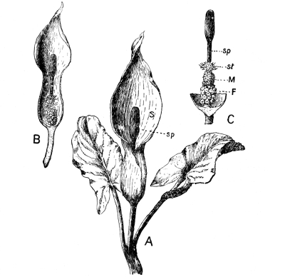
Fig. 97.—A, Cuckoo-pint. (× ¼.)
In B, the front of the leaf is cut away to show
the rod sp (C). F, female flowers;
M, male
flowers; st, undeveloped male flowers.
EXERCISES ON CHAPTER VI.
1. Describe the arrangement of the stamens in the wallflower, the sage, and the primrose.
2. Describe the pistil of the deadnettle, the primrose, and the shepherd’s purse. (1895)
3. In what respects does the flower of the buttercup differ from that of the wild rose?
4. Explain the arrangement and form of the stamens in some flower selected by yourself. Describe the structure and contents of the anther. (1897)
5. Where is the pollen of a flower formed? What is its use? (1898)
6. Name one or two plants which do not ripen seed if insects are excluded, and show why they do not. (1898)
7. Name two plants which would be in flower in each of the months from March to August inclusive, and state in what localities they would be found? (N.F.U.)
8. In what ways are insects attracted to visit flowers? Give examples, with an explanation and drawing in each case, of any special structure which may be a means of attraction. (N.F.U.)
9. Name ten flowering plants which may be found, (a) in a shady wood, (b) in a meadow, in late spring.
10. Name any fresh specimens of flowers which usually could be obtained growing wild in March, June, and October respectively. (N.F.U.)
11. What wild flowers would you expect to find in early April in your part of the country? In what kinds of places would you look for them? (N.F.U.)
12. Name the parts of any flower you have examined which are concerned with the production of seed. (King’s Scholarship, 1904)
1. General features.—Pull up a sod of couch grass or of Yorkshire fog and clear away the earth as well as possible. Notice the fibrous, creeping branches or stolons, and the bunches of fine roots. In summer the plant sends up also erect branches called haulms, which bear leaves and flowers. Examine the shape of the leaves and notice their parallel venation, which indicates (p. 40) that the plant is a monocotyledon.
Observe how the leaves are borne on the haulms. Each leaf arises at a knot (node) which is slightly swollen. The first part of the leaf is a sheath which encloses the haulm up to perhaps the next knot. Then the leaf-blade stands out from the haulm. Turn back the blade slightly and notice at this point a little strap, the ligule (Fig. 98), on the upper surface of the leaf-blade. Notice the variation in the size and shape of the ligules of different kinds of grass.
Pull the haulm until it gives way, and draw the broken part out of the leaf-sheaths. Chew the end, and notice the taste of sugar. Cut across haulms of various ages. The young haulms are solid; the old ones are hollow except at the nodes, where there is a horizontal shelf. Notice this also in a bamboo cane, which is the haulm of a large tropical grass. In spring, mark a young haulm, so that you can recognise it, and measure it day by day. Note the measurements made on various days, and also the date of opening of the flowers. [Pg 126]
2. Different grasses.—Learn to recognise different grasses, not only by the way in which the clusters of flowers are borne on the haulms (Figs. 102 to 110) and by their colours, but also by the general appearance of the plant when it is not flowering; the size and shape of the leaves, the character of the ligules, the manner of rooting, etc.
Again germinate grains of wheat, and especially notice the single cotyledon, the leaves, and roots (p. 21).
Importance of grasses.—It is impossible to over-estimate the value to man of the various grasses. From the earliest times he has obtained his staple food from the grains of such cereals as wheat, barley, oats, rice, and maize; and his flocks and herds have grazed upon the nutritious herbage of the plains. The numberless applications of such large tropical grasses as the bamboo and the sugar-cane are also well known. Sugar occurs very generally in grasses. It can easily be recognised even in ordinary meadow-grass by pulling out the upright “stem” from the sheathing leaves, and chewing the tender end.
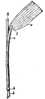
Fig. 98.—Part of a Grass
Stem and Leaf. h,haulm;
s, part of leaf-blade;
l, ligule; v, leaf-sheath;
k, node-like swelling at
the base of the leaf-sheath.
(× ½.).
The general characters of grasses.—In this country, grasses are usually herbs not more than three or four feet high. They have abundant leaves, which are long, narrow, and pointed at the end; they have parallel veins like the leaves of most other monocotyledons. The roots, too, are of the usual monocotyledonous type—springing in bunches from the base of the stem, not branching from a main tap-root. Most grasses spread by prostrate creeping branches or stolons (p. 83), which spring from the axils of leaves and then run along beneath the soil. At some little distance from the parent shoot, the stolon forms roots below and a new shoot above. Other stolons arise in the axils of the leaves of the new shoots, and so the process is repeated. In this way such grasses become thoroughly established in the soil, and—if they [Pg 127] are of the undesirable species which are classed as “weeds”—cause much trouble. This habit of growth is, however, useful in binding together the sand and earth of embankments, etc., into a compact mass. The new shoots of stolon-bearing grasses are of two kinds. Throughout the greater part of the year they consist of tufts of leaves; but, in the spring and summer, there are also produced erect branches—commonly known as stems, but better called haulms—which bear leaves in the “alternate” manner. The point at which a leaf springs from the haulm is called a knot or node; it is usually swollen. The distance between two knots is, of course, an internode. Extra roots are often given off from the lower nodes of the haulm.
The lowest part of the leaf is a cylindrical, and generally split, sheath (Fig. 98), which closely embraces the haulm for some distance—often for the length of an internode. The sheath protects and supports the soft, growing internode, inside it. At the top of the sheath is the blade of the leaf, which stands out from the haulm. At the junction of the blade and sheath, on the upper surface of the leaf, is a little membranous outgrowth called a ligule (Fig. 98, l). It often varies greatly in size and shape in different species of grasses, and is a valuable means of distinguishing between them. Its use is not certainly known. The student should examine the ligule of every grass he studies.
The haulm, or flowering branch, is for some time short and solid, and its nodes are so close together that the sheath of a lower leaf may overlap several upper leaves. The haulm is thus protected from the [Pg 128] rough weather of the early spring. In the meantime, the flowers have been developing at its upper end. When they are almost ready to open, the haulm begins to grow very rapidly; its internodes elongate so quickly that the internal tissues are torn and the haulm becomes a hollow straw, except at the nodes, where there are horizontal shelves. The haulm hardens and stiffens as it grows, so that, when the spikelets of flowers at its upper end open, they are carried high above the leaves on a slender but very strong rod, which bends and dances in the wind, and allows the pollen to be detached and blown to the stigmas of other flowers.
1. The arrangement of the spikelets.—Gather ears (flowering haulms) of several different kinds of grasses and notice the arrangement of the flowers. In the oat (Fig. 99), the meadow grasses (Fig. 103), fescues (Fig. 102), and others, the nodding oval bodies, called spikelets, are borne on delicate stalks which spread outwards from the haulm. In the foxtails (Figs. 104 and 105), timothy (Fig. 106), and sweet vernal grass (Fig. 110) the spikelets are on short stalks, which can, however, be seen on bending the ear sharply on itself. In wheat, barley, couch grass, rye, and rye grasses (Fig. 109) the spikelets are devoid of stalks and are set close along the haulm. Be quite sure you understand what is meant by a spikelet. 39 spikelets are shown in Fig. 99.
2. The arrangement and structure of the flowers.—Take an open spikelet from the ear of a grass (e.g. oat) which has large flowers, [Pg 129] and examine it. At the bottom are two boat-shaped leaves—really bracts—called glumes. Remove them carefully. Above them will be found two or more flowers, each flower enclosed in two other leaves. The outer of these two leaves is called the outer pale; the inner is the inner pale. From the middle of the back of the outer pale of the oat springs a bristle called an awn. Remove the outer pale and make out the three anthers on long filaments, and the ovary with two branching feathery stigmas and short styles. Fig. 100 is a diagram of a spikelet, which makes clear the relation of the glumes, pales, and flowers. Fig. 101, A, is a spikelet of meadow fescue. Its outer pales differ from those of the oat in not bearing awns. Otherwise it is very similar. Fig. 101, B, shows the appearance (magnified) of a flower of meadow fescue from which the outer pale has been removed. The two little scales seen in front of the ovary possibly represent a perianth. Be careful not to confuse the terms ear, spikelet, and flower. The ear consists of a number of spikelets, and each spikelet consists of glumes and one or more flowers.
Similarly dissect spikelets and flowers of other grasses, making notes of the lengths of the stalks of the spikelets, and the presence (and lengths) or absence of awns.
3. The use of the awns.—Try to find out the use of the awns. Are they generally rough or smooth? Have you ever seen grass “seeds” sticking in the wool of sheep? What kept them attached?
4. Grain.—Examine several kinds of grass “seeds” and make out that they not only consist of the entire ripened ovary and are therefore fruits, but that usually the pales also remain attached to them.
5. The embryo and endosperm.—Again cut through soaked grains of maize and wheat, and make out the embryo and endosperm (p. 19).
Grass flowers.—Grasses are true flowering plants; but because they depend on the wind for the transference of pollen to the stigmas, they do not pander to the taste of bees and butterflies by secreting [Pg 130] nectar, and hence have no need to display those advertisement placards which we call petals. For this reason their flowers are not generally recognised as such. It requires a little care to make out the parts of the flower and to understand the manner in which the flowers are arranged among themselves.
The whole group of flowers borne by any one haulm is generally called the ear or panicle. The ear in its turn consists of several bodies called spikelets. The appearance of the ear varies greatly in different grasses (Figs. 102 to 110) according as the stalks of the spikelets are long and spread outwards from the haulm, as in the meadow-grasses (Fig. 103), oats (Figs. 99 and 108), fescues (Fig. 102), and others; short, as in the foxtails (Figs. 104 and 105), timothy (Fig. 106), and sweet vernal grass (Fig. 110); or absent altogether, as in the wheat, barley, rye, rye grasses (Fig. 109), and couch grass.
A single spikelet of a grass is shown diagrammatically in Fig. 100. At the bottom are two boat-shaped bracts called glumes (g, g), which almost or entirely cover the spikelet before it opens. When the glumes have been removed a few flowers remain. Each one is protected by two leaves called pales: outer (p₁) and inner (p₂) respectively. The flower (B), as a rule, has three stamens, the anthers being borne on long filaments and dangling out to the wind (Fig. 101), and a pistil with two branching feathery stigmas. In many grasses the outer pale bears a bristle called an awn, well seen in “bearded” wheat, barley, and upright brome grass.
Fig. 101, A, represents a single spikelet of meadow fescue, with two [Pg 131] open flowers. The outer pales of this grass are not awned. Fig. 101, B, shows a single flower of the same grass from which the outer pale has been removed. The two little scales seen in front of the ovary (and at e in Fig. 100) possibly represent all that is left of a perianth (p. 121).
Fertilisation.—By the time the flowers have opened, the haulm has usually grown so tall that they are lifted well above the leaves. The stamens hang their anthers loosely out to the wind, and, as the slender haulm sways in the breeze, the pollen is readily detached and carried to other flowers, perhaps miles away.
It is in order to catch the wind-borne pollen-grains that the stigmas of grasses are branched and feathery. A great waste of pollen is obviously entailed by this method. But on the other hand it must be remembered that insect-pollinated flowers have to pay for their privileges by storing nectar in cunningly hidden pockets, and by advertisement-expenses, all of which the grasses avoid.
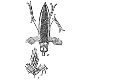
Fig. 101.—Meadow Fescue.
A, spikelet with two open flowers (× 1½);
B, a flower from which the outer pale has
been removed (× 6).
The ovule is fertilised in the usual way, by a pollen tube growing down the style from a grain on the stigma. As a result of fertilisation the ovary becomes a fruit containing one seed which fills it. In many cases (e.g. barley and oats) the pales still remain in position and adhere to the fruit. When the awn is present it may play an important part in distributing the “seeds” by clinging to the hides of animals, etc. [Pg 132] Sometimes the awn is of use in fixing the grain in position on the ground until the seed has germinated.
The structure of a grass seed has already (p. 19) been studied in the wheat and maize. The developing embryo (p. 21) lives upon a store of food called the endosperm (Fig. 16) until its roots and leaves are sufficiently advanced to make food for themselves.
1. The fescues.—Gather plants of meadow fescue, and examine the habit of growth (in tufts), the broad leaves, and the nodding panicles and the flowers. Also examine the “seeds.”
Compare sheep’s fescue (Fig. 102), and notice the very fine leaves. The outer pales are awned.
2. The meadow grasses.—Examine the various species of meadow grass (Fig. 103). They can be distinguished from each other by the characters of the ligules. Notice the tree-like habit of the panicles and compare them with the fescues. The meadow grasses never bear awns. Notice the woolly “webs” at the bases of the “seeds.”
3. The foxtails.—In meadow foxtail (Fig. 104) notice the prostrate stolons and large succulent leaves. Does the grass grow in tufts? Why not? What is the earliest date on which you have seen it in flower? Double the ear on itself to see the short stalks of the spikelets. How many flowers are there in each spikelet? Notice the silky awns of the flowers.
Compare the slender foxtail (Fig. 105), which is a troublesome weed.
4. Timothy.—Compare the leaves and flowers of timothy or “meadow catstail” (Fig. 106) with the meadow foxtail. The ear is green and rough, and the flowers are awnless. What is the date of flowering?
5. Yorkshire fog.—Notice the woolly covering of this weed (Fig. 107). This covering and its bitter flavour make it distasteful to cattle. Observe the “kneed” awn of the flower. Dig up and shake off the earth from a sod to see the stolons. [Pg 133]
6. Wild oat (Fig. 108). Examine the large spikelets and make out the long twisted awn of the flower. Compare this weed with the cultivated oat (Fig. 99) and with the yellow oat grass.
7. The perennial rye grass.—Notice that the ear is flattened, and that the spikelets are without stalks and have only one glume. The leaf is glossy and has a prominent midrib and a flattened sheath.
8. Sweet-scented vernal grass.—Notice the tufted habit of growth and the characters of the leaves. Chew the stalk and notice the sweet odour of new-mown hay. What is the date of flowering? Make out that the flower has two stamens only.
9. Rushes and sedges.—Rushes and sedges are sometimes mistaken for grasses. Examine the stems, the flowers, and the leaves (look for ligules), and tabulate as many differences from the true grasses as possible. [Pg 134]
The student should learn to recognise the common grasses and to distinguish the useful species from the weeds. When the grasses are in flower there is not much difficulty in doing this, but the habit of growth—the characters of the leaves and roots, and of the stolons of the perennial species—should also be noticed carefully, as during the greater part of the year these alone can be depended upon.
The fescues fall into two groups according as their leaves are broad or narrow. The meadow fescue is a good example of the former group. It is found in meadows and pastures and has long broad leaves. Sheep’s fescue (Fig. 102) is a good example of the second group of fescues. It has very fine—almost bristle-shaped—leaves. Like the meadow fescue it grows in tufts; it inhabits high lands and downs, especially in limestone districts. The [Pg 135] nodding spikelets are borne on fairly long stalks, and the panicles are somewhat like those of the meadow grasses (Fig. 103). The flowers of sheep’s fescue, however, bear short awns.
The meadow grasses much resemble the fescues in general appearance, but the panicles (Fig. 103) are rather more tree-like—the stalks of the spikelets spreading more horizontally—and the flowers are never awned. The various species can be distinguished by their ligules; for example, the ligule of the smooth-stalked meadow grass (Fig. 103) is blunt, while that of the rough-stalked species is long and pointed. The annual meadow grass is a weed to be found almost everywhere.
The foxtails are easily recognised by the tail-like appearance of the ears (Figs. 104 and 105). The meadow foxtail (Fig. 104) is a valuable grass with broad, long, and succulent leaves. It spreads by means of prostrate stolons. The spikelets have short stalks, as can be seen on bending the ear on itself, and each spikelet contains only one flower. The silky awns give the ear a silvery grey colour. The grass flowers in early spring. The slender foxtail (Fig. 105) is a most injurious weed of cornfields. It can be distinguished from the meadow foxtail by its less vigorous appearance, by the thinner and more pointed ear, and by the black patches on the ear. It is often called “black bent.” [Pg 136]
Timothy grass or “meadow catstail” (Fig. 106) has a general resemblance to meadow foxtail, but its ears are rough to the touch and green in colour. It also flowers much later in the year (July) and its pales are awnless; the two grasses are therefore easily distinguished. Timothy grows abundantly in clay soils, forming fairly close tufts.
Yorkshire fog (Fig. 107) is a rank weed which is distasteful to cattle, partly because it is covered with hairs and is difficult to wet, and partly because it has a bitter flavour. It is pale in colour and soft. It spreads rapidly by means of creeping stolons.
Oat grasses are of several species; one is cultivated as a cereal, others are valued as forage for cattle, while others still are [Pg 137] troublesome weeds. The wild oat (Fig. 108) is a common weed in cornfields. It is an annual, from two or three feet high, with large spikelets forming a loose panicle, the flowers having awns twice as long as the spikelets. The cultivated oat is supposed to be a variety of the wild oat. The panicle of the yellow oat grass is oblong, and has erect spikelets.
The perennial rye grass (Fig. 109) is very easily recognised by its flattened ears of alternate, stalkless spikelets. Each spikelet has only one glume. The leaves are dark-green and glossy, and the leaf-sheaths are flattened instead of being round, as is the case with most grasses. This is one of the most valuable of forage grasses.
Sweet-scented vernal grass (Fig. 110) does not occur very [Pg 138] abundantly in meadows and pastures, but the fragrance of new-mown hay is almost wholly attributed to it. The odour can be distinctly perceived when a stalk of the grass is chewed. The leaves are somewhat hairy and are broad and flat. The spikelets are borne on short stalks. The flowers open early and are remarkable in possessing only two stamens instead of three.
Rushes and sedges are monocotyledonous plants which are often erroneously called grasses. The stems of rushes are vivid green, round, and pointed, and contain a distinct pith. The flowers (Fig. 111) are never in spikelets; they contain six stamens, and the pistil has three long stigmas. The sedges, like grasses, have narrow, pointed leaves, but the ligules are either very small or absent, and the leaf-sheaths are not split. The flowers are often in spikelets with glumes, as are grass flowers. The stems of sedges are solid and triangular.
The flowers of rushes in many respects resemble those of the lily family. Indeed it is supposed that the ancestors, not only of the lily family, but also (along a different line) of the grasses and sedges, were primitive and now extinct rushes. The lilies have developed the rush perianth more and more as they have increasingly depended on insects for pollination; while in the grasses and sedges the perianth has gradually dwindled because these plants found that wind-pollination was sufficient for their needs. [Pg 139]
EXERCISES ON CHAPTER VII.
1. What grasses are grown as corn in your part of the country? How can you recognise the grasses (a) before flowering, (b) in the ear, (c) by the straw?
2. What forage grasses are most cultivated in your part of the country? Make a list of the earliest dates on which you have seen each in flower.
3. Examine grains of wheat, barley, oats, and rye, as harvested. In which of these are the pales present? What is the “beard” of barley?
4. Make a collection of “seeds” of various grasses, and write down the characters by which they may be distinguished from each other.
5. What are the commonest weeds you have seen in corn fields? Do the corn and the weeds flower at the same time or not? Is the time of flowering an advantage (a) to the weeds, (b) to the farmer?
6. In what important respects do wind-fertilized flowers differ from insect-fertilized flowers? Give examples of each. (1898)
1. The oak.—(a) Habits of growth.—Examine an oak tree growing in an exposed situation. What is its approximate height? Estimate the diameter of the trunk at (a) the ground level, (b) at heights of 1, 2, 3, 4, etc., feet. At what height do the principal boughs come off? About what angle do the boughs make with the trunk? Are they straight, evenly curved, or zig-zag? Contrast in these respects an oak growing in a plantation. Try to account for the differences observed.
(b) The bark.—Examine the barks in oaks of various ages. Do the ridges and furrows caused by the splitting of the bark form any definite pattern, or are they arranged anyhow? Find a piece which shows the pattern well, and make a drawing of it.
(c) Method of branching.—Carefully observe the tree from a distance, and notice how the great boughs spring from the trunk, and in their turn give rise to smaller and smaller branches. Notice the shape of a tuft of the smallest twigs against the sky; then close your eyes and try to recall the picture. A still better way is to make a careful drawing of the tree, not attempting to put in details, but paying special attention to the trunk and main branches, and to the general massing of the twigs. What is the general “expression” of the tree? Would you call it, e.g., graceful, formal, sturdy, delicate, stiff, or sombre? The method of branching is best seen in the winter or spring, when the tree is leafless. [Pg 141]
Examine a twig in winter or spring, and notice the position of the buds. Round the tip there may be three or more crowded buds. The one at the tip generally dies, and those just behind grow out into lateral branches. Can you see any trace of this having been the case with the big boughs? On a twig make out—from the marks on the outside—the growth of last year, two, three, and four years ago. Cut these lengths across with a sharp knife, and count the rings of wood.
(d) The leaves.—Watch a marked twig from spring to summer, and notice how short are the new shoots which come from the buds, and consequently how close together the new leaves are. Does this account for the crown of foliage on the tree being so dense? Make a drawing of an oak leaf. At what time of the year do (i) the leaf-buds open, (ii) the leaves fall from the tree? In winter, try to find an oak which has not shed its leaves; is it a young tree or an old one?
(e) The flowers.—In May notice (i) the hanging catkins, each of which is a bunch of male flowers containing stamens, but no pistil; (ii) the groups of small female flowers which arise in the axils of two or three of the upper foliage leaves; each contains a pistil, but no stamens. Notice the cup of scaly bracts, which surrounds the lower part of a female flower. Does the tree flower before or after the leaf buds have expanded?
(f) The fruit.—Trace the development of the acorn from the female flower, and the change of the covering of scales into the woody cup. If possible, compare for some years the yield of acorns by a selected old oak. Germinate an acorn.
(g) Associated animals.—Make as full a list as possible of the animals which you have seen obtaining food or shelter from the oak. What was each doing when you saw it? Look for galls—the so-called “oak apples”—and cut them open to see the insect inside. Also examine the other kinds of galls found on the leaves and catkins.
[Make similar observations, sketches, and notes of other forest-trees, in addition to the special observations mentioned below, and learn to recognise their “expressions,” methods of branching, bark, leaves, flowers, and fruit.]
[Pg 142] 2. The beech.—Notice the smooth, olive-grey bark, and the “fluted column” appearance of the base of the trunk of many beeches; the long, wavy boughs; the brown, sharp leaf-buds; the smooth, silky-fringed leaves; the scarcity, or absence, of vegetation beneath the tree; the hanging, globular catkins of male flowers; the pairs of female flowers, surrounded by prickly scales. Make notes of the dates of flowering and opening of the leaf-buds. Trees may be considered in flower when the stamens can be seen. Trace the development of the fruit or “mast.” Each pair of flowers gives rise to two three-sided nuts, enclosed in a woody cup or husk. It is covered with hard bristles, and splits into four parts when ripe.
Compare the sweet chestnut.
3. The birch.—Observe the graceful appearance of the tree; the slender trunk, with its bark streaked with brown, yellow, and silver; the purple-brown colour and wiriness of the young twigs; the shape and size of the dark green leaves, and the cylindrical, many-flowered catkins. The male catkins appear in the autumn, the smaller female catkins in the following spring. Examine the two kinds of flowers. Find an old birch-cone and break it open to see the winged fruits. What is the use of the wings?
4. The hazel and alder.—In what situations do these trees grow? Compare them with the birch. Notice that in both hazel and alder the male flowers are in long, dangling catkins. The groups of female flowers of the hazel look almost like leaf-buds, but can be recognised by the spreading red stigmas. The female flowers of the alder form distinct cones. Is the flowering part of the twig of this year’s or last year’s growth? Look in the autumn for the cones and catkins which will expand next spring. Trace the development of the fruit.
The oak.—The student cannot better commence the study of forest trees than by selecting one as a type, and making himself thoroughly familiar with its life-history, and with its appearance at all times of the year. Other trees should then be compared and contrasted point by [Pg 143] point, is and with each other. The oak answers admirably as such a central type. It is perhaps the best-known of all British forest trees, not only from its wide distribution, but also from its historical and legendary associations.
The oak (Fig. 112) flourishes in exposed and sunny situations, especially where the soil is well drained. It can be recognised by the zig-zag and wide-spreading boughs, which often spring from the trunk almost horizontally. A very strong form of trunk is plainly necessary to support such branches, and in an old solitary oak it may often be seen that the bole is very thick at the ground line, and then rapidly narrows, until at a height about equal to the base-diameter it may be only half or one-third the thickness. Above this point it is practically cylindrical up to the origin of the boughs, where it is swollen. An oak growing in a wood, or plantation, is of very different form. Its trunk is tall and straight, and the larger boughs are not [Pg 144] given off until near the top. To secure the necessary light, the tree used its energy in growing in length, and in keeping pace with its neighbours rather than in spreading laterally. In such a crowded oak the lateral buds generally remain undeveloped, while the end of the shoot pushes onwards and upwards to the light. The opposite is the case when there is plenty of room and light on all sides. Then it is usually the terminal bud which dies, while the lateral buds grow out into branches, forming the “knee-joints” which were once so greatly valued for shipbuilding.
The bark of the oak is very rugged, with ridges and furrows running almost vertically.
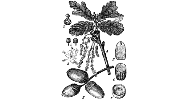
Fig. 113.—The Oak; leaves, flowers and fruit.
A, flowering branch (× ¼);
B, a male flower (magnified); C,
stamens (magnified); D, a female
flower (magnified); E, acorns;
F, cup of acorn; G, H, seed.
The young shoots which are formed when the buds expand in spring are short, and the leaves are set closely together. As a result, the oak’s foliage is characteristically dense and thick. The leaves are very distinctive; their general shape is oval, and the margin is deeply and irregularly lobed (Fig. 113).
If in early spring we go out in the woods and fix on an old oak tree (the oak hardly ever flowers before it is 50 years old), we shall probably see the flowers on some of the young twigs. The female flowers—one to five on each flower-stalk—are near the end of the twig, while the male flowers arise lower down. The female flowers are destitute of stamens, each consisting practically of a single pistil, partially enclosed in two envelopes, the lower of which ultimately becomes the familiar “cup” of the acorn. The stigma has three spreading [Pg 145] lobes for receiving pollen. The male stalks or catkins, hang down from the lower part of the twig, and every stalk bears about a dozen flowers. The male flowers have each from 5 to 12 stamens, but they have no pistil. The stamens produce pollen in the usual way, and when they burst, the wind blows the loose pollen from the stamens and scatters it in the air. Some of the pollen dust is almost certain to be wafted to the stigmas of the female flowers, and the pollen grains put out tubes, and in due course fertilise the ovules.
[Pg 146] Most of our forest trees resemble the oak in being pollinated by the aid of the wind. It is evident that for the process to be successful the trees must flower early in the spring, before the foliage has become so thick as to be in the way of the pollen and prevent it from reaching the stigmas of the female flowers. It is also important that the pollen may be easily detached, and it is for this purpose that the male flowers of the oak and similar trees hang down in the familiar catkin fashion.
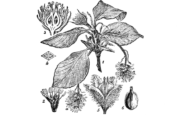
Fig. 115.—The Beech. 1, flowering branch
(× ⅔);
2, a male flower (× 4); 3, a female flower cut through
longitudinally (× 2); 4, cross section of ovary;
5, cup and fruits
(× ⅔); 6, fruit.
After fertilisation the female flower changes into an acorn (Fig. 113, E), a nut enclosed in a cup. The nut contains a single large seed, which on germination grows up into a new oak.
Galls of various kinds are to be found on most oaks. These are excrescences caused by certain insects having laid their eggs in the [Pg 147] soft tissues beneath the surface. More than fifty species of insects obtain their food from the oak. Some of these will be referred to in a later chapter.
The beech.—The beech ( Fig. 114) is easily recognised by the olive-grey, smooth bark, and by the shape of the base of the trunk, which usually has the appearance of being formed by the union of several separate columns. When well grown the tree is lofty, and bears a wide-spreading crown of branches which, when clothed with leaves in summer, casts a dense shade. The ground beneath the tree is generally [Pg 148] destitute of other vegetation. The winter-buds are long and pointed; they expand in May, the new shoots at first drooping, but straightening out in a fortnight or so. The leaves (Fig. 115) are broad, thin, and glossy, and are fringed with fine, silky hairs. Young beeches, like young oaks, often retain their leaves through the winter.
The tree is in flower by the time the foliage has fully developed. The male flowers are borne in small, rounded catkins (a, Fig. 115). The female flowers (3, Fig. 115) are in pairs. Each pair is surrounded by prickly scales, and gives rise after fertilisation to two three-sided and pointed nuts enclosed in a woody cup or husk. The husk is covered with hard, blunt prickles, and when ripe splits into four parts.
Beeches do not harbour many insects, but squirrels frequent them for the sake of the nuts. The prickly husks protect the fruit from being eaten before it is ripe.
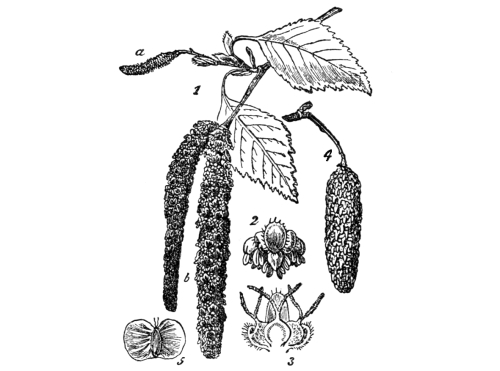
Fig. 117.—The Birch; leaves, flowers and
fruit.
1, branch with male (b) and female (a) catkins (× ⅔);
2, bract with three male flowers (× 2);
3, bract with three female flowers
(× 4);
4, ripe cone (× ⅔); 5, fruit
(× ⁴⁄₃).
The Spanish, or sweet chestnut is allied to the oak and beech. It has long, narrow leaves; it flowers in July. Its fruit is enclosed in a prickly husk, which splits into four when the nuts are ripe in October.
The birch.—The birch (Fig. 116) is a slender, graceful tree, with bark streaked with brown, yellow, and silvery patches. Its leaves (Fig. 117) are rather small, and their glossy, dark-green colour contrasts pleasantly with the brownish-purple of the young twigs. Both [Pg 149] male and female flowers are arranged in catkins (Fig. 117). The male flowers appear in autumn, but do not open until spring, when the newly-formed female flowers are ready. The female catkins (a, Fig. 117) are much smaller than the male (b).
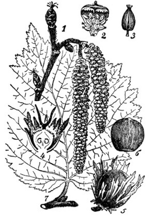
Fig. 118.—The Hazel; leaf, flowers and fruit. 1, a flowering branch (× ⅔); 2, a male flower (× 2); 3, a stamen (× 4); 4, a female flower cut through longitudinally; 5, fruit with cup (× ⅔); 6, fruit without cup; a foliage leaf.
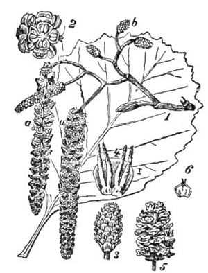
Fig. 119.—The Alder; leaf, flowers and fruit. 1, Branch with male catkins (a) and female cones (b) (× ⅔); 2, male flowers (× 2⅔); 3, female cone; 4, two female flowers; 5, ripe cone (× ⅔); 6, a fruit (× 1).
The hazel and alder develop their flowers in the year preceding their opening. The female flowers of the hazel (Fig. 118) look somewhat like buds, but may be distinguished by the red stigmas. The fruit is a nut, enclosed by a sheath of soft bracts. The seeds are largely dispersed by squirrels. The alder grows on the banks of streams; its female flowers (Fig 119, b) are arranged in short catkins, which may be called cones. The ripe cone (5, Fig. 119) contains two nuts at the base of each scale. The fruits fall into the stream, and float away, perhaps to germinate at a considerable distance. [Pg 150]
1. The willow.—About the third week in March, examine willow trees, and notice the soft, round, silky bodies which spring alternately on the young twigs. On some trees these are broad and yellow; they are male catkins (Fig. 120). Pick off a flower and see the stamens (generally two stamens to each flower) inserted on a small silky bract (Fig. 121, B). With a lens look for the honey cup at the bottom of the bract.
On other trees notice the long, narrow, silvery female catkins. Pick off a flower to see the single pistil with the forked style, also on a small, honeyed bract. Have you ever seen bees visiting the flowers? Are the catkins as conspicuous when the trees are in leaf? Is it an advantage to the trees to flower before the leaves come out? In June, examine the ripened female catkins. Pull out a tuft of the hairy seeds and dry it in the sun, noticing how they form a fluffy mass. Blow the mass of seeds. How do you think the seeds are dispersed?
2. The poplar.—Find male and female poplars and examine their flowers. Are the female flowers pollinated by insects or by the wind? Is self-fertilisation ever possible with willows and poplars? Why not? Which appear first, the flowers, or the leaves of poplars? Why?
Examine the leaves of the English poplar. Why do they turn over so easily, even with a very slight breeze? Are both sides of the leaf of the same colour?
Compare the Lombardy poplar with the English poplar.
The willow.—Many species of willow are known, but the sallow willow—called the saugh tree in Scotland—is common in coppices [Pg 151] and hedges. It has purplish brown branches, and large, broad, downy leaves. In the willow the male and female flowers are produced on different trees, so that self-fertilisation is obviously impossible. The flowers of the male willow form broad yellow catkins which cling closely to the twig (Fig. 121). They are often called “golden palms.” Each flower consists of two stamens, borne on a silky scale which has a tiny honey cup at the base. The female flowers form long, narrow, and silvery catkins; each flower is merely a single pistil with a forked stigma, and, like a male flower, is supported on a small honeyed bract. The flowers appear in March, before the leaves, and the catkins are very conspicuous. They are visited by bees for the sake of the honey and pollen, pollination being thus effected. In June, the female catkins are ripe. Each ovary has now become a fruit, which opens and liberates the silky seeds. When the seeds dry, their fine hairs cling together so that a light fluffy mass is formed, which can be blown to great distances by the wind.
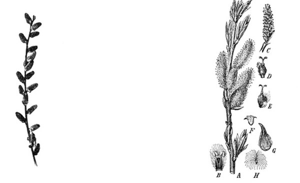
Fig. 120.—Twig of male Willow, with catkins. (× ⅙)
Fig. 121.—The Willow. A, flowering male-shoot (× ⅔);
B, male flower with bract (magnified); C, female cone;
D, E, female flowers (magnified); F, fruit (× ⅔);
G, the same magnified; H, seed (magnified).
The poplar.—The various poplars are also completely unisexual, that is, any one tree bears either male or female flowers, but not both. In this case also the flowers appear before the leaves; but the flowers are pollinated by the aid of the wind, not by insects, and nectaries are therefore not formed. The catkins are not very much like those of the willows in appearance, but the fruit and seeds are [Pg 152] very similar. Both willows and poplars are fond of the banks of streams. The English poplars are graceful trees which can be recognised at a distance by the manner in which the broad leaves turn on their long stalks, exposing the lighter-coloured under-surface at the slightest breath of wind. This is especially noticeable with the aspen or trembling poplar (Fig. 122). The Lombardy poplar is a tall, stiff tree.
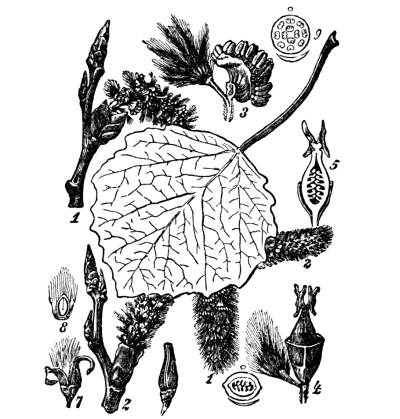
Fig. 122.—The Poplar. 1, male catkin (× ⅔); 2, female catkin (× ⅔); 3, male flower (× 2); 4, female flower (× 4); 5, the same in longitudinal section; 6, fruit; 7, the same after opening; 8, seeds (× 3); 9, diagram of male flower.
1. The elm.—Notice the straight trunk, rough bark, and slender branches of the common elm. Examine the flowers in early spring and notice that each contains both stamens and pistil. When do the flowers open? When do the leaves expand? Draw a leaf and a fruit. What is the use of the broad plate of the fruit?
2. The lime.—Observe the straight trunk, smooth bark, and general pyramid shape of the tree. Draw a leaf, and notice that it is pointed and is larger on one side of the midrib than on the other. Notice that the flower stalks spring from leaves (bracts) differing in shape (Fig. 126) and colour from ordinary leaves. Draw one of these [Pg 153] leaves with its attached flower stalk. When do the flowers open? Try to discover whether bees haunt the flowers. Find out what is the use to the fruit of the long leafy bract.
3. The ash.—Look in winter for the black buds and the flattened tips of the twigs. When do the flowers open? Notice their rich purple colour. Examine a flower and make out the two stamens and the pistil. Trace the formation of the fruit, which is winged and hangs in bunches called keys. What is the use of the wing? When do the leaves expand? Are they simple or compound? Draw one. What is its colour? When do the leaves fall?
[Pg 154] The elm.—The elm (Fig. 123) is usually a lofty tree, easily recognised at a distance by its straight trunk, slender branches, and rounded masses of foliage. The bark is rough and very corky. The tree flowers early in the spring before the leaves are expanded. The flowers are purple and contain both stamens and pistil. Each fruit (Fig. 124) is a flat plate with a rounded seed box in the middle; it is distributed by the wind. The seeds of the common elm do not often ripen in this country. The leaves (Fig. 124) are rough to the touch, and have very prominent veins. They do not fall until late autumn.
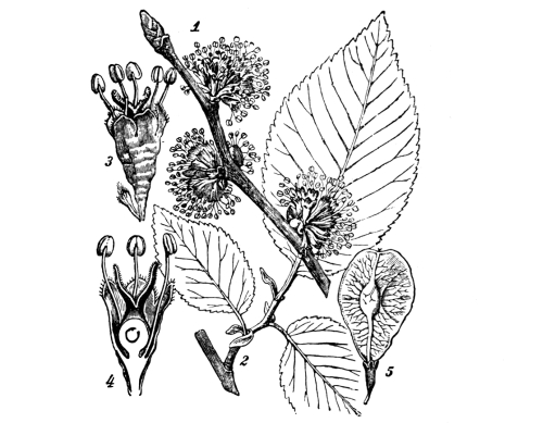
Fig. 124.—The Elm; leaves, flowers and fruit. 1, flowering branch (× ⅔); 2, branch with leaves; 3, a flower (× 2); 4, the same, cut through longitudinally; 5, a fruit (× ⅔).
The lime.—The lime tree (Fig. 125) has a straight smooth trunk. The tree generally spreads at the base, and tapers to a blunt apex. The leaves are bright green, heart-shaped, and pointed, and plainly larger on one side of the midrib than on the other. The yellowish-green flowers are in bunches, carried on a stalk which springs from the middle of a long, narrow bract (Fig. 126). The flowers are complete—calyx, corolla, stamens, and pistil being all present—and are pollinated by bees, which visit them for the sake of nectar, being attracted by the sweet scent. [Pg 155]
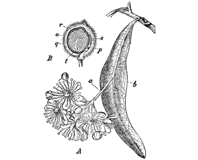
Fig. 126.—The Lime.
A, group of flowers
on stalk (a),
springing from bract (b) (× ⅔);
B,
longitudinal section of fruit (magnified).
The ash (Fig. 127) is a very graceful tree, and its compound leaves, with leaflets springing from the sides of the midrib, give the foliage a characteristically feathery appearance. The bark is ashen-grey in colour. The tips of the twigs are curiously flattened, and the winter buds are jet black. The flowers are small, with two purple-black stamens and a pistil. They open in April before the leaves appear, forming close clusters. The fruits are long and flat, and hang together in bunches which are popularly called keys. They are [Pg 157] only detached by high winds, and are then blown to considerable distances. The leaves appear rather late—about the end of May—and are shed early. They are compound, each consisting of seven or more leaflets arranged in opposite pairs along the midrib, with a single leaflet at the end.
The mountain ash or Rowan tree has leaves somewhat like those of the ash proper, but in other respects it is quite different, as it belongs to the rose family (p. 103).
1. The sycamore.—Observe the size and general shape of the tree. Examine the twigs in winter and watch the buds open in spring. Draw a leaf. Why are the leaves usually so sticky in warm weather? Notice the hanging sprays of green flowers. Are both stamens and pistil present in the same flower? Are the flowers visited by bees? Why? Follow the development of the fruit. Draw a pair of fruits. What is the use of the wing on a fruit? Do the fruits fall off themselves or are they torn off by gales? Look for seedling sycamores in the woods; also germinate seeds in garden soil and again observe the various stages.
Compare the plane, and notice the pyramidal shape of the tree. The leaves are very similar in outline to those of the sycamore, but the flowers are clustered into balls which dangle on long stalks. In summer, pull off a leaf and observe how the axillary bud is covered by the cup-shaped base of the leaf-stalk.
2. The horse chestnut.—Notice the general shape and method of branching. These characters, the large buds in winter, and the shape and size of the leaves in spring and summer, render the tree easily recognisable at all seasons. Trace the connection between the method of branching and arrangement of the leaves and buds on the twigs. Note the date of flowering, and of the unfolding and shedding of the leaves. Examine the flowers; are they pollinated by insects or by the wind? Trace the development of the fruit. [Pg 158]
The sycamore (Fig. 128) is one of the best-known trees in this country, and often grows to a great height. Its leaves (Fig. 33) are large and five-pointed, the main veins spreading from the top of the leaf stalk. When young they are of a beautiful red colour. In hot weather the leaves become very sticky with a sugary syrup called honey-dew. The sycamore is indeed a species of maple, and is closely related to the sugar maple of North America. In May, the flowers hang from the twigs in drooping green clusters. They contain [Pg 159] both stamens and pistils, and are visited by bees for the sake of the honey in which they abound. The fruits or keys (Figs. 137 and 4) occur in pairs, or sometimes three together. Each has a flat, membranous wing, by means of which it is easily transported by the wind.
In Scotland the sycamore is often called the plane. The leaves of the plane and sycamore are somewhat similar in shape, but the trees belong to different families. The plane can be identified by its globular heads of flowers and subsequent balls of seeds, which hang on long stalks from the twigs.
The horse chestnut.—The horse chestnut differs from all our other forest trees of equal size in bearing brilliantly coloured and [Pg 160] conspicuous flowers. The flowers are complete, containing not only stamens and pistil, but also a calyx and a beautiful pink or white corolla. The tree has a striking appearance at all seasons of the year. In winter, the thick twigs, the large terminal buds, and the opposite lateral buds already described on pp. 62-64 are very conspicuous, and give an instructive clue to the method of branching. The buds open early and the new shoots rapidly lengthen, so that the tree is a mass of foliage whilst most neighbouring trees are still bare. The leaves (Fig. 26) are compound, consisting of seven large, spoon-shaped leaflets which spring from a common point at the end of the leaf-stalk. The flowers open in May. The fruit is ripe in October and then falls to the ground, its prickly husk splitting into three parts to liberate the rounded seeds. The leaves fall early, leaving large scars (Fig. 38), which have some resemblance to the hoof-marks of a horse.
1. The Scotch pine.—Notice the shape of the tree: the tall straight stem and rugged bark, and the dark tufts of foliage. Does the tree bear leaves all the year round? Does it ever shed its leaves? Are they shed at any special time of the year? Examine a leafy twig; observe that the leaves are needle-shaped and come off in pairs. At the apex is a terminal bud which will continue the length of the twig next season; the lateral buds below will grow out into twigs at the same time. Try to make out which parts of the twig grew during last year, and which during the two previous years.
Notice the cones. The young female cones (b, Fig. 130, 1) are erect, and their scales separate slightly in spring to allow the pollen to enter. Afterwards they hang down (c) whilst the seeds are ripening. Three years after pollination the scales come apart again to let the seeds fall out. Examine the seeds from a ripe cone and notice the [Pg 161] attached wings (Fig. 130, 4). Examine cones of various ages. In spring look for the pointed cones of male flowers (Fig. 130, 1, a) which produce the abundant pollen.
2. The spruce fir.—Compare the spruce fir. Notice the conical Christmas tree shape and the large spreading branches near the ground. Compare the leaves and cones with those of the pine.
3. The larch.—Compare the larch with the pine and spruce. Notice the drooping boughs, the alternate tufts of leaves, and the small cones arranged in a row along the twig. Is the larch an evergreen?
Cone-bearing trees.—The cone-bearing trees, such as the pines and firs, are true flowering plants, but of a type which is very different from any hitherto described. The flowers are peculiar, and form cones, the male flowers producing pollen and the female flowers ovules. The ovules, however, are not enclosed in ovaries, but are naked, so that the pollen gains access to the ovule directly, and is not received on a stigma. The female cone consists very largely of smooth scales, a pair of ovules being borne by each scale near its base. The pollen grains of these trees are rendered particularly buoyant by being blown out at the sides into little air-filled bladders, and are thus easily carried by the wind. When the pollen falls on the female cone, the grains slide down the smooth scales and very likely come in contact with the ovules at the bottom. Each ovule has a sticky drop of gum at its end, and the pollen is caught in the gum. Such pollen grains as roll off the upper scales are almost certain to fall on a lower one and reach the ovules.
The Scotch pine.—This is a large tree, with a dome-shaped crown of foliage. The bark is rough and scaly. Its foliage leaves (Fig. 130) are long and needle-shaped, and occur in pairs, each pair being carried by a very short branch. The leaves do not fall off each winter, as do those of most forest trees, but remain on the branches for three years or more, so that the younger twigs are clothed with foliage at all [Pg 162] seasons. The cones of male flowers (Fig. 130, 1, a) are found at the base of some of the shoots of the current year. The female cones (Fig. 130, 1, b) are formed round the ends of the young twigs. They consist mainly of overlapping woody scales, each being knobbed on its exposed surface and bearing a couple of ovules near the base, where it springs from the axis of the cone. The young cones are erect, and in spring (when clouds of pollen are blowing about) their scales separate slightly to admit the pollen in the chinks between them. After pollination the scales close again and the cone hangs down (Fig. 130, 1, c). The ovules receive the pollen in May, but the actual union, or fertilisation, does not take place until June of the following year. The seeds become mature two years after fertilisation. When they are ripe each bears a thin wing which has split off from the upper surface of the scale. The scales now separate, and the winged seeds fall out, to be distributed by the wind. When all the seeds have fallen, the empty cones drop off the tree.
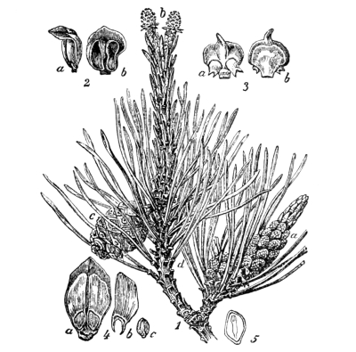
Fig. 130.—The Scotch Pine. 1, branch with male (a) and female (b, c) cones; c, cone (× ⅔): 2, two views of a stamen (× 2): 3, scales bearing two ovules (× 2): 4, scale with two seeds (a), wing (b), seed (c): 5, seed in longitudinal section (× 1½).
Owing to the length of time necessary for the ripening of the seeds, cones of various ages may be found on the tree all the year round.
[Pg 163] The spruce fir.—The spruce has a characteristic conical shape, which is familiar in Christmas trees. The branches are long, and spread horizontally. The foliage leaves are needle-shaped and four-sided in section, but are shorter than those of the pine, and are borne singly. The tree is “evergreen” in the sense that the pine is so, that is, the leaves are not shed all together, but gradually.
The method of pollination, fertilisation, and distribution of the winged seeds is similar to that of the pine. In the spruce, however, the seeds are ripe in October of the year in which the cones are pollinated.
The larch.—The larch is a cone-bearer, and a near relative of the pines and firs, but it differs from them in shedding its leaves annually. The leaves are (as is usual in cone-bearing trees) needle-shaped, but they are very thin and they grow in tufts on short alternate spurs. The cones are small and are arranged in a row on the twig. Their scales do not fit so closely together as those of the pine and fir cones. The larch tree has a conical form, but can readily be distinguished from the spruce by its drooping boughs, and absence of leaves in winter.
Gymnosperms.—The pines, firs, and larches are neither dicotyledons nor monocotyledons, but belong to a class of plants which botanists call gymnosperms, in allusion to the fact that their ovules are not enclosed in ovaries, like those of other flowering plants, but are naked. This is the most ancient and primitive group of flowering plants known. In fact they form a connecting link between higher flowering plants and the group to which the ferns and horsetails—which are still more primitive—belong. [Pg 164]
EXERCISES ON CHAPTER VIII.
1. What trees are the first to put on leaves in England? Which flower before leafing? Which trees are the latest to come into leaf? When do evergreens, for the most part, change their leaves? (N.F.U.)
2. Describe the fruit and seed of four of our commonest trees, explaining in each case how the seeds are distributed. (N.F.U.)
3. Make observations to find which of the following trees endure shade most readily—oak, beech, elm, sycamore, chestnut.
4. What do you suppose are the advantages and disadvantages of shading and crowding trees, as regards the quality of the resulting timber?
5. Study young pines and firs, and find out during which period of life they grow most rapidly.
6. Which trees have you found growing (a) in heavy clay soils, (b) in sandy soils?
7. Describe any observations upon six common British trees which could be made during a walk in early spring. (N.F.U.)
1. The fruit of the wallflower.—Examine wallflower fruits and make out that each consists of the ripened pistil. Does the fruit open of itself? How many chambers does it consist of? Where are the seeds attached? Are they blown off at last by the wind? Draw the fruit. It is called a siliqua.
2. Compare with this the fruit of shepherd’s purse (Fig. 62), and penny cress (Fig. 132), and notice that they are of the type of the wallflower fruit, but are much broader in proportion to the length. Such a fruit is called a silicula. Draw.
3. The fruit of the pea.—Examine a ripe pea-pod and compare it with (a) the pistil of an unfertilised flower, (b) a half-ripe pod. How many carpels have taken part in forming the pod? How many seeds (peas) does the pod contain? Leave a pod on the plant until the shell becomes dry, to find out how the fruit opens. Does it open along one edge only, or along both? How are the seeds attached? Such a pod is called a legume. Draw it.
Compare and draw the legumes of the broad bean, French bean, scarlet runner, laburnum (remember its seeds are poisonous), and bird’s foot trefoil. The legume of the bird’s foot trefoil bursts open suddenly and scatters the seeds in the air. Is the scattering of the seeds any advantage? Why?
4. The fruit of the field geranium.—Make out that five carpels [Pg 166] are grouped around a central rod. Examine fruits which have opened. About noon on a bright, sunny day gently touch a ripe fruit with a small brush, and watch the carpels spring back from the rod and jerk the seeds into the air. Compare and contrast this fruit with a siliqua, silicula, and legume respectively.
5. The fruit of the poppy.—Examine a poppy head. The top of the fruit is the stigma. Observe below this a line of small holes running round the fruit. Draw. Shake the fruit, and notice that seeds fall out through the holes. Cut the fruit across to see the large number of small seeds inside. How does the fruit hang on the growing plant? Does the wind shake it and liberate the seeds?
6. The fruit of the pansy and violet.—Watch the ripening of the fruits on the plants. Observe that the ovary swells up into an egg-shaped body which afterwards splits into three boat-shaped valves containing seeds. Try to make out why the seeds are one by one shot out as the sides of the valves dry. Put a ripe fruit before the fire and watch the process. Imitate it by placing a pea between two flat rulers and pressing the rulers together.
The origin of a fruit.—When the ovules of a flower have been fertilised (p. 92) by the pollen tubes they change into seeds which have the remarkable power of growing up—in favourable circumstances—into plants resembling that which produced the seeds. This is not, however, the only result of fertilisation. Whilst the ovules are changing into ripe seeds, those parts of the flower—the stamens, corolla, and calyx—which have finished their work wither and fall off, though the calyx sometimes remains. Other parts—the pistil and sometimes the receptacle (p. 90)—take on new duties, and become gradually modified in order to protect or scatter the seeds.
Thus, the tender wall of the pistil often becomes a woody, leathery, or juicy seed-case; while the receptacle, or top of the flower stalk, may [Pg 167] become fleshy and swollen with sugary pulp, as a bait for birds and other animals. In any case we give the name of fruit to all such altered and persistent parts together with the seeds which accompany them. A pea pod, for example, is as truly a fruit as a plum, and a poppy-head as a strawberry.
What a fruit is like depends to a great extent upon the characters of the pistil which gave rise to it. If, for example, the pistil consists of several separate carpels, the ripened carpels or fruits will also be separate. When, on the other hand, the pistil is composed of united carpels, these will remain united, at least until the fruit is ripe. Then in some cases they come apart.
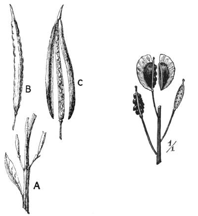
Fig. 131.—A, fruits of wallflower (× ⅕); B, siliqua (× 1); C, siliqua open. |
Fig. 132.—Fruits (siliculae) of Penny Cress. (Nat. size.) |
The part of a fruit which is derived from the walls of the pistil is called the pericarp.
The fruit of the wallflower.—After fertilisation the stamens, petals, and sepals of the wallflower drop off, leaving the pistil alone on the top of the flower stalk. The pistil increases greatly in size (Fig. 131, B) during the ripening of the seeds. At last its wall (the pericarp) splits into two flaps. These become free at the bottom, exposing a central plate which bears the rows of seeds. When the seeds are quite ripe, a slight breeze is sufficient to shake them off, and they fall to the ground to take their chance of finding a place favourable for germination.
[Pg 168] A fruit with a dry pericarp, which opens of itself when the seeds are ripe, is called a capsule. This particular kind of capsule—consisting of two carpels which come apart at maturity, leaving a central partition bearing seeds—is known as a siliqua. When it is short in proportion to its length, as in the shepherd’s purse (Fig. 62) and penny cress (Fig. 132), it is distinguished as a silicula.
The fruits of the pea tribe.—The pod of the pea (Fig. 3) and its relatives is a capsule of another kind. It consists of one carpel only, and opens, when ripe, along both back and front margins to liberate the seeds. Such a fruit is called a legume. In the young fruit the pericarp is somewhat fleshy and succulent, but it becomes dry during ripening. The legume of the bird’s foot trefoil bursts open suddenly, and throws the seeds to a considerable distance.
The fruit of the field geranium (Fig. 133) is a long capsule composed of five carpels arranged round a central column (A). When the seeds are ripe, the carpels suddenly spring from the rod, remaining attached only at their upper ends (B), and fling the seeds into the air. The method may be watched by stroking the fruits very gently with a small brush, when they open in the manner described. The experiment is most likely to succeed in dry, sunny weather, about the middle of the day.
The fruit of the poppy.—The pistil of the poppy swells during ripening into a large, globular capsule known as the poppy head. The top of the fruit (Fig. 134) is the persistent stigma. Just below this, a line of small holes like windows runs round the head. As the fruit [Pg 169] hangs inverted on the top of the flower stalk it is shaken about by the wind, and the tiny seeds fall out through the windows.
The fruit of the violet or pansy.—The arrangement by which the violet (Fig. 134) or pansy sows its seeds is most interesting. After fertilisation the ovary swells into a great egg-shaped capsule, in the inside of which the seeds are arranged in three rows. When the seeds have become ripe and hard, the capsule splits down the side along three lines, and is thus divided into three parts. These open outwards, and [Pg 170] bend back as shown in Fig. 135. Each is a boat-shaped valve. The seeds inside are thus exposed to the air, and they soon dry and become ready for scattering.
Then the curved sides of each valve begin to straighten and come together, and naturally allow less and less room for the hard, smooth seeds inside. The pressure of the sides of each valve on the seeds inside it becomes greater and greater, until one by one they are shot out to a considerable distance. If a ripe violet ovary be warmed before the fire the whole operation may easily be watched. The process may be imitated by putting a pea between two flat rulers and pressing the rulers together, when the pea may be shot to a distance of several yards.
Why fruits are scattered.—A flowering plant is practically confined for life to the place where it first sprang up, so that it is unable to go about and select favourable situations for its offspring. On the other hand, if the seeds simply fell to the ground beneath the parent plant, the seedlings would generally be so crowded together that they would interfere very much with each other’s growth. In addition, they would often be under a great disadvantage, because the parent plant would keep so much light from them. Hence, very many plants have some special arrangement for scattering their seeds, so that some at least of the seeds will have a chance of falling in a place where they will obtain plenty of light, air, and good soil. [Pg 171]
In this section we have studied examples of devices by which plants sow their own seeds. We shall see next that other plants call in the help of the wind and of animals.
1. The fruit of the dandelion.—Examine the manner in which the tiny flowers of the dandelion are grouped together to form the head (p. 113); and make out the various parts of the flower, especially the ovary—a little white knob at the bottom of the flower, and the calyx-tube—forming a tuft of fine hairs above the ovary. Trace the development of the fruits: the withering of the corollas and stamens, the elongation of the calyx-tube, and the expansion of the tuft of hairs to form a parachute. Blow a dandelion “clock” (the head of fruits), and notice how the parachutes float the fruits in the air.
Examine “thistle down” and contrast it with the dandelion fruit.
2. The fruit of the willow.—Examine the ripened catkins of a female willow in June, and notice how each fruit has split into halves, which come apart and expose the silky seeds inside. Pull a tuft of seeds out and dry them in the sun. Notice that they wriggle and writhe about and gradually become entangled together into a woolly mass, which is easily blown away.
3. The elm fruit.—About the end of April look for elm fruits. Notice the flat green plate (wing) with the rounded swelling near the middle (Fig. 124, 5). Cut open the fruit and see that the swelling is caused by a single seed. How does the flat wing aid in the distribution of the seed?
Compare the winged fruits of the ash and sycamore (Fig. 137).
Do these winged fruits drop from the trees easily, or are they torn off by gales?
Take a pair of sycamore fruits. Cut off the wing from one and let it and an uninjured fruit fall at the same instant from a height. Which reaches the ground first? Why?
4. Pine and fir cones.—Examine and compare pine and fir cones [Pg 172] of various ages. Break open a ripe cone and see the scales with the pairs of naked seeds, each seed bearing a thin, papery wing which has split off from the upper surface of the scale.
Wind-sown seeds.—Many plants depend on the wind for the dispersal of their seeds, and consequently the seeds are provided with outgrowths of various kinds, which increase the surface greatly without adding much to the weight, and, acting like parachutes, offer increased resistance to the air and thus prevent the seeds from falling quickly to the ground. In some cases the outgrowth is part of the pericarp, in others it is an appendix carried by the seed itself, while in the lime it is the bract upon which the flowers were formerly borne.
The dandelion fruit.—The fruit of the dandelion (Fig. 136) affords one of the best possible examples of wind-dispersal. It will be remembered (p. 113) that what is commonly called the flower of the dandelion is really a head of perhaps 300 complete flowers: each with a hairy calyx-tube, a yellow, strap-shaped corolla, five stamens, and a pistil. When the flowers have been fertilised, the yellow corollas and the stamens wither, the ovary increases in size with the ripening of the single nutlet in its interior, and each calyx-tube elongates until it is about an inch in length, the tuft of fine hairs being still at its upper end. The attachment of the fruit (Fig. 86, 4) to the disc (receptacle) is so slight when the seed is ripe that a very gentle puff of air is sufficient to overcome it. The tuft of hairs at the upper end has by this time expanded until it acts like a parachute, which supports the tiny fruit for a long time in the air. [Pg 173]
The common thistle—a relative of the dandelion—also distributes its fruit by means of a tuft of fine hairs derived from the calyx. In this case the hairs radiate from the seed. Such “thistle down” is commonly found floating through the air in summer.
The willow fruit.—The catkins of the female willow are ripe in June. Each catkin consists of a large number of tiny pods derived from the ovaries of the flowers (p. 151). Each fruit splits into halves, which bend back from each other (Fig. 121, F), exposing the silky seeds within to the warmth of the sun. The seeds (H) turn and twist about as they dry, and gradually entangle themselves together into a light, woolly mass, which is easily blown to great distances by the wind. The willow and the dandelion, therefore, use very similar devices to ensure the dispersal of their seeds, although these plants are not at all nearly related.
The fruits of the elm, sycamore, and ash.—These common forest trees bear fruits with the seed attached to a flat plate, which is an outgrowth of the pericarp (p. 167). In the elm fruit (Fig. 124, 5) the plate is green and oval, and the seed forms a rounded swelling at, or near, its middle. The fruits of the sycamore generally grow in pairs (Figs. 33 and 137). Each half consists of a single seed with an attached membranous plate, and the two seed-boxes of each pair are in contact. The fruits of the ash hang from the twigs in bunches called “keys,” each fruit on a separate little stalk. The plates which bear the seed are long, narrow, and oval in shape.
It is plain that such plates, or wings, expose a relatively large [Pg 174] surface to the air and prevent the fruit from falling to the ground as quickly as it otherwise would. The fruit is thus often blown to a great distance before it finally settles and the seed germinates. The action of the wing may be well shown by cutting off the plate from a sycamore fruit and letting it and one with an attached wing fall at the same instant from a height. The seed without a wing comes straight down as a pea would; but the winged seed spins in the air and settles more slowly.
In the case of the round downy fruits of the lime, a similar service is performed by the bract (Fig. 126), upon which the flowers were carried.
Pine and fir cones.—If a ripe pine, or fir cone is broken open, it will be seen that each seed is attached to a thin, papery wing (Fig. 130, 4), which has split off the upper surface of the scale bearing the seeds. The winged seeds are shaken out of the cone by the wind, and blown away.
Trees alone bear winged seeds.—Winged seeds would be useless to any but fairly high trees, because if they were formed on the low plant they would fall to the ground long before the wind could catch them properly. It is also interesting to find that such seeds are generally attached so firmly that they are only broken off by gales strong enough to carry them a considerable distance.
On the other hand, the tiny plumed fruit of the dandelion or thistle is so very light in comparison with the surface exposed to the air that it takes quite a long time to fall even a few inches.
1. Hooked fruits.—Examine plants of herb bennet (wood avens) in summer and autumn, and find the fruits. Brush your sleeve against the fruits, and notice how they cling to the cloth. Examine them with a lens and observe the hooks at the ends of the styles. [Pg 175]
Compare goosegrass (cleavers) and find the hooks on the fruits.
2. Nuts.—Examine a hazel nut. Notice the sheathing bracts at the base of the fruit. Crack the nut and examine the broken edge of the shell (pericarp) with a lens. Make out the three layers which compose it. How many seeds are present? Cut the seed (kernel) across and see that the bulk of it consists of two cotyledons (Chap. I.). Does the fruit open of itself, if undisturbed?
Compare the acorn of the oak. Trace the development of the acorn from the female flower, noticing that the cup is developed from a wrinkled disc surrounding the lower part of the flower. Cut across the ovary in June and notice that there are six ovules in it. In the ripe fruit observe that the cup separates easily from the nut. Remove the shell (pericarp) of the nut. How many seeds does it contain? What do you think has become of the other five ovules? Cut through the seed and observe the two cotyledons.
Compare the fruits of the beech. Notice that they are three-sided nuts (each being a seed enclosed in a woody pericarp); and that two nuts occur together, surrounded by a bristly woody cup which splits, when the nuts are ripe, into four valves.
3. Stone fruits.—Examine a ripe plum. Cut it open and crack the stone to see the single seed. Notice that the pericarp consists of three layers as in the hazel nut, but that here the middle layer is soft and fleshy, and the outer layer is the “skin.” The stone is the inner layer of the pericarp. Is there any special means of liberating the seed? Compare the cherry, and examine the seed and the three layers of the pericarp.
Examine the fruits of the blackberry and raspberry, and observe that each consists of several small stone-fruits arranged on the receptacle.
4. Berries.—The gooseberry.—Notice the stalk at the bottom of the fruit, and, at the top, the withered remains of the calyx. Cut across a half-ripe gooseberry and observe the thick, fleshy pericarp enclosing the seeds. Treat a ripe gooseberry in the same way, and [Pg 176] observe that the pericarp has now for the most part become a soft pulp, in which the seeds are embedded. The rest of the pericarp is a membranous skin. Is there any special means of liberating the seeds?
Compare grapes, currants, oranges, and vegetable marrow fruits, and notice that the structure of these resembles that of the gooseberry.
5. The apple.—Cut across the receptacle of the flower (apple blossom) and notice how the five carpels are buried in it (p. 106). Trace the formation of the fruit, and see how the receptacle becomes larger and larger during ripening. At the top of the ripe fruit observe the withered remains of the calyx. Cut the apple through across the middle to see the core. This consists of the horny walls of the five carpels and the contained seeds (pips). From what part of the flower is the fleshy, eatable part of the apple derived? Compare the pear.
6. The rose hip.—Examine fruits of the wild rose. Notice that they are urn-shaped. On the flat rim of the urn observe the five scars left by the sepals (or, in some cases, the sepals themselves). In the opening of the urn see a tuft of greyish hairs. Cut the fruit down from top to bottom through the middle to see the thick, fleshy wall of the urn and the contained nutlets. From what part of the flower is the fleshy wall of the hip derived? Examine a nutlet. What does the tuft of hairs seen in the mouth of the urn consist of? Open a nutlet with a needle and pick out the seed.
7. The strawberry.—Cut a strawberry down the middle and notice at the base the persistent calyx, in the inside the fleshy receptacle, and on the outside the yellowish nutlets. Open a nutlet with a needle and pick out the seed.
The help of animals.—It has been seen in Chapter VI. that insects play a very important part in the fertilisation of many flowers. Very many plants also call in the aid of animals at a later stage for the dispersal of the seeds, and the devices by which this aid is obtained are often very ingenious. [Pg 177]
Hooked fruits.—Sometimes after a country walk the reader has probably found small fruits and seeds sticking to his clothes. They have become attached to the cloth by means of small hooks which they carry. The fruit of the herb bennet (wood avens) is a good example of this device. When the stigma of the fruit breaks away, a little hook is left at the top of the style (Fig. 138). Goosegrass or cleavers—a common hedgerow plant—and many others have also hooked fruits. Sheep or cattle, grazing near such plants are very likely to brush against them and carry off the fruits and seeds in their hair. They may not again reach the ground until they have been carried far from the place where they grew.
The position of hooked fruits.—It is evident that these little hooks would be quite useless if the fruits grew out of the reach of animals. Such hooked fruits are never found, for instance, on high trees.
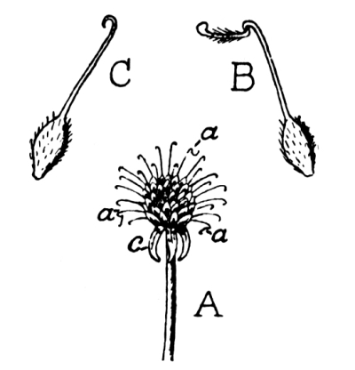
Fig. 138.—A, Aggregate Fruit of Wood Avens (nat. size); most of the stigmas have broken off. a, a, a, fruits with stigmas still attached; c, calyx; B, single fruit with stigma still attached (magnified); C, single hooked fruit after loss of stigma (magnified).
Nuts.—A fruit which has a dry, woody pericarp and does not open of itself is called a nut. The fruits of the hazel, oak, and beech are good examples. The shell (pericarp) of the hazel nut (Fig. 139) is composed of three layers, and encloses a single seed, or kernel. The nut of the oak (Fig. 113) is called an acorn. Its lower part lies in a cup developed from a wrinkled disc by which the lower part of the female flower was surrounded. When the fruit is ripe the nut easily separates from the cup. The acorn contains one seed only, which consists largely of two swollen cotyledons. If the ovary of [Pg 178] the female flower of the oak is cut across in June six ovules are found in it. As in the case of the hazel only one ovule is allowed to reach maturity; the rest are sacrificed in order that the remaining one may be more perfectly developed. In the beech, two three-sided nuts occur together, surrounded by a bristly, woody cup, which splits into four valves when the nuts are ripe.
The dispersal of nuts.—Squirrels and other nut-eating animals are instrumental in the dispersal of the seeds in a somewhat indirect manner. They have a habit of storing up nuts and seeds in holes; but in their active life these animals often forget where their larder is. The seeds, thus left to themselves, sprout and grow into trees. The unripe nuts of the beech are protected from the attacks of squirrels by the hard bristles on the outer husk.
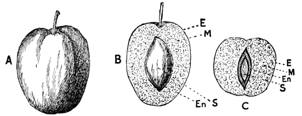
Fig. 140.—A, Plum; B, longitudinal section; C, cross section; E, M, En, outer, middle, and inner layers of pericarp; S, seed. (× ½.)
[Pg 179] Stone fruits.—A ripe plum (Fig. 140) or cherry (Fig. 77), like a nut, consists of a seed and a pericarp; but here the pericarp is specially modified to tempt animals and at the same time to protect the seed from them. The inner pericarp-layer is a hard, woody shell called the stone; the middle part swells up to form a juicy, sweet mass; while the outer layer constitutes the “skin,” and is often beautifully coloured.
Blackberries (Fig. 141 F) and raspberries consist of several small fruits of the plum type, which are arranged round an axis derived from the receptacle of the flower.
Berries.—The gooseberry (Fig. 142) is a type of this class of fruit. It contains several seeds. The pericarp is thick and fleshy before the fruit is ripe, but during ripening the greater part of it becomes a soft, sweet pulp in which the seeds are embedded. The rest of the pericarp is a membranous skin. Grapes, currants, oranges, and vegetable marrow fruits are also berries. The vegetable marrow has obviously a general resemblance to a half-ripe gooseberry but is on a much larger scale.
The apple.—In the apple (Fig. 143) we have an example of a fruit in the formation of which the receptacle has taken a large share. Even in the flower, the five carpels of the pistil are buried in the [Pg 180] receptacle, and as the seeds (pips) ripen, the receptacle swells until it composes the greater part of the fruit and at last becomes sweet and fleshy. The carpels with the contained seeds constitute the core. The withered sepals are still to be seen at the top of the fruit. The structure of the pear is quite similar to that of the apple.
The fruit of the wild rose.—The fruits of the wild rose are called hips. They are urn-shaped, and on the flat rim of the urn five scars show the former position of the sepals.
In some cases the sepals themselves have remained. The narrower mouth of the urn is filled by a tuft of greyish hairs which, when the fruit is cut open, are seen to be the styles of the carpels. The carpels are [Pg 181] hairy, and stand on the bottom and sides of the urn-cavity. Each carpel contains a seed. Comparison with the flower shows that the red, fleshy urn is the developed receptacle.
The strawberry.—In the strawberry (Fig. 144) the eatable part of the fruit is again the swollen and juicy receptacle. In this case it has grown up on the inside of the carpels—the little, yellow nutlets (carpels) lying on the surface. Each carpel contains a seed.
Why some fruits are sweet.—The delicious flavours of sweet fruits have been developed as baits for the allurement of such animals as are likely to scatter the seeds to the best advantage. Consider such fruits as cherries, currants, and rose-hips. Birds—such as thrushes—find them very nice to eat, and are ready to carry them away to consume at their leisure. The birds eat the sweet, fleshy part, but in the case of a stone-fruit they drop the stone and leave the seed to germinate. A seed which is in danger of being swallowed is either hairy like the rose-carpels (and therefore rejected because it irritates the mouth unpleasantly), or it is enclosed in a hard or horny case, upon which the animal’s digestive juices are unable to act. When the seed is dropped it is no worse for its experience, and with good luck grows up into a plant.
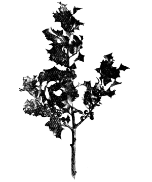
Fig. 145.—Holly. The red colour of the fruits
contrasts strongly with the dark-green
of the leaves. (× ⅙.)
[Pg 182] Eatable fruits generally conspicuous.—In order that birds and other animals may easily find the luscious fruits which they may eat as a reward for scattering the seeds, such fruits are almost always displayed very conspicuously, and are brilliantly coloured. Oranges, plums, red currants, apples, etc., illustrate this fact well. Red is perhaps the commonest colour of eatable fruits, because it contrasts so strongly with the green colour of the foliage (Fig. 145).
EXERCISES UPON CHAPTER IX.
1. Name and define the different kinds of self-opening fruits. From what common plants could you collect examples of these during an Autumn walk? (N.F.U.)
2. Describe any rough, prickly, or hairy seeds or fruits, explaining the form and nature of the outgrowths and their use. (N.F.U.)
3. What wild fruits and wild flowers would you expect to find in September in your part of the country? In what kinds of places would you look for them? (N.F.U.)
4. Describe and draw the fruit of a field geranium, and point out the uses of some of its peculiarities. (1898)
5. Describe the seed vessels of a pansy. Draw one entire, and also burst open. How does it scatter the seeds? (1898)
6. Shortly describe the fruit of an apple or pear, and of a cherry or plum. Point out the chief differences between them. (1901)
7. How are the seeds of cherry, field geranium, and pine or birch dispersed? (1901)
8. Give examples of seeds which are dispersed by the aid of birds or other animals, explaining in each case how the dispersal is effected. (1893)
9. Why is it an advantage to some fruits to be (a) brightly coloured, (b) sweet? Give examples.
10. A school museum contains, among other things, some dandelion fluff, a dish of marrowfat peas, a few nodules of garlic, and some hawthorn berries. How could you employ these to illustrate a lesson on plant germination? (Certificate 1903)
1. The male-fern.—(a) Habit of growth.—In summer dig up a plant of the common male-fern (Fig. 146) and wash the soil from the roots. Make out the short, stumpy, creeping stem, covered with the hairy bases of old leaves; the slender matted roots springing from the leaf-bases; the large, compound leaves or fronds of the current year, and the coiled young leaves which have not yet expanded.
(b) The stem.—Remove the large leaves, leaf-bases, and roots from the stem, and examine it. Notice how the youngest leaves are grouped round the apex of the stem. Cut across the other end of the stem, and notice the cut ends of the conducting strands embedded in a softer ground-tissue. Cut the stem in halves lengthwise and carefully scrape away the ground-tissue of one half to see how the harder conducting strands are connected together. If you spoil this, try again on the other half, after boiling it until it is softened.
(c) The leaves.—Make a drawing of one of the expanded leaves. Notice that the leaf consists of a number of leaflets, and that these are again cut up into segments, or are at least deeply lobed. Notice the brown hairs clothing the leaf-stalk and the midrib.
Notice the crozier-like coiling of the young fronds, and make out that when they uncoil the inside of the coil becomes the upper surface of the leaf. [Pg 184]
(d) The reproductive organs.—Examine the lower surface of full-grown fronds, and notice the brown rounded patches. Dry some leaves bearing these patches and shake them over a sheet of paper. Collect the brown dust which falls from the patches; it consists of minute grains called spores.
(e) The prothallus.—Sow some spores on damp soil sheltered from the direct rays of the sun. Keep the soil moist, and notice that in a few weeks the spores have grown into small, flat, green, heart-shaped plants. Each of these is called a prothallus. Pick off prothalli of different ages with a needle, float them in water, and compare the various stages of growth. In old prothalli notice a young fern plant springing up from the lower surface. Observe that as these become larger the prothalli which bear them shrivel up and die.
2. The bracken, fern.—Dig up a bracken fern, wash the earth from the roots, and compare it, point for point, with the male-fern. Notice:
(a) The long cylindrical, branching, underground stem. Does it lie deeper in the ground than the stem of the male-fern? Cut the stem across and compare the section with Fig. 148, noting (i) the two concentric rings of separate vascular strands (s). (ii) The strengthening material, arranged as a brown zone (lp) just below the skin, and as an incomplete ring (ll) between the two rings of vascular strands. (iii) The softer ground-tissue (R), in which the vascular and strengthening strands are embedded. Draw the external appearance of the stem (natural size), and also the cross section (4 times natural size).
(b) The roots.—Describe the roots. Do they appear to arise from any particular region of the stem?
(c) The leaves.—From what part of the stem do the leaves arise? How many come up each year? What is the appearance of a young leaf before it expands? How does it differ from that of the male-fern? Draw a young leaf, and also an expanded one. Is the leaf simple or compound? Does it branch? Does the leaf of the male-fern branch?
(d) The spores.—Examine the under side of the leaf, and notice the absence of the brown patches seen in the male-fern. Notice that the [Pg 185] edge of the bracken frond is folded over like a hem. Dry a frond, and then run the point of your pencil under the fold and observe the brown dust (spores) which is removed.
(e) The prothalli.—Sow some bracken spores on damp earth and keep in a shaded place. Notice that the prothalli produced are very similar to those of the male-fern. Watch their development and the growth, on old prothalli, of a new generation of ferns.
3. The hart’s tongue fern.—Examine the hart’s tongue fern, and notice how it differs from the two previous types. Are the leaves simple or compound? Observe the brown trenches on the lower surface of the leaf. Try to get out some spores by running the point of a pencil along the trenches of a dried leaf. Sow the spores and try to raise prothalli.
The daily life of a fern.—The everyday life of a fern is very similar to that of a flowering plant; for the organs by which it obtains food are—broadly speaking—of the same type. Ferns are lovers of damp and shady situations. The plant obtains its mineral food from the soil by means of its roots; and its spreading green leaves, or fronds, enable it, with the help of the sunlight, to decompose the carbon dioxide of the air and to build up starch, sugars, and other carbonaceous compounds.
The male-fern.—The common male-fern (which, by the way, has no sex whatever) may be taken as a type of the group. It occurs abundantly in woods and hedgerows. The stem, or rhizome, is short and stumpy; it grows obliquely upwards, and does not branch. It is covered with old leaf-bases, which are clothed with brown, scaly hairs. The stem consists of a rather soft ground-substance, in which is embedded a hollow cylindrical network of conducting strands. In a cross section these appear as a somewhat irregular ring of dots (Fig. 146, 2, a). When the soft ground-tissue is carefully scraped away, the network of strands is left as a skeleton, with large diamond-shaped meshes. [Pg 186]
The thin wiry roots spring from the bases of the leaf-stalks, just where these come off the stem.
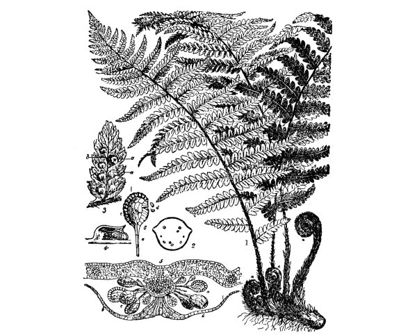
Fig. 146.—The Male-Fern. 1, Illustration showing general habit (× ¹⁄₁₀); a, young leaves: 2, cross section of stem showing conducting bundles a: 3, portion of leaf; b, spore-boxes; a, covering scale: 4, longitudinal, and 5, cross section of group of spore-boxes; a, leaf; b, scale; c, spore-boxes: 6, a single spore-box; a, stalk; c, spring; d, spores.
The leaves or fronds of the male-fern arise near the growing-point (the upper end) of the stem. Leaves of almost all ages are present. Those which surround the growing-point are mere rudiments; while slightly older ones (Fig. 146, 1, a) are tightly rolled up into a coil. During their growth the coils unfold, the inside of the coil becoming the upper surface of the leaf. A fully expanded leaf is roughly triangular in shape. The leaf-stalk and the midrib are covered [Pg 187] with brown, scaly hairs; and the blade of the leaf is divided into several distinct leaflets, which are arranged on the midrib in two rows. The leaflets are also in many cases sub-divided into separate segments.
The spores.—On the lower surface of many of the segments of a male-fern frond may be seen a number of small, brown, kidney-shaped scales (Fig. 146, 3, a). Each scale is attached to the leaf by a short stalk, and the structure thus bears a rough resemblance to a little umbrella. Attached to the bottom of the short “handle” of the “umbrella” are several tiny boxes, somewhat like pill-boxes. These are shown at b (Fig. 146, 3), and highly magnified at 4 and 5. Fig. 146, 6, represents a single box, more highly magnified. When ripe, each box contains about fifty minute grains, which may thus be likened to the pills in the pill-boxes. These “pills” are called spores. Summing up thus far, we may say that the spores are formed in spore-boxes, which are attached by stalks to the lower surface of the frond, each group of spore-boxes being covered by a protective scale.
The scattering of the spores.—When the spores are ripe, each box (Fig. 146, 6) becomes dry, and is ultimately burst by the sudden straightening of a spring (c) which is coiled round its edge. The force of the uncoiling of the spring is sufficient to jerk the spores (d) out of the box into the air, and they may be carried for some distance by the wind before they at length reach the ground. Once there, however, each spore, under favourable conditions, begins to grow, and gives rise to a plant which, curiously enough, is not in the least like the parent fern plant which produced the spore.
The difference between a spore and a seed.—It is important to notice that the spore is produced by a purely non-sexual process. In this respect it differs widely from the seed of a flowering plant, [Pg 188] which, it will be remembered (p. 92), results from the union of the living matter of a pollen grain with that of an ovule.
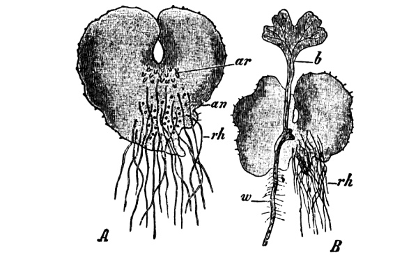
Fig. 147.—The Male-Fern. A, Prothallus seen from below; an, male organs; ar, female organs; rh, hairs. B, Prothallus with young fern attached to it; b, the first leaf; w, the primary root (about 5 times nat. size).
The prothallus.—The new plant, which is produced when a fern spore germinates, is called a prothallus (Fig. 147, A). It is a flat, filmy little plant, of the form which is generally called heart-shaped. It has neither stem nor roots, but, as it contains green colouring matter like that of leaves, and as it puts out on its lower surface little hairs (rh) which take up watery solutions from the soil, it is in no danger of starvation, and is quite capable of taking care of itself and leading an independent existence. This tiny plant (a large fern prothallus is perhaps half the size of a 3d. piece), in contrast with its parent, produces sexual organs. Some of these organs (an) give rise to male cells and others (ar) to female cells. The male cells are excessively small, and can only be seen by high powers of the microscope. When they are ripe they swim about in a drop of rain or dew, as if they were little animals, and find their way to the female cells, which they fertilise. The embryo which results from the union grows up (Fig. 147, B) into an ordinary fern plant, one being borne by each prothallus. In its young stages it is parasitic on the prothallus, i.e. it depends entirely upon the prothallus for its nutrition; but it soon develops a little leaf (b) and a root (w), and henceforth feeds itself. When the young fern is well established in [Pg 189] independent life, the prothallus shrivels up and dies. The subsequently-formed roots of the fern are not branches of the primary root, but spring from the bases of the leaves.
Alternation of generations.—In the life-history of the fern there are thus two very different generations. The first generation—the ordinary fern, which is non-sexual—produces, by means of spores, the sexual generation, called the prothallus. The prothallus gives rise, in its turn, by a sexual process, to the obvious fern plant. Each generation therefore resembles, not its parent, but its grandparent.
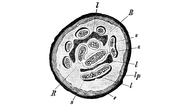
Fig. 148.—Cross section of underground stem (rhizome) of Bracken Fern. s, Conducting strands; l, lp, strengthening tissue; R, ground-tissue; e, skin. (× 7.)
The bracken or brake fern differs in several respects from the male fern. Its cylindrical stem (rhizome) creeps along horizontally beneath the ground, branching at intervals, by a division of its growing point into two. Conducting strands run along the stem and into the leaves and may be seen in cross section (Fig. 148, s) [Pg 190] to form two somewhat irregular, concentric rings. Strands and plates of strengthening tissue (l) accompany them, and a cylindrical zone (lp) of similar material also occurs just beneath the outside skin (e) of the stem. The great advantage of having supporting structures arranged as hollow cylinders has already (p. 72) been referred to. The rest of the stem consists of softer packing or ground-tissue, in which starch is often stored.

Fig 150.—Hart’s
Fig. 151.—Hart’s Tongue Fern. Part of a section through the fertile
Tongue Fern. (× ⅙.)
portion of a leaf; sg, spore-boxes; i, i, protecting flaps. (× 25.)
The sappy stalk of a young bracken leaf is very sturdy; when it is only six inches high the stalk may be already half an inch across at the bottom, and half that thickness where it curls over at the top to form a crook. The end of the leaf-stalk divides into three, each bearing a frond which, at this stage, is coiled up tightly, looking somewhat like a green caterpillar. At a later stage, the middle one of the three branches commonly divides again into three, and the branching may continue until the fully expanded leaf (Fig. 149) is very complex. [Pg 191] The spore-boxes and contained spores of the bracken are very similar to those of the male fern, but they are not collected in patches, like those shown in Fig. 146, 3. Instead, they are arranged in a row along the margin of the lower surface of the frond-segment; and the margin is turned over—like a hem—so as to cover them in.
The spores are liberated in the usual way (p. 187) when ripe; and each germinates, under favourable conditions, to form a sexual prothallus. Each prothallus gives rise to an embryo, which is at first parasitic upon it, but presently grows up into an ordinary bracken.
The hart’s tongue fern (Fig. 150) has simple and undivided leaves, which, as usual, bear spore-boxes upon the lower surface. In this case the spore-boxes are produced in trenches, which appear to the naked eye as oblique brown lines. Each trench is covered in by a pair of thin flaps (i, Fig. 151). The life-history closely resembles that already described for the male-fern and bracken.
1. Habit of growth.—Carefully dig up a plant of the common horsetail. Notice (a) the deep, creeping, underground stems; (b) the thin wiry roots springing from the stem; (c) the two kinds of upright shoots or haulms: one kind (Fig. 152)—to be found in summer—being thin, and giving rise to tiers of green branches, which come off like the ribs of an umbrella; the other kind (Fig. 153), coming up in March, being paler in colour, without branches, and bearing little cones at the apex.
2. The underground stem.—Make out that the stem is distinctly divided into nodes—marked by toothed leaf-sheaths—and internodes. Does it branch? Notice that some of the branches are swollen, forming tubers.
3. The roots.—These are thin and wiry, and come off from the stem. Do they come off at the nodes or at the internodes?
4. The branched haulms.—Observe that each is divided into [Pg 192] nodes—marked by toothed leaf-sheaths—and internodes. Are the internodes smooth or ridged? From what parts of the haulms do the lateral branches arise? Carefully tear down a leaf-sheath to see its relation to the branches. Notice that the branches themselves branch repeatedly. What is the colour of the branches?
5. The cone-bearing haulms.—Do these haulms appear earlier or later than the others? What is their colour? Does a cone-bearing haulm give rise to branches at the nodes? Examine the cone with a lens and see that it is covered with hexagonal scales. Does the hexagonal shape allow the scales to be more closely packed? Take off a scale with a needle and notice, with the help of a lens, the spore-boxes attached to its inner surface.
6. The prothallus.—Dry a ripe cone and shake it over paper to collect the spores. Sow these on moist soil, and try to raise prothalli. [Pg 193]
The general appearance of the horsetail.—The common horsetail (Fig. 154), which may be found in hedgerows and cornfields, is not in any respect showy. It rarely exceeds a few feet in height, and does not attract attention by any display of bright colours. The plant has stiff, jointed stems or haulms, standing gracefully erect and bearing rings of small united leaves at intervals. The branches arise just above the leaves, and alternate with them: several coming off at each level, somewhat like the ribs of an umbrella. The whole plant has thus a rather stiff and formal aspect.
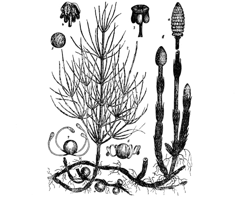
Fig. 154.—Common Horsetail. (× ⅙.) 1, Fertile haulms, terminating in cones (a): 2, a sterile haulm; a, tubers: 3, scale of cone with spore-boxes (mag.): 4, scale with ruptured spore-boxes (mag.): 5, 6, 7, spores (mag.).
[Pg 194] The haulms are very plainly of two kinds; for some, which are of a pale colour, do not branch, and are developed merely to bear the pretty little cones (Fig. 154, 1, a) which appear at their upper ends. As the purpose of the cones is to give rise to the next generation, the haulms which bear them may be called the fertile haulms. They will be described presently, when the reproduction of the plant is considered.
The sterile haulms.—The erect branching shoots (Fig. 154, 2), bear no organs of reproduction, and are hence referred to as the sterile haulms. Their work is to provide the whole plant with carbonaceous food, and it is on this account that their branches are of the characteristic green tint. In most green plants the manufacture of carbonaceous food (Chapter III.) takes place mainly in the leaves. In the horsetail, however, the leaves are small and of very little importance in this respect, and the work is carried on by the sterile haulms and their branches.
The underground stem and roots.—A large part of the horsetail plant is hidden beneath the surface of the ground, and often penetrates to a great depth. In ordinary language these underground parts are called roots. They are, however, subterranean stems, as is shown quite plainly by the fact that they bear small leaf-sheaths like those on the stems above ground. In the common horsetail the underground stems often become swollen in parts by the formation of tubers (Fig. 154), as is the case with the potato. The true roots are very slender and thread-like, and spring from the nodes (p. 45) of the underground stems. They penetrate the soil in all directions, seeking for water, which they take up by the fine hairs which clothe them, like velvet-pile, a little behind their points. The stem and its haulms, like those of grasses, are stiffened by silica, which is deposited in [Pg 195] the outer layers. This is of course obtained from the soil. It is not absolutely necessary to the life of the plant, but it is nevertheless very useful, as it enables the haulms to stand upright and spread out their branches to the light and air.
The sterile branches are thus concerned with the horsetail’s daily life. The roots provide it with water and mineral food, and the green branches supply the necessary carbonaceous matter. Stem, branches, and roots are permeated with a complete system of canals, through which the food substances find their way to the various centres of activity.
The reproduction of the horsetail.—It has been seen that some of the haulms do not bear branches, but are set aside for the production of the cones. These fertile haulms come up in March, before the green sterile haulms appear. When the cones are carefully examined they are seen to be covered by a number of shield-shaped scales. To the inner side of each scale (Fig. 154, 3, 4) are attached from five to ten boxes, each containing a large number of little grains or spores. The boxes burst open when they are ripe, about the end of March, liberating the spores; and the fertile haulms, having performed the one duty for which they were developed, at once die down.
If the spores fall in a favourable situation they germinate, and each gives rise to a new plant. The new plant is, however, not a horsetail, but a small filmy prothallus, somewhat like the prothallus of a fern. Some of the prothalli produce male cells, while others give rise to female cells. When the minute male cells are ripe, they are set free, and are able, by means of fine, lashing threads, to swim towards the female cells of a neighbouring prothallus, through a drop of dew or rain. The two cells fuse together and give rise to a little embryo, which in due course grows up into a new plant—an ordinary horsetail with stems and branches and roots like its grandparent.
[Pg 196] The advantage of an alternation of generations.—The life-history of the horsetail is evidently very similar to that of a fern, each exhibiting a well-marked alternation of generations. There is reason for believing that the prothallus-generation is the original form, and that the ordinary fern and horsetail were developed simply to scatter spores at intervals, and so to give new plants the advantages of fresh soil.
Living and extinct horsetails.—None of the British horsetails is of great height, although one species may attain to six feet. Some of the tropical members of the family, however, are very much larger than this, reaching even forty feet. In spite of the last-named fact it is quite plain that the horsetails have had their day, and are dying out. If we wish to form an idea of what they were at the height of their prosperity, we must carry our minds back to the long distant age when our coal was being formed; when so much of this country as then existed was low-lying swamp, covered with exuberant vegetation. Then the horsetails and their relatives were stately forest trees, and at the head of the vegetable kingdom. Some of them towered to a height of over ninety feet. Nor did mere height constitute the only difference between them and their degenerate descendants. Many of these old-world giants had already found out the device, since invented afresh by more modern plants, of thickening their stems and roots with secondary wood, and of giving rise to bark like that of our present-day forest trees. The wood of these Calamites (Fig. 155), as they are called by [Pg 197] geologists, had already reached much the same stage of development as is found to-day in such a tree as the yew. The cones often show great variation from the comparatively simple form found in the modern horsetail, but they are essentially of the same type.
Flowering and flowerless plants.—Botanists divide plants into two great groups—flowering plants, which reproduce themselves by means of pollen grains and ovules; and flowerless plants, which still retain the primitive marriage customs of their ancestors. The flower is a comparatively recent invention in the history of plant life, and its success is shown by the dominant position in the plant world which the flowering plants now occupy.
Many of the flowerless plants—like the horsetails—have fallen behind their competitors. The ferns, in spite of their conservatism, still hold their own, and seem in no danger of extinction. Many other flowerless plants maintain their position by sheer force of numbers. Yet others have become completely extinct, and can now be known only by their fossil remains. But these latter are sometimes so distinct that stems, roots, and spore-boxes can be seen with all their sharpness of outline unimpaired; the delicate tracery of frond and leaf is visible, as clear and fresh as if made yesterday. And such rocky herbaria tell us in unmistakable terms that our forest trees and other flowering plants are after all mere parvenus and upstarts.
EXERCISES ON CHAPTER X.
1. Explain the formation of the green scales which are frequently seen on the surface soil of a fernery. Whence do they arise? What happens to them if they are allowed to grow? (King’s Scholarship, 1902)
2. Point out the differences between the fronds of a fern and the leaves of most flowering plants. (1901) [Pg 198]
3. Show how the spore-producing plant of a fern is attached to the prothallus, and trace the early development of the former.
4. Make a list of the ferns which you have seen growing wild, and state exactly in what kinds of places they were growing.
5. Make experiments to prove that the prothallus of a fern is capable of manufacturing starch.
6. Make drawings of the spore-bearing leaves of all the ferns you can find, marking in each case the position of the spore-groups.
7. In what situations have you found horsetails growing? What other plants were growing near?
8. What is the difference between a seed and a spore?
9. Describe the situation in which the main stem of the bracken is found, its mode of growth, and the simpler facts of its structure. (1904)
10. In what respects do you consider the rhizome of a fern (a) similar; (b) dissimilar to the root of a tree? (Certificate, 1905)
11. Where are the spores of a fern formed, and how are they dispersed? What do they produce on germination? (1905)
1. A common liverwort.—Look along the sides of a brook or a well, and try to find a flat green plant with numerous lobed and overlapping branches. Each branch is perhaps half an inch across. This is one of the commonest liverworts (Pellia). Notice the prominent midrib running along each branch. In spring, observe the “frilled” appearance of the end, caused by the small new branches.
Pull the plant up, and notice that it is attached to the soil by a large number of fine hairs which spring from the lower surface of the midrib.
In February, or March, examine the upper surface of the growing plant with a lens, and notice the small, dark-green balls, mounted on short, thick stalks. Examine these at intervals until May, and notice that the stalks then grow rapidly until they are two or three inches long. Each is white, and still bears the black ball (the capsule) on its summit. When the stalk is full-grown, the capsule opens—its wall splitting into four parts—to liberate the spores. How soon, after the liberation of the spores, do the capsule and its stalk die down?
2. A common moss.—Separate a single plant from a tuft of the common moss (Funaria) which grows, almost everywhere, on the ground and on walls. Notice that the plant consists of a stem perhaps half [Pg 200] an inch high, thickly covered with small simple green leaves. The moss is fixed in the soil by a tuft of fine hairs.
On some of the plants notice a thin stalk (about half an inch long) springing from the top of the stem; and on the end of the stalk an ovoid capsule or spore-box. In cases where the stalk is not yet full-grown, notice that the capsule is covered by a conical hood, somewhat like a candle-extinguisher.
Select a plant bearing a ripe capsule, and warm it gently before the fire—holding it over a sheet of paper—to dry it. Examine the mouth of the capsule with a lens, and try to see the teeth which surround the opening. Then shake the capsule over moist soil in a small flower-pot to scatter the spores; cover with a sheet of glass and keep in a warm room. In a few days notice that the soil is covered with fine green threads. Ultimately new moss plants will grow from these.
The life-history of a liverwort.—One of the commonest and simplest of this class of plants is known to botanists as Pellia. It may generally be found growing by the sides of streams or old wells. It has neither stem, leaves, nor root, but consists of flat, green, overlapping lobes which fork at their ends. It branches very freely, and in spring the new branches give the ends of the lobes a frilled appearance. A rather prominent midrib runs along each branch. The plant is attached to the soil by a large number of fine hairs which spring from the lower surface of the midrib.
The whole appearance of this liverwort is very suggestive of the prothallus (Fig. 147) of a fern; and, indeed, it corresponds to a prothallus, not only in its general structure and mode of life, but also in bearing sexual organs of a very similar type. It was seen in Chapter X. that a fern prothallus at length produces an embryo, which grows up and forms the spore-bearing generation which is what people usually understand by a “fern”.

Fig. 156.—A Moss. A,
a plant with spore-case
still covered by hood (c)
(× 1). B, a plant with ripe
spore-case (k); s, stalk;
d, lid; rh, hairs
(× 1). C,
mature spore-case with
lid (d) removed; p, fringe
of teeth (× 3).
The liverwort also gives rise to a spore-bearing generation, but in [Pg 201] this case it consists merely of a small, round, spore-box, dark-green in colour, which is carried on the summit of a white stalk—the whole looking somewhat like a stout pin. In February, or early March, the spore-boxes may be seen upon the upper surface of the plant as small balls, perhaps one-sixteenth of an inch in diameter, protruding from the mouth of a little pocket in which their early stages are passed. About May, their stalks lengthen so rapidly that in a few days the spore-boxes are lifted to a height of two or three inches. Then each box opens by its wall splitting into four, and the spores are liberated to germinate and form new liverworts. Having thus shed the spores, the box and stalk die down.
The complete life-cycle of a liverwort thus includes two unlike generations, as that of a fern does; but it is a different generation which attains the greater development in the two cases. The ordinary fern is the spore-bearing generation; the “ordinary” liverwort is the sexual generation, and its sporing offspring is not a separate plant at all, but a mere stalked box, almost entirely dependent upon its parent.
The mosses.—A moss-plant appears at first sight to be very similar in its general features to one of the higher plants; for it has a little stem, bearing flattened green leaves which build up carbonaceous food in the usual way. It has no true roots, but fine hairs penetrate the soil and do the work of roots by taking up solutions of mineral food. It is all the more remarkable, therefore, to find from its method of reproduction that the moss-plant belongs to the generation which corresponds to the prothallus stage of a fern’s life-history, and not to the leafy, sporing generation which it somewhat resembles superficially.
The sexual organs of a moss are essentially of the same type as those [Pg 202] which a prothallus bears, and here also the tiny male cells gain access to the female cells by swimming through a drop of rain or dew. The result of fertilisation is an embryo which, however, grows up to form, not a plant with stem and leaves, but a stalked spore-case only.
In a tuft of the moss Funaria, which is so common in all country places, several stages of development of the spore-cases may usually be seen. Fig. 156 represents a moss which is very similar to Funaria but larger. In A, the spore-case is still covered by a conical hood (c). In B, the stalk (s) has grown much longer, and the hood has dropped off. The spore-case or capsule (k) is now seen to be a pear-shaped body, closed by the lid (d). When the spores are ripe, the lid becomes detached. The mouth of the nodding capsule is still blocked, however, by a number of teeth (Fig. 156, C, p) which remain close together in damp weather. In dry weather the teeth separate, and allow the spores to fall out. When a moss spore falls in a favourable situation it germinates to form a fine, branching network of green threads, from which new moss-plants arise as buds.
The obvious plant of a moss—like that of a liverwort—therefore corresponds to the prothallus generation of a fern or horsetail. Its sporing generation, corresponding to the obvious fern-plant or [Pg 203] horsetail, is a stalked spore-case, which is always more or less dependent on its parent and dies down as soon as its work of scattering the spores is accomplished.
1. Habit of growth.—In what situations do you find mushrooms growing? In what kinds of weather and at what time of the year are they most abundant? Take up a mushroom with a trowel, and carefully wash the earth from the lower part to see the tangle of white threads (called the mycelium), which is the underground part of the plant. See that several of these run into the bottom of the stalk. On the underground mycelium look for young mushrooms in the “button” stage. Look around, and try to get a series of mushrooms showing all stages from the smallest buttons to fully-opened specimens.
2. Structure.—Draw a side view (natural size) of a full-grown mushroom, showing the stalk and cap. Running round the stalk notice a ragged flap, called the collar. What is the height of the collar from the base of the stalk? Examine younger mushrooms to find what the collar really is. Notice that in young specimens a membrane or veil stretches from the edge of the cap to the stalk, and that as the mushroom grows larger this veil is torn away from the cap (Fig. 157), and remains as a ragged flap (the collar) on the stalk. In young specimens, therefore, the lower side of the cap is completely shut in by the veil, while in fully-grown ones it is exposed.
3. The gills and spores.—Cut the stalk across at the top, and make a drawing of the lower surface of the cap. Notice the radiating vertical plates (the gills), and their flesh-coloured or dark-brown tint. Be careful to show the exact arrangement of the gills in, say, a quarter of the drawing. Cut off the cap of a mushroom which has brown gills and lay it, gills down, on a sheet of paper; cover it with a tumbler to shield it from draughts, and leave it for a day. Then take off the cap (being careful not to smear it along the paper), and observe the radiating brown lines. On touching them, it will be seen [Pg 204] that the lines consist of fine brown dust. The particles of dust are spores. They have evidently fallen from the gills.
4. The source of the mushroom’s food.—What is the colour of the mushroom? Cut through the stalk and cap in various directions and notice the pure white “flesh” of the interior. Do you think it likely that the plant can obtain carbonaceous food from the air? Why not? Dry a mushroom and burn it carefully. Does it contain carbon? Where must the carbon have come from, if not from the air. Can you find any decaying vegetable or animal matter in the soil in which mushrooms grow?
5. Toadstools.—Carefully compare the common mushroom with other gilled fungi which look somewhat like it. Notice particularly the following characters: the colour, shape, and texture of the surface of the cap; the colour and arrangement of the gills; the proportions of the length and thickness of the stalk to the diameter of the cap; the presence or absence of a collar on the stalk; the presence or absence of a cup, or scaly swelling, at the base of the stalk.
The following precautions are necessary in selecting mushrooms for food: Never eat a “button” mushroom; in this stage wholesome and poisonous mushrooms cannot be properly distinguished from each other. Reject all mushrooms which show signs of a cup or a scaly swelling at the base of the stalk—especially if they have also white spores and a collar.
The common meadow mushroom.—The common mushroom (Fig. 157) may easily be found in meadows, especially in autumn and after damp weather in summer. The part which rises above the surface of the ground consists of a stout stalk, about three inches long and perhaps three-quarters of an inch thick. On the upper end of the stalk is a circular horizontal cap, convex and smooth above and concave below; with a diameter about equal to the length of the stalk. The interior of the stalk and cap is composed of a firm white fleshy substance. The underground part of the plant consists of a tangle of [Pg 205] fine white threads, most of which are woven together into strands; and it is easy to see that several of these strands run into the base of the stalk. Each of the fine threads is called a hypha, and the network which they together compose is known as the mycelium. It has indeed been found by microscopic examination that the whole plant is built up of such hyphae—closely packed together in the stem and cap, more loosely aggregated in the underground strands. The stalk of the full-grown mushroom bears, a little above its middle, a ragged ring of tissue—the collar. This ring is the remains of a membrane or veil which in the younger stages reached to the edge of the cap, completely enclosing its lower surface; but which was torn off the cap (Fig. 157) as the stalk of the “button” mushroom elongated.
The reproduction of the mushroom.—The lower side of the cap of a mature mushroom bears a very large number of radiating plates called the gills. These are at first pink, but the colour afterwards changes to a dark brown. Each gill produces an immense number of extremely fine spores, too small to be seen individually by the naked eye. When, however, the cap of a mushroom is laid, gills down, on a sheet of paper and protected from draughts, a “spore print” is obtained; the dust of the fallen spores marks the positions of the gills in brown radial lines. Quite recently it has been found possible to raise new mushroom plants by germinating the spores, and there can be no doubt that this is the natural method of reproduction.
The method of life of the mushroom.—The mushroom, like all the [Pg 206] plants we have studied, thus consists of parts which perform one or other of two duties; that of feeding the plant, and that of continuing the race. In this case the food is supplied entirely by the underground mycelium; while the aërial part, consisting of stalk and cap, is concerned only with the work of scattering the spores. There is nothing corresponding to the leaf-green of the higher plants, and the mushroom is therefore unable to make use of the carbon dioxide of the air (p. 34) as the source of the carbonaceous food which is necessary for its life. Carbonaceous and mineral food alike are obtained from the soil by the underground mycelium. The plant is, in short, dependent upon carbonaceous food which has been previously built up by some other plant or animal, and can therefore grow only in soil containing decaying animal or vegetable matter. It would be quite unable to subsist upon the nutritive solution of salts which was seen (p. 29) to suffice (when supplemented by fresh air) for green plants.
Fungi.—The mushroom belongs to a class of plants called the Fungi, all of which obtain their carbonaceous food ready-made from some other plant or animal, living or dead. For this reason many fungi are parasitic upon other living plants. Most of the diseases of crops and of forest trees are due to fungi. The moulds which destroy food are fungi, and the dry rot which ruins timber is caused by organisms of the same class.
Toadstools.—The common meadow mushroom is a justly-esteemed article of food, but some other gilled fungi, which are intensely poisonous, bear a sufficiently close resemblance to mushrooms to render them dangerous. The student should therefore take every opportunity of examining these, and of comparing them with the edible mushroom in the manner described on p. 204. [Pg 207]
1. The growth of moulds.—Damp three small slices of bread, and cover them with tumblers to prevent them from drying. Expose one slice to ordinary daylight; keep another in a dark place at about the same temperature; and submit the third to the direct rays of bright sunlight as much as possible.
2. White mould (Mucor).—In one or two days observe that the bread is covered with white fleecy threads. Some of these may grow to a height of an inch or more, and end in small black knobs which can be seen with the naked eye. The knobs contain spores. Examine the mould with the help of a lens. On which of the three pieces of bread is there most mould, and on which the least?
Mix freshly-boiled and strained juice of stewed fruit (preferably colourless or nearly so) with an equal quantity of water. Half fill a small glass with the mixture, and with the point of a needle add a few spores from the mould on the bread. Cover the glass, avoid shaking it, and examine day by day. Notice the delicate threads (hyphae) of the mycelium which spreads over the liquid and sends down branches below the surface. Observe also the hyphae which grow up into the air and bear spore-knobs at the end. Similarly, sow some of the spores on a little of the nutritive solution of salts (p. 27) contained in a clean glass. Do these spores also develop into moulds? Why not?
Scratch through the skin of a ripe fruit (e.g. plum, apple, grape, etc.) with a needle, and lay the fruit aside with the scratch upward. Scratch a similar fruit in the same way, and rub into the scratch a few mould-spores before laying it aside. With these put a third fruit which has a perfectly whole skin. Compare the fruits after a few days.
3. Blue mould (Penicillium).—After some days the white mould on the bread will probably be crowded out by a blue or greenish velvety mould called Penicillium. Examine it with a lens. As with Mucor, make experiments as to the action of light upon this mould; the [Pg 208] germination of its spores (which form a greenish powder on the ends of the short aërial hyphae) in fruit-juice; and the action of the spores upon ripe fruits.
Common moulds.—In the dust floating about in the air the spores of certain moulds are almost always present. When these spores fall upon materials which—like bread, fruit, old leather, etc.—are capable of affording them suitable food-substances, they germinate and form the woolly growths which are familiar to everyone.
The white mould (Mucor).—What is known familiarly as white mould, and botanically as Mucor, is very convenient for study on account of its abundance and large size. If a piece of bread is kept in a damp atmosphere it generally becomes covered, after a day or two, with a fleecy growth of the white threads of this mould. These may attain a height of an inch or more, and many of them bear at their ends a small black knob, in which the spores are formed. When mature, the knobs burst open, and the fine spores are scattered in the air. The various parts of the mould are best seen in position by scattering some of the spores, or a little dust from a shelf, upon the surface of clear, colourless fruit-juice in a glass vessel, and following the stages of growth with a lens. It then becomes clear that the mould—like the mushroom—consists of (a) a buried tangle or mycelium of hyphal threads which take in the plant’s food, and of (b) an aërial part which scatters the spores. The mould also resembles the mushroom in not containing the green colouring matter possessed by the higher plants, and therefore in being dependent upon ready-made organic food. Provided with this, it can grow freely even in the dark.
Blue mould (Penicillium).—It is generally found that the fleecy hyphae of Mucor, which first cover the damp bread, are crowded [Pg 209] out in a short time by the growth of another mould, which is known to botanists as Penicillium. This also consists of a mycelium, which penetrates the nutritive substance, and of aërial hyphae which produce spores. Here, however, the spores are not borne in cases, but break off from the ends of brush-like branches of the aërial hyphae (Fig. 158). The spores of Penicillium are greenish-blue in colour, and it is from this circumstance that the mould receives its common name. The colour is, however, quite different from the leaf-green which gives the higher plants the power, in sunlight, of decomposing the carbon dioxide of the air and building up their own carbonaceous food—a power which neither Penicillium nor any other fungus possesses.
The smallest plants.—The description of microscopic organisms is beyond the scope of this book, but the student ought to realise that by far the greater number of plants are quite invisible to the naked eye. Some of these—to be found in every pond and ditch—are green, and, though they are of very simple structure, they obtain their food in a manner substantially resembling that adopted by a flowering-plant or a fern. Others, like the yeast plant, are fungi, and are subject to the limitations in food-supply which are characteristic of that class. The smallest of all plants are the bacteria; they are almost inconceivably minute, yet they possess an influence upon the health and wellbeing of mankind which it is impossible to over-estimate. [Pg 210]
EXERCISES ON CHAPTER XI.
1. Explain in what respects a moss plant resembles and differs from the prothallus of a fern.
2. What structure in a moss corresponds, as regards reproduction, to an ordinary fern plant?
3. What do you mean by the term “alternation of generations”? Explain your answer by references to ferns and mosses.
4. How does a mushroom resemble and differ from a green plant in its method of obtaining food?
5. Why does a piece of damp bread become mouldy when it is exposed to the air?
6. Describe simple experiments which prove that the dust of ordinary air contains living particles.
7. Mention common plants which are wafted about by currents of air in a dwelling-house, and point out changes which such plants may set up in articles of human food. (1905)
1. The habits of the wild rabbit.—In what places have you known wild rabbits to have a warren? In what kind of ground is a rabbit warren generally found? How can you recognise it? Are all the holes of the warren of similar size, or can you distinguish between main entrances and “bolt-holes”? Look for smooth paths, perhaps nine inches wide, which lead to the main entrances and intersect each other, so as to form “runs.” Watch the animals feeding and playing; to do this successfully it will be necessary to keep very still and silent; avoid walking on the runs. What do the rabbits eat? Do they walk, or hop? How do they run? Notice how conspicuous is the white tail of a running rabbit. In June look for a nest of young rabbits. The position of such a nest may often be recognised, when the doe is away from home, by a smooth patch of earth with which she has covered up the hole. Dig up this very carefully and notice how the nest is lined. Examine the young ones without hurting them, and then cover them up again.
What is the colour of a wild rabbit? Does the colour render the animal less conspicuous? Are tame rabbits so often of this colour? Why [Pg 212] not? Watch a tame rabbit, noticing especially the movements of its nostrils, whiskers, and ears, and its method of feeding. Try to see how it gnaws the bars of its hutch.
2. Fur.—Examine a dead rabbit. With what is the skin covered? What are the differences between the fur of a rabbit, the hair of a dog, and the wool of a sheep? Is fur curly? Does it lie flat on the skin? Does it consist of different sizes of hair—short, fine hairs and long, thicker ones? What other animals do you know which have fur?
3. The head.—(a) The whiskers.—On what parts of the head are the longest hairs found? How do these differ, apart from length, from ordinary hairs? What other common animals have whiskers? Do such animals often make their way through narrow passages? What do you suppose is the use of the whiskers?
(b) The skull.—Feel the bones of the head through the skin, and make out the rounded brain-case, the ridge above and the arch below each eye, and the positions of the jaws.
(c) The external ears.—Examine the large ear-flaps. Notice that the upper parts are thin and almost transparent, and that the lower parts are gristly and lead into the interior of the head.
(d) The eyes.—Notice that the eyes are at the sides of the head. Is this position an advantage to the rabbit? Examine, in each eye, the upper and lower lids; and also, in the angle of the eye next the nose, the third eyelid—a fold of white skin. Take hold of this fold with the forceps and see that it is easily pulled over the eyeball. Look in a mirror and see the little fleshy body which occupies a similar position in your own eye; this corresponds to the rabbit’s third eyelid. In the visible part of the rabbit’s eyeball notice the round dark pupil in the middle (contrast the pupil of a cat’s eye); the coloured ring (the iris) surrounding the pupil; and the “white” (called the sclerotic) surrounding the iris.
(e) The lips and nostrils.—Notice how the upper lip is split in the middle line, so as to show the front teeth. What is the use of the split? Notice the grooves passing from the upper lip to the nostrils. [Pg 213]
(f) The inside of the mouth.—Open the rabbit’s mouth and notice:
(i) The two gnawing or incisor teeth in the lower jaw, and the pair of strongly-grooved incisors which are so conspicuous in the upper jaw. Just behind the large, upper incisors two smaller incisors may be felt with the finger. Separated by a wide space from the incisors are the grinding teeth—six on each side in the upper jaw, and five on each side in the lower jaw.
(ii) The tongue, lying between the halves of the lower jaw.
(iii) The hard ridges running across the fore half of the roof of the mouth.
(iv) The hairiness of the inside of each cheek, between the incisor and grinding teeth.
4. The neck.—Notice how the neck enables the head to be turned freely in various directions without the body being moved. Feel through the skin and identify:
(a) In front of the neck, the trachea or windpipe, with the larynx or “voice box” at its upper end between the halves of the lower jaw.
(b) The bones of the neck-part of the spine.
5. The trunk.—Feel through the skin and make out the spine, the breast-bone, and the curved ribs which connect these. The spine, breast-bone, and ribs together form a bony cage which encloses the fore-part (the thorax) of the body. The hinder and larger part of the body (the abdomen) is not protected by ribs, but the bones of the spine are largest and stoutest in this part of the body.
6. The limbs.—How many limbs has the rabbit? Which pair is the longer? Is the difference in length an assistance in leaping? Make out the main divisions of each limb, and, by feeling through the skin, the manner of attachment of its bones to the bones of the body (Fig. 161).
7. The tail.—Notice the length, shape, position, and colour of the tail. The chain of bones inside it is a prolongation of the spine.
Methods of studying animals.—In studying animals, methods [Pg 214] similar in principle to those described in previous chapters for plants should be employed. A real knowledge of the habits of animals can only be obtained by watching them as closely as possible in their natural surroundings. It is often very difficult to get sufficiently close to shy animals to see them distinctly without causing alarm, and a good field-glass is a valuable help in such cases. A great deal of first-hand knowledge of wild life can, however, be gained without such aids if the student will learn to move quietly and silently, and to remain motionless as soon as he is in a good position for observation. The books of such masters of woodcraft as Richard Jefferies, William J. Long, Ernest Thompson Seton, and W. H. Hudson give charming descriptions of the methods of tracking and studying wild animals, and should be carefully read by every field-naturalist. Observations made in the field should be at once recorded, with the date, in a note book.
Many animals can be kept for some time, without cruelty, in confinement; and a more intimate knowledge of certain of their habits can be thus obtained. The practice of keeping pets is, however, to be encouraged only when every possible care is taken to secure the comfort of the captives.
To understand the internal structure of an animal, dissection is necessary. This consists in exposing and separating the internal organs of the dead animal from each other, in order to notice their mutual relation. A careful outline drawing of every dissection should be made and preserved. The student should make it a rule never to kill any animal unless for some useful purpose, and then to do so in the quickest and most painless manner.
The habits of the rabbit.—Wild rabbits live in burrows or underground passages which they excavate, by means of their strong feet, in the soil of sandbanks, fields, woods, etc. The animals are sociable, and the burrows belonging to any one community are [Pg 215] collectively known as a warren. The passages of the warren communicate with the outside world by means of openings, some of which are in common use, while others seem to be used mainly as “bolt-holes” in cases of sudden alarm. A rabbit which is bolting to its burrow exposes the white underside of its tail, and thus acts as a danger signal and guide to its fellows. When a rabbit is startled, or puzzled by seeing some unusual object, it generally thumps the ground smartly with its long hind-foot; other rabbits in the neighbourhood are thereby warned.
It is common to find, outside a rabbit warren, a number of intersecting paths, perhaps nine inches wide, and worn smooth by the patter of little feet. These are the highways, or “runs,” which lead from the holes of the warren to the various feeding-grounds.
Rabbits breed very rapidly; it has been estimated that in five years a single pair might have about a million descendants, were it not for the countless mishaps to which rabbits are exposed. The young are born and [Pg 216] suckled in a special shallow burrow, which the doe excavates and lines with dry leaves, fur, etc. When she leaves the nest for any purpose she covers up the entrance with soil.
The proportions of the parts of the body of a very young rabbit are markedly different from those of the adult. The head is relatively larger, the tail longer, and the ears shorter; while the hind limbs and fore limbs are of almost equal length. During the first six months of its life the animal gradually takes on the proportions of the adult—with small head, long ears, large hind legs with long feet, and small, upwardly-turned tail.
A rabbit is able to stand upright on its hind legs, and to maintain itself in this position for a considerable time. It thus obtains a wider view and a greater choice of food.
The great difference in the length of the fore and hind legs gives the animal a characteristic gait. “In a freely moving rabbit,” says Jefferies, “both fore-feet stop when the hinder come up—one hinder foot slightly behind the other, and rather wide apart.” Rabbits are exclusively vegetarian feeders, living on green herbs and on the tender shoots and bark of shrubs and young trees.
The external characters of the rabbit.—The outside of the rabbit’s body is almost entirely covered with fur. This consists of two kinds of hair—coarse and fine. The coarser hairs are fewer in number and longer than the fine ones, which they protect from wet. The fine hairs are extremely closely-set, and stand straight out from the skin. In a seal-skin coat only the short hairs of the fur are to be found; the longer hairs have been removed by the dressing process. The great warmth of furs is due to the air which is entangled between the fine, close hairs. It is generally the case that fur-bearing animals are exposed to either mud or wet by their method of life; in spite of this fact, while the hair of a dog soon becomes wetted by rain, and the [Pg 217] wool of a sheep retains a great deal of dirt, furred animals are noted for keeping their coats clean and dry.
The longest hairs of the rabbit are the stiff whiskers which stand out from the upper lip, the cheeks, and above the eyes. They are extremely sensitive to touch, and are of great assistance to the animal in finding its way through the dark burrows.
The colour of the wild rabbit is greyish brown, except on the belly and under the tail, where it is white. This colour harmonises well with the surroundings, and renders the animal much less noticeable. Wild rabbits are exposed to so many enemies, that individuals which happen to be born with conspicuously coloured fur have generally but a poor chance of surviving and leaving offspring to inherit their disadvantages. There is thus in each generation a natural selection of the animals which are best protected, by their colour, from observation. Among tame rabbits, on the other hand, protective colouration is of very little importance, and one variation of colour is as likely as another to be transmitted by heredity to the next generation.
The regions of the body.—For convenience and precision in describing animals it is customary to use the words anterior and posterior to indicate the fore, or head, end, and the hind, or tail, end respectively. The belly-surface is said to be ventral, and the back dorsal.
The body of a rabbit obviously consists of head, trunk, a short tail, and four limbs. The general arrangement of the bony framework, or skeleton, (Fig. 161) which supports the softer parts may be felt through the skin. The skeleton consists of (1) the skull; (2) the spine or vertebral column (generally spoken of as the backbone), placed dorsally, and reaching from the anterior end of the neck into the tail; (3) the ventral sternum or breast-bone, which is connected [Pg 218] with the vertebral column by means of curved ribs; (4) the bones of the two pairs of limbs, with the shoulder-and hip-bones to which they are attached. The bones will be studied in more detail in the next chapter.
The trunk is divided into two regions—an anterior thorax, or chest, (enclosed in the bony cage formed by the ribs, sternum, and the adjoining part of the vertebral column), and a posterior abdomen. The two cavities are divided from each other by a fleshy partition called the diaphragm.
Organs of special sense.—In addition to the sense of touch which is possessed by the whole surface of the body, the rabbit has organs which enable it to distinguish objects by sight, sounds, scents, and taste. Its sense of smell is so keen that, to be successful in snaring rabbits, “in walking to the spot selected for the snare it is best to avoid even stepping on the run, and while setting it up to stand back as far as convenient and lean forward. The grass that grows near must not be touched by the hand, which seems to impart a very strong scent. The stick that has been carried in the hand must not be allowed to fall across the run; and be careful that your handkerchief does not drop out of your pocket on or near it. If a bunch of grass grows very tall and requires parting, part it with the end (not the handle) of your stick.”[8]
The shape of the ear-flaps, which can be turned in different directions (Fig. 159), enables the rabbit to catch very slight sounds.
The eyes are placed on the sides of the head, so that the animal has a wide field of view, and an enemy approaching from behind is not likely to be unnoticed. The eye of the rabbit is very similar in structure to our own, but it possesses one useful adjunct which ours has lost, in the shape of a third eyelid—an opaque flap of skin which lies in the inner angle, and can be drawn over the eyeball at [Pg 219] will. The little fleshy nodule in the corresponding position of the human eye is a rudiment of a similar structure.
The rabbit a gnawing animal.—Rabbits are much addicted to gnawing young trees for the sake of the bark and the softer juicy tissues between bark and wood. Even tame rabbits exhibit the same instinct by gnawing their wooden hutches, although these are of no use as food. The gnawing is done by the sharp teeth, called incisors, which are conspicuous in the middle of both upper and lower jaw. In the upper jaw two incisors (which are so deeply grooved that they look like four) are visible even when the mouth is closed, owing to the split in the middle of the upper lip. The hardest part of the tooth (a substance called enamel) is at the front. Behind this the tooth is composed of a softer, bony material called dentine; and the back of the tooth consists of still softer dentine. The result of these differences in the composition of the various parts of the tooth is that the gnawing of hard substances wears away the back of the tooth most, the middle part next, and the front least of all; and thus a sharp chisel-edge is always maintained. Moreover, the teeth of the rabbit never stop growing, so that they never become appreciably shorter through use. Immediately behind the two visible incisors of the upper jaw, another and smaller pair can be felt by the finger; the enamel-faces of these are directed backwards towards the cavity of the mouth. The softer and more easily worn faces of the two pairs of upper incisors are thus in contact, and are continually worn down to form a groove. Into this groove the two incisors of the lower jaw bite. The incisor teeth stand well out from the jaws, and the split upper lip can be drawn back, so that the lips are not injured by gnawing. In the hinder part of the mouth, where the chewing, or mastication, of the food takes place, are six flatter but cross-ridged grinding teeth [Pg 220] on each side of the upper jaw, and five on each side of the lower jaw. The insides of the cheeks are protected from sharp splinters of wood by a patch of hair on each side, which extends from the region of the incisors to the grinding teeth behind. The roof of the mouth is protected by hard cross-ridges, and the tongue by tough skin.
Rodents.—Rabbits and hares, rats and mice, and squirrels are said to be rodents (Lat. rodo, I gnaw), as they not only agree in the gnawing habit, but also in other very important respects.
Mammals.—Rodents, and all other animals which suckle their young, are included by naturalists in the class Mammalia. These animals agree further in breathing air, in having warm blood, and in being more or less completely covered with hair.
Vertebrates.—Mammals, birds, reptiles, amphibians, and fishes are grouped together to form the sub-kingdom Vertebrata. They are given this name because they all possess a spinal, or vertebral, column (which usually consists largely of a chain of bones), running below the dorsal surface (p. 217) of the body, from neck to tail.
The position of the rabbit in the animal kingdom.—It is clear from the above that the rabbit is, in the first place, a vertebrate animal; it belongs, secondly, to the mammalian class of vertebrates, and thirdly, to the rodent-order of mammals.
EXERCISES ON CHAPTER XII.
1. In what respects does the hind-foot of a rabbit differ from the fore-foot? What is the use of the difference?
2. Describe the way in which a rabbit runs. What precautions does it take when feeding in an open place? (1901)
3. Make observations of the habits and external characters of hares, and compare them, point for point, with those of rabbits. [Pg 221]
4. Describe the inside of a rabbit’s mouth, and explain the advantages of any peculiar features to be seen in it.
5. Mention the two chief constituents of a rabbit’s tooth. Describe by what means the edges of a rabbit’s incisor teeth are kept sharp.
6. Make a list of all the vertebrate animals with which you are acquainted. Why are they called vertebrates?
7. By what external characters would you recognise that an animal was a mammal?
1. The examination of the bones.—In a boiled rabbit clear away the flesh from the bones. Before separating a bone, notice carefully how it is attached to neighbouring bones. Notice also, especially in the limbs, the attachment of the bundles of flesh (muscles) to the bones, the ends of each muscle being fixed to separate bones. Notice that the places of attachment of the largest muscles are marked by ridges or roughnesses on the bones.
2. The skull.—Observe the two rounded knobs at the back of the skull, which fit into hollows on the first bone (vertebra) of the vertebral column. In the skull notice the saw-like boundaries of the various bones, and make out: the great, rounded brain-case; the external openings of the ears; the eye-sockets; the snout, with the two nasal chambers; and the upper and lower jaws. Before separating the lower jaw observe carefully how it is hinged on the arches which run below the eyes, and notice the great muscle on each side which moves it. Examine the teeth in detail, and remove them one by one from their sockets. Notice the bony shelf or palate separating the nasal chambers from the cavity of the mouth. Draw a side view of the skull.
Shave off the top of the brain-case with a sharp knife, and examine the brain.
3. The vertebral column.—Make out that each of the bones [Pg 223] (vertebrae) which compose the vertebral column is really a ring, and that the whole column is therefore a bony tube. In this tube is enclosed the spinal cord, a backward continuation of the brain. Examine and draw vertebrae from the various parts of the column, and notice how they vary in shape and size. Compare vertebrae from (a) the neck, (b) the region of the chest (notice the attachment of the ribs), (c) the abdominal region, (d) the region of the hips (four united vertebrae; leave these for the present in position between the hip-bones), (e) the tail.
4. The ribs and sternum.—Examine the manner of attachment of the anterior (p. 217) pairs of ribs to the sternum or breast-bone. Some of the more posterior ribs are free at their ventral ends.
5. The bones of the fore-limb.—Examine the bones in order and make out: (a) the triangular shoulder-blade, (b) the single long bone of the upper arm (its upper, rounded end fitting into a socket in the shoulder-blade; its lower end forming, with the bones of the fore-arm, the elbow joint), (c) the two bones of the fore-arm (these lie side by side; notice the peg which makes it impossible for the hinged elbow-joint to be bent back beyond a straight line), (d) the small bones of the wrist, (e) the bones of the hand.
6. The bones of the hind-limb.—Notice that the limb as a whole is divided into parts—upper leg, leg, ankle and foot—which correspond to the divisions of the fore-limb. Make out: (a) that as the bone of the upper arm fits into a socket in the shoulder-blade, so the bone of the upper leg has a ball-shaped end which works in a socket of the hip-bone. Notice that the two hip-bones are joined together ventrally, and that they are fixed to the welded vertebrae of the hip-region. Does this give increased strength to the hind-limb? Identify: (b) the two bones of the leg (between knee and ankle) and notice that they differ from the corresponding bones of the arm in being fused together in the lower part; (c) the bones of the ankle, and (d) the bones of the foot.
7. The structure of a long bone.—Take the long bones of the upper arms and upper legs, and examine them further. Break one across [Pg 224] the middle. Is it solid or hollow? What is the advantage of its being hollow? Is the tube empty or does it contain marrow? Are the ends of the bone hollow or solid? Place one of the bones on a bright hot fire. Is there any sign of burning? Does all the bone burn away? Carefully remove what is left, and compare the brittleness of the “burnt” bone with that of an unburnt bone. Place another bone for several days in a cupful of water to which has been added about two teaspoonfuls of strong hydrochloric acid. Then take it out, wash it; and try to bend it. Continue the treatment until the bone is soft enough to tie in a knot. Can you tie an ordinary bone in a knot? Why not? Put a “burnt” bone in similar dilute acid. Does it dissolve? What do you think gives a bone its hardness?
8. Bones moved by muscle.—Stretch out one of your arms and grasp the middle of the upper arm firmly with the other hand. Now bend the elbow, and notice that the great muscle (the biceps) of the front of the upper arm thickens and becomes shorter. Straighten the arm again, and notice that the muscle becomes thinner and longer again. Examine Fig. 160, which shows how the upper end of the biceps is attached at a to the shoulder-blade and its lower end to one of the bones of the fore-arm at P. If the biceps muscle shortens, the fore-arm must be pulled up, because the shoulder remains stationary.
Carefully notice the various movements of which your arm is capable—e.g. extension of the arm; bending (flexure) on the elbow-joint; rotation of the fore-arm, so that either the palm or the back of the hand can be turned upwards; and grasping. Observe your power of touching the tip of the little finger with that of the thumb. If a human skeleton is accessible watch how one (which?) of the two long bones of the fore-arm rotates when the hand is turned over. How many of these movements can the rabbit make?
The uses of the skeleton.—The bodies of vertebrate animals (p. 220) are supported, and their more delicate parts protected from injury, by an internal framework called the skeleton. In some of the simpler fishes this is composed of gristle or “cartilage”; but in more highly developed animals it consists almost entirely, in the adult [Pg 225] state, of bone. Further, almost all the bones are connected with strands or bundles of red flesh called muscle, which the animal is able to shorten or “contract” at will. When a muscle shortens, the bones to which its ends are fixed are of necessity pulled nearer to each other. If the bone at one end of the muscle remains stationary, that at the other end is moved into a new position. This is, generally speaking, the manner in which the numberless movements of the limbs, head, etc., are made.
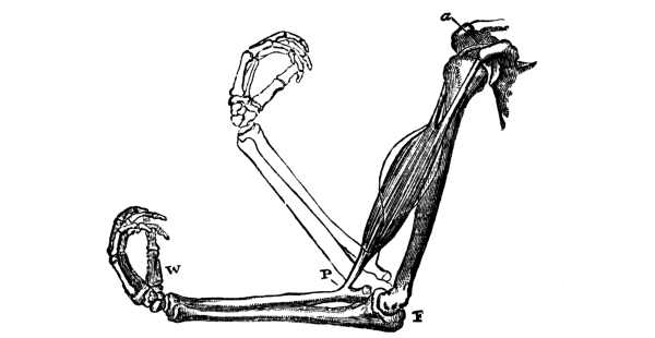
Fig. 160.—The action of the Biceps muscle.
a, the attachment of the muscle to the shoulder-blade;
P, attachment of the muscle to the fore-arm;
F, elbow; W, hand.
The movements of the limbs.—An excellent illustration of this relation between the bones and muscles is seen in the bending of the arm at the elbow. When the arm is bent, a great mass of flesh, the biceps muscle, in front of the upper arm may be observed to become much thicker. The muscle (Fig. 160) thickens because it becomes shorter. Its upper end is attached to the corner of the shoulder-blade (a), which remains stationary; while its lower end is connected at P with one of the bones of the fore-arm. The shortening of the muscle therefore draws [Pg 226] up the fore-arm. The elbow-joint, about which the motion takes place, is a very perfect hinge. Several other forms of joint are also found in various parts of the body. It is seen, when the skeleton is examined, that in every case the characters of the joints and the attachment of the muscles are most admirably adapted to the movements with which they are associated.
The study of the skeleton.—The study of the rabbit’s skeleton is not only highly interesting in itself, but necessary for the intelligent appreciation of the animal’s life. And when it is compared with the skeletons of other familiar animals, a common plan of structure is found which illustrates in the most convincing manner the kinship which often exists between very dissimilar creatures. Such a comparison shows that, almost bone for bone, the skeleton of a rabbit corresponds with that of a man or a horse, and even with that of a bird or a frog. Mounted skeletons are shown in most natural-history museums, and the student should, whenever possible, examine and compare them.[9] He can himself, however, easily separate the bones from a boiled rabbit, and make out their main features and relationships.
The rabbit’s skull and backbone—The bones of the head are collectively known as the skull. This consists of (1) a large brain case; (2) the cavities for the organs of special sense, viz., (a) a pair of nasal chambers (in which the organ of smell is located) in front: their hinder ends open into the top of the throat; (b) the eye-sockets at the sides, and (c) the flask-shaped chambers for the internal ears at the sides of the hinder end of the brain-case; (3) the jaws: the upper jaw is rigidly attached to the brain-case, but the lower jaw is hinged at each side on the hinder end of the bony arch which runs below the eye. Both upper and lower jaws bear teeth, which [Pg 227] are fixed in sockets. On each side the upper jaw contains two incisor teeth (p. 219) and—much further back—six grinding teeth. In the lower jaw are one incisor and five grinding teeth on each side.
The vertebral column or backbone is a chain of bony rings or vertebrae which runs dorsally (p. 217) from the hinder end of the skull to the tail, and forms a long tube containing the spinal cord—a backward continuation of the brain. On the anterior face of the first vertebra are two hollows into which a pair of knobs on the skull fit. Although, with the exception of the first and second, all the vertebrae are formed on essentially similar lines, there is considerable variation in shape and size in the different regions of the spine, as may be seen from Fig. 161. There are seven neck vertebrae, and this number is remarkably constant in mammals (p. 220). Following these are the chest vertebrae, which bear pairs of ribs. The majority of the ribs curve round and join on to a ventral bar of bone called the sternum or breast bone; so that the cavity of the chest, containing those very important organs the heart and lungs, is enclosed [Pg 228] in a protective bony cage. The vertebrae of the abdominal region of the body are very large and stout. Between them and the tail-bones are four fused vertebrae, forming a mass which on each side gives attachment to the large hip-bone.
The bones of the rabbit’s limbs.—The fore and hind-limbs of the rabbit are obviously comparable: the upper arm corresponding to the upper leg, the elbow to the knee, the fore-arm to the “leg,” the wrist to the ankle, and the fore-foot to the hind-foot. This similarity becomes even more apparent when the limb-skeletons are examined. Commencing in each limb at the end nearest the body we find a single long bone. In the fore-limb the rounded, upper end of this bone works in a socket at the anterior angle of the shoulder-blade, a triangular plate on each side which overlies the chest dorsally; in the hind-limb the rounded end of the corresponding bone works in a cup in the hip-bone. Again, between the elbow and the wrist are two bones lying side by side; and between the knee and the ankle are two corresponding bones, although here they are only separate in their upper parts. Similarly, the wrist bones correspond to the ankle bones, and the bones of what may be called the fingers to those of the toes. Certain of the ankle bones are, however, much elongated: obviously a great advantage to an animal which progresses by a series of hops—owing to the increased leverage which is thereby given to the hind-foot. The rabbit’s fore-foot bears five toes, the hind-foot four.
The structure of a long bone.—The long bones of the limbs are hollow except at the ends. Strength and lightness are thus secured by a device (the hollow cylinder), which has already (p. 72) been seen to be adopted by the supporting structures of plants, and is also copied by [Pg 229] human engineers. The cavity of the tube is filled by marrow, which supplies the bone with food. The hard, bony tube itself is partly composed of mineral matter and partly of animal matter. The mineral matter is left as a white, brittle framework when the animal matter is burnt away; while on the other hand the mineral part—which gives the bone its rigidity—may be dissolved out by immersing the bone in dilute hydrochloric acid, leaving the organic tissue as a soft flexible substance having the shape of the original bone.
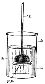
Fig. 162.—Experiment to show
that starch solution will not
pass through a thin membrane.
b, beaker; w, water;
p.p., parchment paper;
t.t., stem of thistle funnel;
s,starch solution in head
of thistle funnel.
1. A solution of starch will not pass through a thin membrane.—Rub up with water as much starch as will lie on a shilling, so as to form a thin “cream,” and then pour on it about a cupful of boiling water. The starch swells up and largely dissolves in the water. Add a few drops of the starch solution to about half a pint of water, stir, and test it by adding a little iodine solution (p.2, footnote). A beautiful blue colour is obtained, showing that the test is a very delicate one. Now take a thistle funnel and with a file cut through the stem about six inches below the head. Wet a piece of parchment paper or thin bladder (having previously held it up to the light to be sure there are no holes in it), and tie it tightly across the mouth of the funnel. Fill the head and about an inch of the stem with the starch solution. This can easily be done by means of a “canula,” such as is used for filling fountain pens. Now put the funnel into a beaker of water, in the manner shown in Fig. 162, and put the arrangement aside for a few hours. After that time add iodine solution [Pg 230] to the water in the beaker. No blue colour is formed, showing that no starch has passed through the membrane.
2. A solution of sugar will pass through a thin membrane.—A delicate test for certain varieties of sugar (not, however, table sugar) is a liquid known as Fehling’s solution.[10] Place a particle of honey in a test tube with a teaspoonful of Fehling’s solution, and put the test tube into a vessel of boiling water. Notice that in a short time the blue colour of the solution disappears and the liquid becomes red and turbid.
Now repeat Experiment 45, 1, but instead of starch solution use honey dissolved in water. To show that some of the honey-sugar has passed through the membrane, take about a teaspoonful of the water in the beaker, and put it in a test tube with twice as much Fehling’s solution. Heat as before, and notice the red turbidity. If table sugar is used it may be recognised by the sweet taste of the water after the experiment.
3. The action of saliva on starch.—(a) Chew a piece of india-rubber to induce a free flow of saliva, and collect the liquid. In one test tube put half a teaspoonful of starch solution; in a second tube put a mixture of equal quantities of starch solution and saliva; in a third put saliva alone. Keep the tubes at blood heat for twenty minutes. Then add a little water to each tube and divide its contents into two parts. Test one part of each for starch with iodine solution, and the other part for sugar with Fehling’s solution. The first tube contains only starch. The second now contains no starch, but shows the presence of sugar. The third contains neither starch nor sugar. Evidently the saliva has changed the starch of the second tube into sugar.
(b) Repeat the experiment, but keep the mixture of starch and saliva in a cold place. No sugar is produced.
(c) Repeat the experiment as in (a), but use saliva which has been heated to boiling in a test tube. No sugar is formed.
The necessity for food.—It is common knowledge that a rabbit, [Pg 231] like every other animal, must have a regular supply of food if it is to continue healthy, and that it would soon die outright if food were withheld. The reason for this is that the living substance of the animal’s body is incessantly wasting away. The rabbit cannot move a muscle except at the expense of the living substance of the muscle, and the more active is its life, the more rapidly does its body waste. It is to counteract this continual waste by continual formation of new living substance that food is taken.
But the succulent plants which the rabbit eats are not suddenly transformed into animal muscle and bone, and so forth, when they are swallowed. They have first of all to undergo a process which is called digestion. This takes place in a tube—the digestive canal—which runs from end to end of the body. The digestive canal of the rabbit is coiled in a somewhat intricate manner. That of the frog is, however, much simpler and more typical of vertebrates (p. 220) generally, and will serve equally well in so elementary a consideration of digestion as the present.[11]
The frog’s digestive canal.—The hinder end of the frog’s mouth opens (Fig. 163) into the gullet (gul.), a short wide tube which leads to a capacious bag called the stomach (st.). The termination of this is continuous with a coiled narrow tube called the small intestine (s. in.), which passes suddenly into a much wider large intestine or rectum. The rectum opens to the exterior, at the hinder end of the body, by an aperture (an.) known [Pg 232] as the anus. In addition to the digestive tube proper, two large digestive glands, the liver and the pancreas, should be carefully noticed. The liver (lr.) is a large, dark red organ, consisting of two lobes which lie at the sides of the stomach. It makes a digestive fluid called bile. The pancreas (pn.) is an elongated body which lies in the loop between the stomach and the first portion—called the duodenum (du.)—of the small intestine. It makes a digestive fluid called the pancreatic juice. In the frog both the bile and the pancreatic juice are discharged into the duodenum by one tube or duct (b.d.). Small digestive glands also occur in the inner wall of the stomach; these discharge a fluid called gastric juice into the cavity of the stomach.
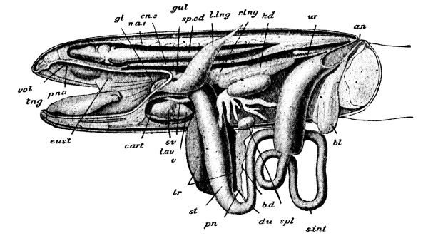
Fig. 163.—The Frog, dissected from the left side. gul, gullet; st, stomach; du, duodenum; s. int, small intestine; an. anus; lr, liver; pn, pancreas; b.d, bile duct; bl, urinary bladder; c.art, l.au, s.v, v, parts of the heart; cn. 3, “body” of 3rd vertebra; eus t. Eustachian tube; gl, glottis, leading to l.lng, left lung, and r.lng, right lung; n.a. 1, arch of 1st vertebra; p.na, internal opening of nostril; sp.cd, spinal cord; spl. spleen; tng, tongue; kd, kidney; ur, ureter; vo.t, vomerine teeth.
The rabbit’s digestive canal—In its main features the digestive canal of the rabbit resembles that of the frog; here also the mouth opens into a long tube consisting of gullet, a bag-like stomach, a [Pg 233] small intestine, and a large intestine. There are also a liver and a pancreas which discharge their fluids—by separate ducts, however,—into the first part of the small intestine; and the stomach is supplied with gastric juice by small glands in its inner wall. There are, however, certain differences besides those of size in the digestive tubes of the two animals. In the first place, a fluid called saliva is poured into the rabbit’s mouth by the ducts of salivary glands which occur near the mouth. Secondly, the coils of the small intestine are very much more complicated than in the frog. And lastly, at the junction of small and large intestines there is given off, in the rabbit and in herbivorous mammals generally (when these have simple stomachs,—p. 261), a great, spirally-constricted tube which ends blindly in a finger-like process.
How the rabbit digests its food.—A rabbit needs food to repair the constant waste of substance which the activities of its life entail; and the same is true of every other living thing, be it plant or animal. Now, every part of a rabbit’s body is irrigated and drained by the finest branches of a system of pipes through which blood is always flowing; and it is in this blood-stream that the food is conveyed to the muscles and other organs which are to be repaired. The food finds its way into the blood when that fluid is flowing through the small vessels which lie in the thickness of the wall of the digestive tube. Before food can gain access to the blood it must be in a condition in which it is capable of diffusing through the thin membrane which separates them. Digestion is the process which renders food soluble and diffusible, and hence capable of passing into the blood. The food of animals is of several different kinds. A few of these are soluble and diffusible at the time they are eaten, but most of them are neither, and therefore require treating according to their nature. This is why so many different fluids are poured into the [Pg 234] rabbit’s digestive canal. Saliva, gastric juice, bile, and pancreatic juice are each capable of acting upon certain constituents of the food and rendering them soluble and diffusible.
It would be beyond the scope of this book to consider the work of these fluids in detail, but the action of saliva is not only fairly typical, but it can easily be imitated outside the body. Starch is a very common constituent of vegetable foods, and its presence or absence is readily determined by the blue colour which it gives with a solution of iodine. Starch is quite insoluble in cold water, but when treated with hot water it swells up and, to a great extent, dissolves. But a mere solution of starch cannot get into the blood, for it is incapable (Expt. 45, 1) of passing through a thin membrane. On the other hand, if starch is mixed with saliva, and the mixture is kept at the temperature of the body, it is found in a short time that the starch has been changed into sugar, which is not only soluble, but readily diffuses through a thin membrane. In other words, starch as such is useless to the rabbit as food; only after it has been digested by conversion into sugar can it be used by the body.
Something very similar often takes place in plants. It was seen (p. 33) that the cotyledons of a pea become sweet during germination. Starch is a convenient form of food for storing in the cotyledons of a pea, the endosperm of the maize seed, the short stem of the crocus-corm, and so on; but before the plant can use it as food the starch must be made soluble and diffusible by being changed into sugar. In plants the change is brought about, not by saliva, but by a substance known as diastase.
The pancreatic juice continues the change of starch into sugar which is commenced by the saliva, but it also digests other food-stuffs as well. Similarly, gastric juice and bile are each concerned with the digestion [Pg 235] of certain foods. The result of the action—separate and combined—of the digestive fluids is that, when a rabbit eats no more food than it requires, all the useful parts of the food are absorbed into the blood, and then distributed to the tissues by the blood as it flows through them.
1. Evidence of the circulation.—(a) The arterial pulse.—Feel your pulse at the wrist and count the number of beats per minute.
(b) The beats of the heart.—Similarly count how many times your heart beats per minute. Put your ear over the heart of a friend, and listen to the sounds of the heart. How many different sounds can you distinguish?
(c) The valves of the veins.—Press your thumb on your arm inside the elbow, and then move it down the arm towards the wrist. Notice the small knots which rise under the skin between the point of pressure and the wrist.
2. The structure of a sheep’s heart.[12]—Procure from the butcher a sheep’s heart with, if possible, the lungs still attached. If the thin transparent bag which naturally surrounds the heart has been left on, carefully cut it away. Make out the main external features of the heart before cutting into it. The shape is conical, the apex of the cone being posterior, the base (where the blood-vessels are attached) anterior. The ventral face is rounded; the dorsal face is flatter. Compare Fig. 164. The line of fat (3) marks the line of division between two chambers, the right (R.V.) and left (L.V.) ventricles. Feel that the right ventricle yields to pressure more readily than the left. Two more chambers, the right and left auricles, are situated at the basal (thick) end. R.A. and L.A. are flaps of the right and left auricles respectively. Identify the great vessels SVC and IVC which discharge into the right auricle. Cut them open to see the entrance. Then lay open the right auricle. [Pg 236] Pass your finger down and notice that the right auricle communicates freely with the right ventricle. Observe that you can from the right ventricle pass your finger into the tube P.A.; make out that this goes to the lungs. Lay open the left auricle, and see that it receives vessels from the lungs. Pass your finger from the left auricle into the left ventricle, and notice that this latter chamber leads into the Aorta, Ao. (A´o´. a branch of Ao.). Notice that the walls of the blood-vessels in connection with the two auricles are collapsed, while those (P.A. and Ao.) leading from the two ventricles are more elastic and remain open. Which of the two auricles has the thicker wall? Which of the two ventricles has the thicker wall? Can you pass your finger from one auricle to the other? From one ventricle to the other? Cut away the auricles and pour water into the ventricles. Then squeeze the heart gently, and notice the flaps which rise to close the openings into the ventricles. Could blood pass from the ventricles to the auricles? Why not? Lay open the bases of the vessels P.A. and Ao. leading from the right and left ventricles respectively, and notice the pockets of thin membrane which are attached there. Open them out gently with the point of a pencil. Put the heart under the tap and let water trickle down the vessels towards the ventricles; notice how the pockets stand out as they fill with water. What would be the effect of blood trying to pass from P.A. or Ao. back into their respective ventricles?
The circulation of the blood.—The blood of such an animal as the rabbit is contained in a system of closed tubes called blood-vessels, through which the fluid is continually flowing. The regular flow of the blood in one direction is maintained by the action of the heart, a four-chambered organ which is situated in the chest, between the lungs. The heart is muscular, and, like other muscles (p. 225), has the power of “contracting” in definite directions. The contraction of the walls of the chambers of the heart lessens their capacity, and therefore drives the blood out of them, the [Pg 237] direction of flow being determined by valves. The vessels which carry blood to the heart are called veins; those which transmit the blood which is pumped out of the heart are called arteries. The arteries branch into smaller and smaller tubes which supply the various organs of the body, and the small arterial branches divide again and again in the organs until their finest ramifications form a close-meshed network of vessels with excessively thin walls, through which diffusion can readily take place between the tissues and the blood. These finest blood-vessels are called capillaries. They can be studied in the transparent web of the frog’s foot with the aid of a low power of the microscope. The blood can then be seen flowing, at a speed which varies with the size of the vessel, its course being rendered obvious by the tiny oval particles (red corpuscles) which are suspended in it. The smallest capillaries are so thin-walled that they appear to be merely channels in the substance of the web, and the corpuscles creep along in single file. But these channels unite to form larger vessels with obvious walls, and these unite again and again until a main vein is formed in which the blood, with its suspended corpuscles, rushes along in a swift torrent towards the heart.
The heart.—The beginner will find the sheep’s heart more convenient for examination than the rabbit’s, on account of its larger size; but apart from some difference in the arrangement of the great blood-vessels opening into them, the two hearts are broadly similar.
The heart (Fig. 164) consists of four chambers. Two of these, the auricles, are receiving-chambers, and are placed at the thick, anterior end of the heart. Into the right auricle open the great veins (SVC, IVC,) which bring blood from all parts of the body except the lungs; the left auricle receives only blood from the lungs. Each auricle opens into a more posterior chamber called a ventricle, the right auricle opening into the right ventricle and the left auricle [Pg 238] into the left ventricle. The ventricles pump blood into the arteries. The blood from the right ventricle is sent into the artery (P.A.) which supplies the capillaries of the lungs; while the blood of the left ventricle is forced into the aorta (Ao.), a great artery which branches and supplies with blood all parts of the body except the lungs. The two auricles contract simultaneously, forcing their contents into the flaccid ventricles. Then both the filled ventricles contract at once, and pump blood into the great arteries, flaps of membrane between the auricles and ventricles preventing a backward flow into the auricles. Similarly the bases of the great arteries are provided with membranous pockets which readily admit the blood from the ventricles when these contract, but entirely prevent a return of blood to the ventricles. The appearance of these four sets of valves, as seen from above, is shown in Fig. 165. After the contraction of the two ventricles there is a short rest, then the auricles contract again and the whole process is repeated.
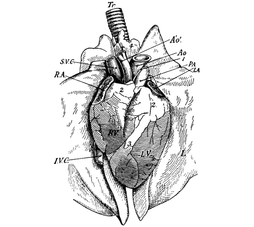
Fig. 164.—Heart of Sheep, in position between the lungs. R.A., appendage of right auricle; L.A., appendage of left auricle; R.V., right ventricle; L.V., left ventricle; S.V.C., I.V.C., great veins from the system generally; P.A., artery to lungs; Ao., aorta; A´o´., branch of aorta; L., lung; Tr., windpipe, leading to lungs; 2, 3, 4, fat.
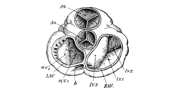
Fig. 165.—The orifices of the heart seen from above, the auricles and great vessels being cut away. PA, valves at base of artery to lungs; Ao, valves at base of aorta; R.A.V., orifice between right auricle and ventricle, with its valves l.v. 1, l.v. 2, and l.v. 3; L.A.V., orifice between left auricle and ventricle, with its valves m.v. 1 and m.v. 2.
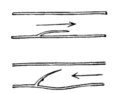
Fig. 166.—Diagrammatic sections of veins with valves. In the upper figure the blood is supposed to be flowing in the direction of the arrow, towards the heart; in the lower, back towards the capillaries.
The sounds which are heard when the ear is placed over another person’s heart are often compared to the syllables lub-dup. The “lub” is partly caused by the contraction of the ventricles; the “dup,” which immediately follows, is caused by the sudden closure of the valves at the bases of the great arteries. The throb of the heart, which can be felt from the outside, is really the thrust of the apex of the heart against the chest-wall at each beat. The sudden forcing of blood into the already-full but elastic arteries causes a wave to travel along these vessels, which can readily be felt, or even seen, at places where a fairly large artery lies just beneath the skin. This arterial wave is called the pulse. Many of the veins are [Pg 240] provided with pouch-shaped valves which permit the blood to flow freely towards the heart, but which bar the passage of blood in the opposite direction. Their action will readily be understood from Figs. 166 and 167.
The importance of the capillaries.—The capillaries are the least conspicuous part of the circulatory system, but they are by far the most important. The heart, arteries, and veins exist merely to renew constantly the blood which flows through these minute channels. The excessive thinness of the walls of the capillaries makes it possible for a ready exchange to take place between the living tissue and the blood, and the vessels themselves form a network of such extremely close texture that it is practically impossible to prick any living part of the body with a fine needle without puncturing some of them and “drawing blood.” The work of the blood in supplying the various organs of the body with food has already been referred to. We have next to see how this all-important fluid is of service in supplying the organs with oxygen.
1. Carbon dioxide is formed when flesh burns.—Dry a piece of meat and attach it to the end of a wire. Then light it, and when it is burning vigorously lower it into a clean glass jar. When the flame goes out remove the charred flesh, and at once pour a little clear lime-water into the jar and shake up. The lime-water turns milky, showing the presence of carbon dioxide gas in the jar. Examine what is left of the meat. It is charred, showing that meat contains carbon. How was the carbon dioxide formed during the burning?
2. Carbon dioxide is formed by the living body.—Breathe through a glass tube into clear lime-water, so that the air you expel from your lungs bubbles through the liquid. Does the lime-water remain clear, or turn milky? Does the air you breathe out contain a considerable quantity of carbon dioxide gas? [Pg 241]
Burning and life.—When a piece of the flesh of any animal has been dried it may easily be set on fire. The burning is caused by the union of the constituents of the flesh with some of the oxygen of the air to form various gases. One of these gases is carbon dioxide, formed by the combination of the carbon of the flesh with oxygen. Carbon is present in all the parts of animals and plants, as is evident from the separation of charcoal (an impure form of carbon) in the first stages of burning; the carbon dioxide gas which is formed may easily be recognised by the milkiness which it produces in clear lime-water. The liberation of heat, and the formation of carbon dioxide, which always accompany the burning of animal and plant tissues, are worthy of very careful attention.
It is well known that the body of a living animal such as a rabbit or a man is always warm; and the experiment (Fig. 168) of passing through clear lime-water the air breathed out from the lungs shows, by the milkiness produced, that the animal is also constantly producing carbon dioxide during its life. Is life, then, always accompanied by a peculiar form of burning, in which the living substance of the body is the combustible material? It seems so, and the experiments of physiologists tend to confirm this view.
The necessity for breathing.—The energy which enables a muscle to contract is derived from the oxidation—the slow burning—of part of its substance, just as the energy which enables a steam-engine to move is derived from the burning of fuel in the boiler fires. The fires soon go out, and the engine stops, unless fresh fuel is added from time to [Pg 242] time and a plentiful supply of air is available. Similarly, a muscle loses its power of contracting, a gland that of secreting, the brain that of thinking, unless the waste matters resulting from previous activities are cleared away and replaced by fresh food and fresh oxygen. Wherever vital action is taking place, whether in a contracting muscle, a secreting gland, or a thinking brain, there is continual consumption of oxygen and continual production of waste material, chiefly carbon dioxide. In the higher animals the renewal of oxygen and the removal of waste material are performed by the blood. Blood-vessels are to the body what rivers and canals are to a country: they act as highways for the transport of material. We may perhaps carry the analogy a little farther and find in the red corpuscles of the blood the rough equivalents of boats or canal-barges, for they carry with them tiny loads of oxygen. As the blood creeps along the narrow channels in an active tissue the red corpuscles relinquish their oxygen, and the fluid portion of the blood takes up carbon dioxide. The blood continues its course, and sooner or later arrives at a place where it can obtain a fresh supply of oxygen and get rid of its surplus carbon dioxide. In the rabbit this exchange takes place as the blood is passing through the capillaries of the lungs. There the blood is separated from the air by a membrane so delicate that gases can readily pass through it; and, hence, on leaving the lungs the blood has got rid of the waste carbon dioxide, and its red corpuscles are laden with fresh oxygen. This exchange of useless carbon dioxide for oxygen constitutes respiration or breathing.
The mechanism of respiration.—In active animals the air inside the lungs soon becomes vitiated, unless there is some means of changing it. Under ordinary conditions a man changes the air in his lungs from thirteen to fifteen times a minute. He does this quite automatically, [Pg 243] and without thinking about it. Every four seconds or so a set of muscles contracts and pulls his ribs upwards and outwards; another muscular contraction pulls down the floor of his chest at the same time. As a consequence the cavity is much enlarged. The lungs follow the movements of the walls of the chest, and some thirty cubic inches of air are sucked in. Immediately the ribs fall back to their former position, the chest-floor rises, and air is driven out. Then after a short pause the process is repeated. It should be noticed that only about thirty cubic inches of air are changed at each respiration, although the capacity of the human lungs averages about 230 cubic inches. All mammals breathe in much the same way.
Plants and animals.—There are considerably more points of similarity than of difference between plants and animals. In every case the vital activities are accompanied by an oxidation of living substance, and from this fact arises the necessity for food and oxygen. The breathing of plants is essentially like that of animals, and consists in taking in oxygen and giving out carbon dioxide; though the mechanism of respiration is—except in the lowest plants and animals—entirely different in the two cases. It is in the sources from which they obtain their food that plants and animals are most unlike. An animal must be supplied with food which has already been prepared by some other living thing; and it is obvious that the food even of carnivorous animals can ultimately be traced back to plants, for the flesh-eater preys upon the vegetarian. Animals are therefore entirely dependent upon plants for food. In this sense the saying “all flesh is grass” is full of significance.
Green plants (Chapter II.) are quite independent of all other forms of life, and can build up their substance from water, mineral matter, and the carbon dioxide of the air. The taking in of carbon dioxide and giving out of oxygen by green plants has nothing whatever to do with [Pg 244] respiration; it is part of their process of feeding. Green plants breathe in the usual manner—by taking in oxygen and giving out carbon dioxide. It should, however, be noticed that the peculiar method by which a green plant obtains its carbonaceous food is of the highest importance to animal life; for by this process the amount of injurious carbon dioxide in the air is considerably lessened, while the proportion of life-supporting oxygen in it is greatly increased.
Fungi (Chapter XI.) seem to be intermediate, as regards their method of feeding, between green plants and animals. They require their carbonaceous food in an organic form, that is, already prepared by other living things; but they can obtain the other elements of their food from mineral salts and water.
The thoughtful student will be increasingly impressed by the extent to which the plant and animal kingdoms are dependent upon each other, and by the manner in which each utilises the waste products of the other for carrying on its own life processes.
EXERCISES ON CHAPTER XIII.
1. Of what parts does the skull of a rabbit consist? What is the use of each part?
2. Describe one of the long bones of a rabbit’s leg. To what features does it owe its strength?
3. The bones of the skeleton are useful (1) as affording points of attachment for the muscles; (2) as affording protection for delicate tissues and organs. Give examples of each of these uses. Do not give the technical names for the various muscles. (King’s Schol., 1902)
4. Draw and describe one of the middle joints of the backbone of a quadruped, and explain the uses of the various parts. (1898)
5. What is meant by “digestion”? Why must food be digested? Where does digestion take place?
6. Give full practical instructions for demonstrating the chief properties of saliva, and its action upon various kinds of food. (1897) [Pg 245]
7. Prove that the action of human saliva upon starch is not due to living particles contained in it. (1898)
8. What are the chief uses of the blood? Why is it necessary that it should be kept in motion? (1901)
9. Where would you look for the Aorta in a sheep’s heart? What valves are found in it? How does it differ in appearance and feel from a large vein? (1901)
10. What kinds of valves are found in a sheep’s heart, and where are they placed? Describe a valve of each kind. (1898)
11. How does air breathed out from the lungs differ from common air? How can the differences be demonstrated? (1898)
12. Describe the process by which a mammal renews the air in its lungs.
13. What is meant by “respiration”? Why is respiration necessary?
14. Point out the most remarkable differences between the nutrition of a green plant and that of an animal. (1897)
15. Name organisms which can derive nourishment from carbon dioxide, from sugar, and from the dead bodies of animals. (1898)
16. What are the simplest functions which distinguish living animal matter from inanimate matter? (King’s Schol. 1903)
17. Explain as fully as you can how food taken into the stomach acts upon organs, such as the brain, which are not closely connected with the stomach. (1904)
18. The uses of bone are, generally speaking, to protect delicate structures, to support weight and to gain leverage. Illustrate this statement by a simple description of one example of each type. (Certificate, 1904)
19. The flow of liquids through the body is regulated in certain localities by valves. Explain the action of a valve, and indicate where they are to be found in the body. (Certificate, 1904)
20. In which kind of blood-vessel can the pulse be felt? In which kinds can it not be felt? Explain the reason of the difference. (Certificate, 1905)
21. How is oxygen conveyed from the lungs to the various parts of the body? Describe what could be observed if a drop of blood were spread out on a piece of glass and examined under a microscope. (King’s Schol. 1905)
1. The external characters of the cat.—Carefully and gently examine a cat, and make notes of the following characters:
(a) Hair.—What is the covering of the body like? Is the hair like that of a rabbit, i.e. fur (p. 216)? Are the whiskers very noticeable? On what parts of the head do they grow? Are the fur and large whiskers in any way connected with the animal’s habits?
(b) Eyes.—Look at a cat’s eyes in a bright light. Is the pupil (p. 212) round or slit-like? Keep the cat in the dark for a few minutes and then look again at the pupil of the eye; has it changed in form? Is the change of any advantage to the cat?
(c) Teeth.—Gently open the cat’s mouth and examine the teeth. Notice the sharp, pointed teeth behind the incisors (p. 219); they are called the canine teeth. Has a rabbit any canine teeth? Why does a cat, and not a rabbit, need such teeth? Feel the remaining teeth with your finger; are they flat like those of a rabbit, or sharp-edged? How are the characters of the teeth associated with the kind of food? Watch a cat eating; does it chew its food or swallow it “in lumps”?
(d) Tongue.—Pass your finger-end over the cat’s tongue; is it rough or smooth? In which direction of motion of the finger does the tongue feel roughest? Would the roughness be of any help to the animal in licking meat from bones?
(e) The limbs.—Measure the legs. Are the fore and hind [Pg 247] limbs of equal length, slightly unequal, or very unequal? How do they compare in this respect with the limbs of the rabbit? Make out the main bones by feeling through the skin, and especially notice where the ankle-joint is. Examine the toes and notice how, when you gently squeeze them just behind the ends, the sharp claws protrude, and go back into a kind of sheath when the pressure is removed. Can the animal put out its claws and draw them back at will?
2. The habits of the cat.—(a) Food.—What kind of food does the cat prefer, animal or vegetable? Does it bolt its food greedily, or does it eat deliberately and daintily? Have you ever known cats to hunt other animals? Do they hunt singly, or do several cats join together to hunt? How do they approach the prey; do they try to run it down by speed, or do they creep up slyly and then spring? What is the use of the claws? How does a cat drink?
(b) Locomotion.—Watch a cat moving slowly; does it walk or hop? Try to find out the order in which it puts its feet down. How does it run? Is it nimble or clumsy? Does the cat walk with the whole sole of its foot on the ground, or does it walk “on its toes”?
(c) Likes and dislikes.—Do you consider a cat sociable, i.e. fond, in general, of the society of other cats? Do cats, as a rule, show much appreciation of the difference between right and wrong? Are they as affectionate as dogs? Have you ever heard of any cat trying to remain in a house after the family had removed to another house? Are cats fonder of warm or of cool places in a house? Do they like getting wet? Do they pay much attention to personal cleanliness? How do they wash themselves? Do you think the tongue is used as a comb? How? Can you know whether a cat is pleased or angry (i) by the appearance and movements of the tail, (ii) by the sounds which it makes?
(d) Intelligence.—Write accounts of cases of great intelligence which you have known cats to show.
(e) Voice.—How do you describe a cat’s voice? How does the voice vary according to the animal’s mood?
(f) Play.—How does a cat play?
[Pg 248] 3. Kittens.—(a) Appearance.—Are kittens helpless or active when they are born? Is there any very marked difference between the proportions of the body and limbs and those of a full-grown cat? At what age is a cat full-grown?
(b) Play.—How does a kitten play? How does it pretend to “stalk” a small object, such as a ball of wool? In the same way that a full-grown cat stalks a mouse? Why does the kitten adopt this method before it has any experience of hunting?
(c) Education.—Watch a cat with its kittens, and describe any actions which seem like education. Have you ever seen a cat teach its kittens to fight? Have you ever seen it punish a kitten for disobeying a call?
4. The external characters of the dog.—Examine a dog in the same way, and compare it point by point with the cat as regards the following characters:
(a) Hair.—Notice that the dog is covered, not with fur, but with rough hair, and that the whiskers are not very large. How are these differences associated with differences in habit?
(b) Eyes.—Notice that the pupil is always round, although it is smaller in a strong light than in a weak one.
(c) Teeth.—Compare the teeth with those of the cat, and notice that they are of similar form—with strong interlocking canines and a sharp-edged tooth on each side of the upper jaw which clips against a similar tooth in the lower jaw—but that the cat’s teeth are more pointed.
(d) Tongue.—Notice that the dog’s tongue is much smoother than the cat’s.
(e) Limbs.—Examine and measure the limbs, comparing them with those of the cat, rabbit, and man. Notice that the claws are blunter than the cat’s, and that they cannot be withdrawn into sheaths. How many toes are there on the feet?
5. The habits of the dog.—(a) Food.—Does the dog prefer animal or vegetable food? Watch a dog gnawing a bone, and observe the use of the clipping teeth. Does a dog eat greedily or deliberately? Does it chew its food or swallow it in lumps? Do dogs hunt singly or in packs? Do they stalk the prey stealthily, as cats do, or do they try to run it down by speed? Whenever possible watch a pack of hounds; upon [Pg 249] what sense—hearing, sight, or smell—do the hounds rely most? How does a dog drink?
(b) Locomotion.—Does a slowly-moving dog hop or walk? Try to find out the order in which it puts its feet down. How does it run? Does a dog walk flat-footed or on its toes?
(c) Likes and dislikes.—Do you consider dogs sociable or otherwise? Do they seem to know the difference between right and wrong? Are dogs affectionate? Are they more attached to the people or to the houses to which they are accustomed? What can you learn of a dog’s feelings, by the movements of its tail? Write accounts of instances, which you know to be true, illustrating its likes and dislikes. What expression of the human face seems to you most like the snarl of an angry dog? What are the resemblances?
(d) Intelligence.—Write accounts of evidence of intelligence, or reasoning power, which you have observed in dogs.
(e) Voice.—What is the ordinary voice of the dog? What other sounds do dogs make, and what do they mean?
(f) Play.—How do dogs play? Do you think dogs have any sense of humour, or are able to appreciate a joke for the joke’s sake? Have you ever seen any expression resembling a smile on a dog’s face?
6. Puppies.—(a) Appearance.—Are puppies blind and helpless when they are born, or are they active? How soon can they see? Are the proportions of the body and limbs markedly different from those of the full-grown dog? At what age is a dog full-grown?
(b) Play.—Does a puppy play in the same manner as a kitten? What differences have you noticed? Have these differences any connection with the methods of catching the prey of the adult animals?
(c) Education.—Have you ever seen a puppy being taught to do anything by its mother? Write full accounts of such cases.
7. Different breeds of dogs.—Make notes of your observations of as many different breeds of dogs as possible, e.g. collies, terriers, retriever, fox-hound, pointer, spaniel, etc., and describe the resemblances and differences in size, form, habits, intelligence, etc. [Pg 250]
The external characters of the cat and dog.—The cat and the dog are so commonly kept as pets that they are perhaps more easily examined than any other animals. In addition, they are so closely related and yet exhibit so many differences that they afford a valuable exercise in the methods of comparison and contrast which are at the foundation of all successful work in Nature-Study. Several differences are at once apparent on even a casual inspection. The body of the cat (Fig. 169) is covered with soft, smooth fur, and its head is provided with long, sensitive whiskers. In both of these respects it resembles the rabbit and other mammals which are in the habit of creeping along narrow and dark passages. It is commonly said that cats can see in the dark. Although this is not altogether true—for no animal can see in total darkness—the cat’s eyes have the power of adapting themselves remarkably to the intensity of the light. In a dim light, the curtain, or iris, which surrounds the pupil (the dark, central window through which light enters the eye) is drawn back so as [Pg 251] to admit as much light as possible; whereas in a very bright light the curtain is so nearly closed that the pupil is merely a narrow, vertical slit. On the other hand, the dog (Fig. 170), which is not fond of dark passages, has its body clothed with rough hair, instead of fur; and its whiskers are not nearly so long as those of the cat. Again, although the iris of a dog’s eye alters in size to regulate the amount of light entering the eye, the pupil is always round, and the change of size is much less marked than in the cat. Another very noticeable difference is in the claws at the ends of the toes, corresponding to the nails at the ends of our own fingers and toes. In the cat these are sickle-shaped and extremely sharp, and are kept drawn completely back into sheaths when they are not required. The dog’s claws are blunt, and cannot be retracted. Both the dog and the cat rest the weight of the body upon the toes—not upon the sole of the foot—when walking or running.
[Pg 252] There is also a very marked difference in the tongues of the two animals. The tongue of the cat is rough, with small points directed backwards. These points are of great help in licking the flesh from bones; and they also serve as a comb when the cat—which is fastidious about the cleanliness of its fur—“washes” itself. The tongue of the dog is smooth and moist.
The inherited habits of the cat and dog.—Very obvious differences are also to be seen in the habits of the two animals, and these would be somewhat difficult to explain if we confined our attention to the domesticated animals only, which live under artificial conditions. When, however, we consider the wild relatives of the cat and dog, many of the differences become full of significance. The wild animals of the cat family, almost without exception, are either solitary or live in pairs; whereas the wild dogs (wolves, jackals, etc.) live in packs. The habits which these respective methods of life entailed have become so firmly implanted in the nature of the race that even now, after thousands of generations of domestication, they may be traced. Such inherited habits, which are not dependent upon, or may be at variance with, present conditions of life, are called instincts.
We will first see how the ancestral custom of living in organised packs has left its impress upon the instincts of the domesticated dog. The first essential to the success of any community of animals—whether these are bees or rooks, wolves or men—is that all the creatures composing it shall conform to certain rules, which have for their object the good of the community and not merely that of the individual. Acts which promote the wellbeing of the society as a whole are good, and are directly or indirectly rewarded. Acts which tend to injure the society as such are bad, and inevitably bring punishment either to the [Pg 253] individual offender or, what is worse, to his pack. A distinction between right and wrong is thus established which would be impossible to any animal living so solitary a life that its acts affected only itself. In this manner were aroused the social instinct, the love of praise and the dread of shame, the lifelong attachment to early friends, and almost all the other qualities which have so endeared the dog to mankind; because these qualities resulted naturally from the ancestral pack-life. Left to itself, the dog loses its nerve, for it is by nature unfitted for a solitary life. What more woe-begone animal is ever seen than a lost dog?
Contrast the cat in these respects. It is at heart an outlaw, like its wild ancestors: recognising, in general, no motives but those of its own ease and gratification. Its social instinct is almost absent; and though it sometimes displays affection to people who pet it, the cat is, as a rule, more attached to places than to persons. It retains, too, the independence and versatility which are developed by a solitary life. A lost cat can usually take care of itself and find sufficient food; and cases are not uncommon of cats leaving comfortable homes and choosing to live wild lives in the woods.
Ancestral methods of hunting also account largely for certain differences in the domestic cat and dog. The solitary ancestral cats, like the lions and tigers of to-day, sprang suddenly upon the prey at the end of silent, stealthy stalking. How firmly this method has become fixed in the character of the race may be seen still in the manner in which a cat stalks a mouse or a small bird, or even, in play, a dead leaf. At the final spring the sharp, sickle-shaped claws are used to hold down the victim. The solitary hunter is able to devour its capture at leisure, and the domestic cat is still distinguished by its dainty and deliberate manner of feeding.
The wild dogs hunt in a quite different manner. The whole pack joins in [Pg 254] the chase, the trail being followed by the sense of smell. There is no attempt at concealment, no stealthy stalking; but an open reliance upon speed, endurance, and numbers, rather than upon cunning. If one dog loses the scent another picks it up and gives the signal. Mutual help is thus the secret of success in hunting. But this is at an end when the prey is killed. The victim is torn to pieces and devoured greedily, each animal eating as rapidly as possible, for in most cases there is not enough to satisfy them all. And the well-fed domestic dog still betrays the ancestral necessity for hurried eating in the manner of bolting his food.
Methods of expressing feeling.—Animals express their feelings in various ways—by the voice, by the face, by the tail, and by the general attitude of the body. Perhaps no animal’s feelings are more readily recognised by man than the dog’s. “It is a remarkable fact,” says Darwin,[13] “that the dog, since being domesticated, has learnt to bark in at least four or five distinct tones. Although barking is a new art, no doubt the wild parent-species of the dog expressed their feelings by cries of various kinds. With the domesticated dog we have the bark of eagerness, as in the chase; that of anger, as well as growling; the yelp or howl of despair, as when shut up; the baying at night; the bark of joy, as when starting on a walk with his master; and the very distinct one of demand or supplication, as when wishing for a door or window to be opened.” The movements of the tail are also full of meaning, and capable of expressing several different moods. It seems likely that in a dog the movements of the tail were originally of use chiefly to signal to the rest of the pack. The use of the movements of a cat’s tail is not very clear, although these also differ according to the animal’s feelings.
[Pg 255] Carnivores.—Dogs and cats, with several other mammals, which are mostly flesh-eaters, are called Carnivores. They have never fewer than four distinct toes on each foot, and the claws (nails) are often capable of being withdrawn into sheaths. The teeth (Fig. 171) are characterised by the large, interlocking canines, which are conical, curved, and pointed; and by one of the cheek teeth on each side having a sharp, cutting edge which bites against the similar tooth of the other jaw, almost in the manner of the blades of scissors. The seals and walruses, however, which are carnivores adapted to living in water, have no such clipping teeth.
1. Habits.—At what time of the year have you seen bats flying about? Do they fly in broad daylight, or only in the evening? How can you distinguish a bat’s flight from that of a bird? Have you ever heard a bat squeak? Upon what does it feed? Are flying insects plentiful in winter? Have you ever seen a bat drink? How does it drink? What does the bat do in winter? Try to find a sleeping bat in winter in a barn, a hollow tree, or a belfry. What is its position?
2. Appearance.—Examine a sleeping bat or a stuffed specimen. What is its body covered with, hair or feathers? Is it a bird or a mammal? Is the hair soft and furry? How large are the ears? Are the whiskers large or small? What are the wings like? Can you see any fingers? How many fingers are there? Do any of the fingers bear claws? Do the fingers support the wings? Which are longer, the fore limbs or the hind limbs? Do the toes of the hind limbs bear claws? What is their use? If possible, put a live bat on the ground; does it walk easily? Apart from the wings, what animal does the bat seem most to resemble? Do you know of any other flying mammal? [Pg 256]
The Bat (Figs. 172 and 184 a).—On summer evenings in most country districts of Britain bats may be seen flitting about catching insects. The flight is peculiar, and somewhat suggestive of that of a butterfly, so that even in the dusk the animal may be distinguished easily from a small bird. “Bats drink on the wing like swallows, by sipping the surface as they play over pools and streams. They love to frequent waters, not only for the sake of drinking, but on account of insects, which are found over them in the greatest plenty.”[14]
The voice of the bat is a shrill squeak, of so high a pitch that many people cannot hear it at all. The animal is probably quite blind, but it has keen powers of scent and hearing, and in avoiding obstacles seems to be greatly aided by patches of specially sensitive skin on the face, and by delicate whiskers. During the day it lurks in dark corners of barns, church-towers, hollow trees, etc., hanging head downward by the hooked claws of its hind feet.
On close examination the body of the bat is seen to be covered with soft fur, a character which at once proclaims the animal to be not a bird—as it has been incorrectly considered—but a mammal. The fur is often of a bright chestnut colour. The ears are large and practically devoid of hair, and are so thin as to be almost [Pg 257] transparent. The wings are thin folds of skin, which are attached in front to the long arms, are supported by the greatly elongated fingers, and reach to the hind limbs. Another membrane passes between the hind limbs, and in common species is also supported by the tail. The thumb is free, and bears a claw which is of some assistance in climbing. The hind limbs are small.
The structure of the bat is obviously but little adapted to walking, and the animal moves about very awkwardly when on the ground, although it can rise on the wing again without much difficulty.
The bat is rarely to be seen abroad after the middle of November, for as the cold weather approaches and insects become scarce, it suspends itself by its hind claws in some dark and sheltered corner, and goes to sleep for the winter, reappearing in early spring.
1. The external characters of the sheep.—(a) Wool.—How does the covering of a sheep’s body differ from the fur of a cat or rabbit, and from the hair of the dog? Examine a lock of “raw” wool; is it at all greasy? Dip it in water; is it much wetted? Are the fibres easily entangled together? Why is woollen clothing so warm?
(b) Teeth.—Obtain a sheep’s head from the butcher, and examine the teeth. Notice the absence of incisors and canines in the upper jaw, and of canines in the lower jaw. Observe the thickened pad on the surface of the upper jaw, against which the lower incisors bite. Are the cheek teeth flat, or sharp-edged like the cheek teeth of carnivores? Watch a sheep feeding, and notice how it bites the grass. How is the lower jaw moved during the chewing of the cud?
(c) Horns.—Which sheep have the largest horns, the males (rams) or the females (ewes)? If you can find a cast horn notice whether it is hollow or solid. What is the use of horns?
(d) Limbs.—Is there any marked difference in the lengths of [Pg 258] fore and hind limbs? Notice the hoofs which cover those parts of the feet touching the ground. How many hoofs are there on each foot?
2. The habits of the sheep.—Are sheep solitary or do they live in flocks? What kind of ground do they seem to prefer, flat or hilly? Are sheep on a hillside easily seen from a distance? Why not? Are sheep nimble or clumsy? Can they run very fast? Would a flock of sheep be safer from, say, wolves on a rocky hillside or on an open plain? In a grazing flock of sheep notice whether most of the animals have their heads turned in the same direction. Has the direction any relation to the direction of the wind? Can you explain this?
In a running flock of sheep does any one animal act as leader? Is the leader a ram or a ewe? Is it a lamb or an old animal? Do the rest of the flock imitate the actions of the leader, and, for example, leap over a wall at the same place in single file? Have you ever noticed that if one animal jumps at a certain place, all the following sheep jump at the same place? Have you ever seen sheep fighting? Were the combatants rams or ewes? How did they fight?
What is the voice of a sheep like?
3. Lambs.—(a) Appearance.—At what time of the year are lambs born? Are they helpless or active? Have they long legs? What advantage to the lamb is length of limb?
(b) Play.—Watch lambs playing. Do they show a preference for any eminence, e.g. a rock, in the neighbourhood? What is the meaning of this preference? How do lambs fight? Have you ever seen a ewe stamp with her fore-feet when anyone approached her lamb? At what age is a sheep full grown?
The sheep.—The sheep (Fig. 173) differs in several important respects from any of the animals previously mentioned. It is of course a mammal, for it suckles its young, and its body is covered by hair; but the hair is of that warm fleecy kind which is called wool. The fibres of wool are seen under the microscope to be rough and scaly. [Pg 259] For this reason they can be spun into loosely-textured threads which entangle a great deal of air; woollen garments are thus very bad conductors of heat. The wool upon the sheep’s body is slightly greasy, from a substance which is given off by the skin and protects the animal from rain. The toes of the sheep are not armed with ordinary nails or claws, but claws are represented by horny masses called hoofs, which encase the ends of the toes, and upon which the whole weight of the body is thrown. The sheep, especially the male (ram), is often provided with weapons in the form of hollow horns, which grow upon its forehead.
Method of feeding.—The sheep is a strict vegetarian, living largely upon grass. It grips the grass between its lower incisor teeth and a hard pad on the upper jaw; there are no incisor teeth in the upper jaw. Neither upper nor lower jaw bears canine teeth; and the cheek teeth, which are used for chewing the food, have their crowns ridged lengthwise. The arrangement of the teeth of the sheep is shown in Fig. 174. During grazing, however, the food is not at once chewed, but is simply mixed with a large quantity of saliva and swallowed, the chewing-process being performed at a later period. It is obviously a great advantage to an animal, which in the wild state is liable at any moment to be attacked by enemies, to be able to stow away its food quickly, and afterwards masticate it at leisure. This is rendered possible, in oxen, sheep, goats, deer, and the few other animals which chew the cud, by the peculiar form of stomach shown in Fig. 175. The hastily swallowed food is passed into the large paunch b and into the compartment c. When the animal finds an opportunity of “ruminating” or chewing the cud, the food is returned to the mouth in small quantities at a time, and is there finely divided by the cheek teeth. In this condition it is again swallowed, and makes its way at once into the compartment d, where it is strained between leaf-like folds and then passed into the last chamber e, and thence to the intestines, to undergo the final processes of digestion. [Pg 261]
Ruminants.—Animals which, like the sheep, oxen, goats, deer, etc., ruminate or chew the cud in this manner, are called ruminants. In all ruminants the weight of the body is supported by the tips of the third and fourth toes of the feet, the remaining toes either having completely disappeared or remaining very small (Fig. 176). The tips of the toes are encased in horny hoofs, which represent greatly enlarged claws or nails. The ruminants therefore belong to what may be called the even-toed hoofed mammals. Pigs are also even-toed hoofed mammals, but they are not ruminants, because, having simple stomachs, they do not chew the cud.
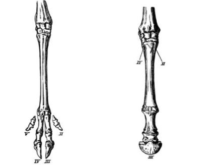
Fig. 176.—Bones of fore-foot of Red Deer.
Fig. 177.—Bones of fore-foot of Horse.
(After Flower.) (After Flower.)
Other hoofed mammals.—In the horse and donkey the reduction of the number of toes has gone still further; for these animals have now only the third or middle toe of each foot left (Fig. 177); and because it has to carry the whole weight of the body, it has become very large and stout; its tip is encased in a hoof.
The hoofed mammals are therefore divided into two groups:
(1) the odd-toed, including the horse and ass (they have simple stomachs, and therefore do not chew the cud); and
(2) the even-toed, including (a) ruminants like the sheep, etc., and (b) such non-ruminants as the pigs and their relatives. [Pg 262]
The inherited habits of the sheep.—As in the cases of the dog and cat, so in the sheep, the true explanation of several curious habits is to be found in the manner of life of the wild ancestors; for it must be remembered that domestication, however kindly an animal may take to it, is an artificial condition of life. Wild sheep live in flocks, as a rule in cold and mountainous districts; and some organisation is necessary if they are not to be at the mercy of savage carnivores. An old and experienced ram is generally in charge of the flock, and in case of alarm he leads the way to a more inaccessible position. The rest of the flock follow in single file, closely imitating his every movement, leaping without hesitation—and therefore saving valuable time—wherever he has leapt. The survival of this instinct in domestic sheep may be observed whenever a flock is travelling along the road. Even young lambs still display a decided preference for rocks, hillocks, and other elevated positions. Wild sheep, being so liable to sudden interruption when grazing, are enabled by their compound stomachs to swallow food quickly and postpone the chewing process to a more favourable opportunity. Sentinels usually keep watch, and warn the flock of approaching enemies by stamping their hoofs on the ground, an action which may still be seen whenever a ewe fears danger to her lambs.
The play and education of the young.—The young of mammals are usually under the care of the mother for education and protection until they are nearly adult; and it is generally found that the longest infancy (in proportion to the natural life of the animal) occurs in the most intelligent races. The importance of the play and education period is very great, for it not only gives the young animal an opportunity of training its natural faculties in comparative safety, by exercise of various kinds and by games with companions of its own age and strength; [Pg 263] but it allows the mother to impart, by direct instruction, some of the experience which she has personally gained during her life. The extent of this maternal education is greater than has been generally supposed; and every opportunity should be taken of observing and recording cases of it.[15]
The play of young animals also in many cases gives important clues to lost ancestral habits, which are not now to be seen in adult life. The inherited tendency of animals to repeat, during their own development, the history of their race is very great; some striking cases of this will be considered in later chapters.
EXERCISES ON CHAPTER XIV.
1. Make a list of cases which you have observed of protective colouration in mammals, specifying (a) the colour of the animal, (b) the colour of its surroundings.
2. Describe any cases which you have observed of mammals having differently-coloured coats in summer and winter. Of what use is the change in colour?
3. Make a list of the mammals you know from observation to walk (a) flat-footed, (b) on their toes, (c) on the tips of their toes.
4. Make a list of the mammals which habitually hop, walk, fly, and swim respectively; and find out, by observation if possible, how the structure is adapted to the method of life.
5. Study the habits of the mole, and try to discover by what modifications it is enabled to burrow so rapidly.
6. What is the difference in the ways in which cows and horses get up and lie down?
7. For what purposes do the following mammals use their tails—cows, squirrels, rabbits? What mammals do you know which are without tails? [Pg 264]
8. Observe and describe the differences—apart from speed—between walking, trotting, and galloping, in the case of the horse.
9. Describe how you have tamed any wild animal. Why is it easier to tame a young animal than an old one?
10. Describe cases of mammals showing antipathy to certain colours. How do you explain the dislike?
11. Which domestic mammals are (a) most shy, (b) most inquisitive, (c) most gentle, (d) most suspicious, (e) most intelligent?
12. Of what shape is the pupil of the eye in a dog, a cat, and a sheep? Does the shape of the pupil change from time to time in any of these animals? (1898)
13. Describe the fore-foot of a cat, a dog, and a sheep. The bones of the foot need not be described in detail. Draw a footprint of each animal (fore-foot), and show how each part of the print is produced. (1898)
14. Give a short account of the life of a bat. When and where does it seek its food? How does it pass the winter? (1898)
15. Give illustrations (from your own experience if possible) of the curiosity of dogs and cats. Show that their curiosity is necessary to their welfare. (1901)
16. Mention animals which are nocturnal (only coming forth at night), animals which burrow in the ground, animals which are solitary, and animals which are social. (1901)
17. How are young cats treated by their mother when they are helpless, when they first run about freely, and when they are able to get their own food? (1903)
18. Mention some of the peculiarities which serve in most cases to distinguish the hind limb of a quadruped from the fore limb. (1905)
19. The habits of an animal can be inferred from its teeth. To what extent is this statement true of (a) the cat, (b) the rabbit? (King’s Scholarship, 1905)
1. General observations upon the dovecote pigeon.—Watch a group of pigeons. What is the shape of the body? With what is the body covered? What is the colour of the feathers? Does the bird walk or hop? How many walking limbs has it? Has it any other means of moving from place to place, in addition to walking? How many wings has it? Are the wings anterior or posterior (p. 217) to the legs? Watch a pigeon preening its feathers; why does it apply its bill so frequently to its tail? Can the bird bend its neck easily in all directions? On what do pigeons feed? How do they pick up their food? Do they chew the food? What is the voice of the pigeon like? At what time of the year do pigeons moult? How many eggs does the hen-pigeon lay? What is the colour of the eggs? What are newly-hatched pigeons like? How do the parent birds feed them?
Try to find a wood-pigeon’s nest. What is its shape? What is it made of? Is the top open or closed-in?
2. The external characters.—Closely examine a dead pigeon in respect of the following features:
(a) Feathers.—In what direction do the free ends of the feathers point? Where are the longest feathers?
(b) The head.—What is the general shape of the head? Notice the horny bill; is it blunt, pointed, or hooked? Does it bear teeth? Observe the cere, a whitish patch of swollen skin at [Pg 266] each side of the base of the upper beak, and surrounding the nostril. Examine the large eyes, and notice the upper and lower eyelids and the transparent third eyelid. Find the ear-opening, a little below and behind the level of the eye, and hidden by the small feathers of the neck. Open the mouth and see the pointed tongue.
(c) The neck.—In the feathered bird this appears short; it will be better seen after the removal of the feathers (contour feathers) which cover it.
(d) The trunk.—What is the apparent shape of the trunk? Where is its heaviest part?
(e) The wings.—Open out the wings fully and measure the distance from tip to tip; compare it with the length of the trunk. Which surface, upper or lower, of the expanded wing is rounded (convex) and which is hollowed (concave)? Put up an umbrella and, holding it by the handle, move it quickly (i) away from you, (ii) towards you. In which direction of motion does the air offer more resistance? Is it an advantage for the expanded wing to be convex above and concave below? Why? Feel the wing-bones through the skin, and notice that the wing-skeleton consists of three parts corresponding to the bones of your upper-arm, fore-arm, and hand (with the wrist) respectively; and that they are bent on each other in the form of a Z when the wings are folded. The large quill-feathers attached to the segment which corresponds to the hand and wrist are called primaries; count them. Those attached to the fore-arm are called secondaries; count them. Find the tuft of smaller feathers which spring, at the front edge of the wing, from the thumb; this tuft is called the thumb-wing. The primaries and secondaries collectively may be called the rowing feathers. Notice the smaller feathers which, on both the upper and lower surface of the wing, cover the quills of the primaries and secondaries; these smaller feathers are called the wing-coverts.
(f) The legs.—Feel the leg-bones through the skin, and make out the various segments. Is the whole of the hind-limb feathered? Which part is devoid of feathers? With what is this part covered? How many [Pg 267] toes are there? Do any point backwards? Stretch out the legs and notice that the toes open. Bend the ankle-joint and notice that the toes close automatically. Of what use is this in perching?
(g) The tail.—Examine the tail and notice the large quill-feathers attached to it; count them. As they are largely used for steering during flight they are sometimes called the steering feathers.
3. The feathers.—Examine the arrangement of the feathers. The short feathers which clothe the body generally are called contour feathers because they determine the contour of the unplucked bird; remove one or two and place them aside. The feathers which cover the bases of the large quills are called coverts—wing-coverts or tail-coverts according to their position. Remove one or two for future examination. Examine the arrangement of the quill-feathers of the wings, and notice how the vane or web of one partially overlaps the next. The vane is supported by a shaft. Are the two sides of the vane of equal width? Is the narrower side directed forwards or backwards? Pull out a quill-feather from each wing and compare them. How could you recognise, if you did not know, whether any particular feather had come from the right or from the left wing? Pull out a quill-feather from the tail; are the sides of the vane of equal width?
4. Examination of various feathers.—Examine in detail a quill-feather from the wing. Make out:
(a) The quill.—Is it hollow or solid? Try to bend it and notice its great strength. Observe the small hole at its base.
(b) The shaft.—This is the prolongation of the quill, and carries the web or vane. Is the shaft hollow or solid? Notice the small tuft of down on the inner face of the feather, at the junction of the quill and shaft.
(c) The vane.—Hold up the feather to the light and examine the vane with a lens. Notice that the vane consists of a number of laths which spring from the shaft; these are called barbs. Try to separate the barbs, and observe that they offer considerable resistance to the pull, as if they were somehow fastened together. Examine a barb with a strong lens and observe the finer branches, called barbules, which it [Pg 268] bears. The barbules on one side of the barb carry little hooks, while those of the other side bear flanges, on which fit the hooks of barbules carried by the next barb below. The hooks and flanges cannot be seen without a microscope.
(d) Other feathers.—Compare the covert and contour feathers with the quill feathers. Observe, in the contour feathers, the less perfect interlocking of the barbs of those parts which are covered by other feathers.
Pluck the pigeon, and notice the hair-like structures left in the skin. They are called filoplumes. Pull one out and examine it with a lens; it bears a few barbs at its upper end, but these do not interlock.
Examine a nestling-pigeon, and notice the down-feathers which clothe it temporarily. Each is at first covered at the base with a horny sheath. The barbs do not interlock. The down-feathers are pushed off by the growth of the permanent feathers.
5. The plucked pigeon.—Observe that feathers do not grow on all parts of the body, but are confined to definite feather-tracts, which can be recognised by the scars left by the quills. Notice especially the sockets of the large wing-and tail-quills. Make out the oil-gland, a small knob just above the tip of the true tail. It furnishes a lubricating fluid used in preening the feathers. Examine more closely the different regions of the body—the neck, the joints of the limbs, etc., which were disguised by the feathers. The filoplumes have already been seen. Feel the great muscles of the breast, and the edge of the sternum or breast-bone in the middle line. Just in front of this feel the soft crop, with the grain which is probably present in it.
Before boiling the bird in order to separate and examine the bones, open the crop and inspect its contents. Cut away the enormous muscles of the breast (why are they so large?) and open the body-cavity behind and remove the internal organs, being careful not to break any bones.
6. The skeleton.—Boil the bird until the flesh can be easily removed. A small nail-brush will be found useful in cleaning the bones. Keep as many of these in contact as possible, and make out and examine the following parts: [Pg 269]
(a) The skull, with the rounded brain-case, horny beak, and large eye-sockets. Notice, at the back of the skull, the single knob, which fits into a hollow on the first vertebra.
(b) The backbone.—Notice the long neck, the fusion of many of the vertebrae in the trunk-region, and, at the end of the tail, the “ploughshare bone.” This last supports the tail-quills.
(c) The breast-bone, or sternum, produced ventrally into a thin plate called the keel (to which were attached the great muscles of the breast), and connected by ribs to the backbone.
(d) The position of the shoulder joint, and the socket for the bone of the upper-arm. Notice the V-shaped “merrythought.”
(e) The bones of the fore-limb (wing).—Make out the parts belonging to the upper-arm, fore-arm, and wrist and hand, and notice that the bones of the wrist and hand are firmly fused together to give increased strength.
(f) The large hip-bones.—Notice their forward extension and fusion with the joined vertebrae of this region, and the resulting increased firmness of this part.
(g) The bones of the hind-limbs.
7. Pneumatic bones.—Examine the bone of the upper-arm of the pigeon and notice, just below the head of the bone, a small hole which leads into the interior. Break the bone across; is it hollow or solid? Does the inside contain marrow? How does it differ from the corresponding bone of a rabbit?
8. Different breeds.—Examine various breeds of domestic pigeons, and compare them with the common variety, as regards colour, shape, method of flight, extent of feathering of the hind-limbs, etc.
Birds.—It is probable that no class of animals has been more studied than that of the birds; and the reasons for this are not difficult to find. The wonderful power of flight—rivalled only by the insects—and the perfect adaptation of structure to this power; the habits, always interesting and in many cases showing a curious parallelism to human institutions; the beautiful voices of song birds; the grace of movement; the mysteries of migration; and, it may be added, the remarkable story of the origin of birds which has been [Pg 270] revealed by modern zoology,—form a combination of characters, at once familiar and elusive, which has greatly stimulated human sympathy and imagination. It is well to begin the study of birds by first taking one familiar species and examining it somewhat closely, and afterwards comparing and contrasting other members of the class. Such a convenient type for study is found in the dovecote pigeon.
The pigeon.—The body of the common dovecote pigeon (Fig. 178) is ovoid in shape, tapering gently at the neck and appearing, in the living animal, to pass insensibly into the head. The fore-limbs are modified to form a pair of wings, which are set on a little above and in front of the centre of gravity of the body. Except during flight the weight of the body is entirely supported by the hind-limbs or legs, which are fixed almost exactly below the centre of gravity of the body, i.e. in the best imaginable position. Each foot bears four toes, the first of which is directed backwards. Each toe is armed with a claw.
The body generally is clothed with feathers, remarkable structures which are as characteristic of birds as hairs are (p. 220) of mammals. The great majority of the feathers are small, overlapping and plate-like, with their free ends pointing backwards; and they form [Pg 271] a light, warm, and smooth covering which is admirably adapted to the animal’s needs. Certain large feathers, carried by the wings and tail, are used in flight. The legs are feathered to the ankle-joint, but the feet are covered only with scales. The general colour of the dovecote pigeon is a slaty blue.
The head is rounded, and terminates in front in a horny bill, which does not bear teeth. At the base of the upper beak there is, on each side, a whitish patch of swollen skin called the cere, which surrounds the opening of the nostril. The eyes are large and round; in addition to upper and lower eyelids, each is provided with a transparent third eyelid, which can be flicked rapidly across the eyeball. Birds generally have very powerful sight and depend more upon this sense than upon any other. There are no external ears, but when the feathers are separated, a little below and behind the eyes, a pair of apertures leading to the internal ears may be seen.
Habits.—The pigeon lives upon grain, which it picks up by means of its horny bill. The length and flexibility of the neck are a great help in feeding. The food is swallowed immediately and passed into a large bag called the crop, which is really a dilatation of the lower part of the gullet. Here it is macerated for some time before it passes onward to the true stomach. The hinder part of the stomach is called the gizzard; it has very thick walls and a hard horny lining. In the gizzard the food is ground up by the aid of small stones which the pigeon swallows for the purpose. The stomach is followed by a much-coiled intestine, in which the process of digestion is completed.
The pigeon differs from many birds in walking instead of hopping. When perching, it bends its legs at the ankles, an action which automatically closes the toes in such a manner that the perch is grasped behind by the first toe and in front by the second, third, and [Pg 272] fourth. The weight of the body is sufficient to maintain a tight clasp upon the perch, so that the bird is able to sleep comfortably in this position.
The hen-pigeon lays two white eggs, which are sat upon by the [Pg 273] parents for fourteen days and thereby kept at a temperature of about 40 degrees Centigrade. The young birds then break through the shell and are hatched. As the chicks are at first quite helpless and unable to feed themselves, the parents supply them with a milky fluid secreted by their crops. This is sometimes called “pigeon’s milk.” Newly hatched pigeons are covered with fine feathers called down, which are pushed off by the development of the permanent feathers beneath them. The nests of wood-pigeons (Fig. 180) are usually built in trees; they are somewhat rough structures, composed of twigs, and open at the top.
The wings.—A bird’s wings are full of interest, from whatever point of view they are considered. Even now, the exact manner in which they and the tail are used to bring about the many different movements of flight is not fully understood; but a general idea may be obtained by an examination of the bones and feathers and the careful observation of flying birds. [Pg 274]
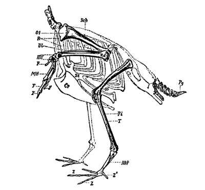
Fig. 181.—The skeleton of the limbs and tail of a flying-bird. OA, bone of upper arm; OS, bone of upper leg; Rd, Ul, bones of fore-arm; T, Fi, bones of leg; HW, MH, bones of wrist; F, F, F, bones of fingers; Z, Z, Z´, MP, bones of foot; Scp, shoulder blade; Cr, keel on sternum (St); Pg, ploughshare bone.
The bones of the fore-limb and the neighbouring regions show a remarkable modification of the primitive plan. The bones of the upper arm OA, (Fig. 181) and fore-arm (Rd., Ul.,) are arranged much like the corresponding bones of the rabbit; but those of the wrist and hand are unlike anything we have yet examined. The fourth and fifth fingers have entirely disappeared; and the first (thumb), second, and third, though they can be still recognised (Figs. 181, F, F, F and 184a), have become consolidated with what remains of the wrist (HW, MH) to form a firm mass which is in no danger of “giving” during the powerful down-stroke of the wing. The head of the bone of the upper arm fits into a socket (gl.cv., Fig. 182) at the junction of the shoulder-blade (scp.) and another and stouter bone (cor.) which runs back and is attached to the breast-bone or sternum (st.). The sternum in its turn is connected by means of ribs with the spinal column, which is strengthened here by the fusion of some of its bones. The ventral part of the sternum is produced into a large plate called the keel (car., Fig. 182), which gives attachment to the great muscles of the breast, used in the movements of the wings. So great is [Pg 275] the development of these muscles in flying birds that in the pigeon they have a weight equal to one-fifth the total weight of the body. The three segments of the wing, corresponding to the upper-arm, fore-arm, and wrist and hand respectively are, in the position of rest, bent upon each other like the letter Z. The large quill feathers of the wing, fixed along the hinder border of the limb, are in two series (Fig. 179). Those attached to the fore-arm are called secondaries; while those attached to the wrist and hand are known as primaries. A smaller tuft of feathers borne by the thumb is called the bastard-wing or thumb-wing. The smaller feathers covering the bases of the wing-quills are called wing-coverts.
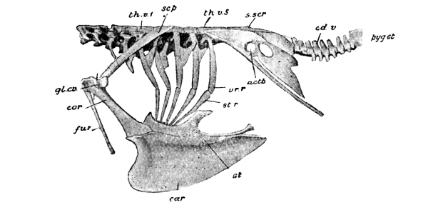
Fig. 182.—Pigeon. The bones of the trunk. actb, socket for bone of upper leg; car, keel of sternum; cd.v, vertebrae of tail; cor, coracoid; fur, merrythought; gl.cv, socket for bone of upper arm; pyg.st, ploughshare bone; scp, shoulder blade; s.scr, fused vertebrae of hip region; st, sternum; st.r, vr.r, rib; th.v.1, first, and th.v.5, last thoracic vertebra. (× ⅓.)
The hind-limbs and tail.—The hind-limbs, or legs, of birds are the sole support of the body except during flight, and a relatively great weight is thus thrown upon them. To provide for this, the skeleton of the hip region is exceptionally strong. Not only are the bones of the spine here welded together into one solid mass, but the [Pg 276] hip-bones extend forward much further than is the case in quadrupeds, and are firmly attached to the fused vertebrae. Each hip-bone possesses a socket (actb., Fig. 182) into which fits the head of the bone of the upper leg. Another fusion of bones is to be noticed at the end of the tail, resulting in the ploughshare-bone (Fig. 182, pyg. st.) which supports the large steering feathers of the tail. The quills of these feathers are covered at their bases by the tail-coverts.
Just above the end of the tail of a plucked pigeon is to be seen a small conical body, the oil-gland, from which the bird obtains a fluid which it applies to the feathers during the preening process.
The feathers.—Feathers do not grow upon all parts of a bird’s body, but are restricted to certain definite areas or “feather tracts” which, however, differ in arrangement in different species.
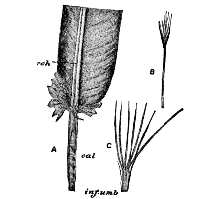
Fig. 183.—Pigeon. A, portion of a wing feather; cal, quill; rch, shaft; B, filoplume; C, nestling-down.
One of the large feathers of the wing of the pigeon is illustrated in Fig. 183, A. It consists of a hollow, horny quill (cal.), prolonged into a solid shaft (rch.) which supports the web or vane. The outer face of the feather is slightly convex and smooth; while the inner surface is somewhat concave and rough, and carries a little tuft of down at the junction of quill and shaft. The growing feather is nourished by a conical projection of the deeper part of the skin, which fits into a small hole (inf. umb.) at the base of the quill. It is easily shown that the vane of the feather is composed, on each side, of a large number of parallel laths which spring from the [Pg 277] shaft. These laths are called barbs. Considerable resistance is felt when an attempt is made to pull the barbs apart, but the manner of their connection cannot be clearly seen without the aid of the microscope. A fairly low magnifying power, however, shows (Fig. 184) that each barb bears extremely delicate threads—barbules—on each side, arranged on the barb much as the barbs are arranged on the shaft. The barbules on one side of each barb (the side furthest from the quill) are seen to carry tiny hooks; while the barbules of the other side of the barb are furnished with flanges. The hooked barbules of one barb cross the flanged barbules of the next and interlock with them; so that relatively great force is required to pull the barbs apart and destroy the continuity of the web. The quill-feathers of the tail, and the coverts, have webs of similar structure.
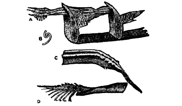
Fig. 184.—Structure of a Feather. A, small portion of a feather with pieces of two barbs, each having to the left three hooked barbules, and to the right a number of flanged barbules; B, hooklet of one barbule interlocking with flange of another barbule; C, two adjacent flanged barbules; D, a hooked barbule. (After Pycraft.)
The contour-feathers are used for protection and warmth rather than for flight, and hence have less-perfectly interlocking barbs. The hair-like structures seen on the skin of the plucked bird are extremely simple feathers called filoplumes (Fig. 183, B). Each consists of a long, slender axis, bearing at the end a few barbs which do not interlock.
The down-feathers (Fig. 183, C), which form the temporary covering of nestling pigeons, also bear barbs which do not interlock.
At regular intervals, either once or twice a year, birds moult, [Pg 278] that is, shed most or all of their feathers and grow new ones. The moulting usually takes place gradually and symmetrically: a flight feather from each wing, for example, being dropped at the same time.
Flight.—The outstretched wing of a pigeon has a relatively great area; and is markedly convex on the upper surface and concave on the lower, resembling in this respect an open umbrella. The great difference in the resistance which the inside and outside of an open umbrella respectively oppose to the wind is familiar to everyone, and illustrates the advantage of the dome-shape of the outstretched wing. The force which the great breast-muscles put into the down-stroke of the wing is enormous in comparison with the weight of the body, and is sufficient to push the bird upwards and forwards in the air. The gently tapering shape and smooth surface of the feathered bird diminish the resistance of the air. When the wing is raised again for the next stroke, the quill-feathers are turned a little edgewise, so that the air slips between them; just as an oarsman “feathers” his oar to lessen the resistance to the blade in the return-stroke. The direction of the wing-stroke can be altered in accordance with the direction of air currents, and the fan of tail-feathers is capable of being opened or closed, raised or lowered, and turned at various angles to act as a rudder.
But the method of flight which perhaps most of all excites the observer’s admiration is soaring, in which the bird seems to remain almost passive, with outstretched wings and spread tail, and mounts automatically in a spiral course. Exactly how the soaring is performed, only the birds themselves know; but it must be similar in essence to the arrangement of the sails of a yacht so as to select that component of the force of the breeze which will drive the boat in the required direction, even though this be almost “in the teeth of the [Pg 279] wind.” Such spiral soaring may be seen to perfection, among common British birds, in the skylark (p. 316). A breeze is necessary for soaring.
In hovering, the motion of the wings is extremely rapid, and the bird remains poised in one place. The kestrel (p. 330) derives its common name of “windhover” from its habit of using this method of flight when looking for food.
The pigeon’s air supply.—We know that a man breathes more quickly when he is taking active exercise than at ordinary times; and we might, from this and similar observations, expect to find the most perfect breathing organs in animals which lead the most active lives. Of all vertebrate animals, birds probably perform the most work in proportion to their size; and it is not surprising, therefore, to learn that they have special facilities for quickly replacing fouled air by fresh.
The lungs are comparatively small, but they are not the only organs of respiration (p. 242); for the windpipe opens also into several air-sacs, which supply nearly all parts of the body with air, and even communicate with the interior of certain bones. The bone of the pigeon’s upper-arm, for example, is hollow and contains air. By means of the system of air-sacs, the air in the lungs is completely renewed at each respiratory act, and thus the waste carbon-dioxide in the blood can be exchanged for fresh oxygen much more completely than it is in mammals. One result of this is that the blood of birds is much warmer than that of mammals. The air-sacs are also an assistance to flight by rendering the body more buoyant.
Different breeds of pigeons.—The various breeds of domestic pigeons furnish one of the best examples of the changes in structure which breeders can bring about, after several generations, by careful crossing. In spite of the great differences between, say, the [Pg 280] tumbler, with its habit of turning head-over-heels in the air; the pouter, with its exaggerated crop; the Jacobin, with its neck-feathers reversed; the fantail, with its large number of erect tail-feathers; and others,—there can be no doubt that all these varieties have been artificially produced from the rock-pigeon; and it is curious to observe occasional reversions to the characters of the ancestral stock. “The rock-pigeon is of a slaty blue, with white loins.... The tail has a terminal dark bar, with the outer feathers externally edged at the base with white. The wings have two black bars. Some semi-domestic breeds, and some truly wild breeds, have, besides the two black bars, the wings chequered with black. These several marks do not occur together in any other species of the whole family. Now, in every one of the domestic breeds, taking thoroughly well-bred birds, all the above marks, even to the white edging of the outer tail-feathers, sometimes concur perfectly developed. Moreover, when two birds belonging to two or more distinct breeds are crossed, none of which are blue or have any of the above-specified marks, the mongrel offspring are very apt suddenly to acquire these characters.”[16] This is a striking instance of the tendency exhibited by living things to revert to ancestral characters, even though these latter may have lain dormant for hundreds of generations.
EXERCISES ON CHAPTER XV.
1. Why does a bird require a long neck, and a keel upon the breast bone? (1898)
2. Draw the bones of a bird’s wing, and mark the places of insertion of the largest quills. (1898)
3. What bones carry the primary and secondary quills of a bird’s wing, and the quills of the tail? Illustrate your answer by a drawing. (1901) [Pg 281]
4. Point out the chief differences between the skulls of a quadruped and a bird. (1901)
5. Describe one of the large feathers of a bird’s wing. (1898)
6. Draw general plans of the fore-limbs of a mammal and a bird. Letter the principal bones, so as to show how they correspond in the two animals. (1901)
7. If the skull of a bird were placed before you, what features would enable you to recognise it with certainty? (1901)
8. How does the fact that a bird stands on two legs affect the skeleton of the trunk? (1897)
9. Point out the chief differences between the wing of a bird and that of a bat.
10. Explain how the barbs of a quill feather are attached to one another. Describe a feather at the time when it is forcing its way through the skin. (1905)
11. What peculiarities distinguish feathers which aid in flight from feathers which merely prevent loss of heat? (1906)
12. Describe the wing of a bird, both as to skeleton and as to external appearance. Compare the skeletal parts with those of the corresponding structures in a Mammal. (1908)
Obtain three or four hen’s-eggs, and make the following observations upon them:
1. External appearance.—What is the colour of the egg? What is its shape? How does the shape differ from that of a cricket ball or other sphere? The shape of an egg is said to be ovoid. [What is the difference between an ovoid and an oval?] Measure, with a tape, the length and breadth of the egg, and also its distance round, (i) in the direction of its greatest length, (ii) in the direction of its greatest width. What advantages has this shape? Could a hen sit so comfortably upon her eggs if they had sharp corners? Put an egg upon the table and roll it gently. Does it roll in a straight line? Does it roll far before coming to rest? Compare the rolling of a cricket ball or other sphere. Is it an advantage that an egg is not likely to roll far, or in a straight line? Why?
2. The strength of the shell.—Hold an egg with your thumb at one end and your first or second finger at the other, and press exactly in the line of the length of the egg. You cannot break the shell.
3. Structure.—(a) The shell.—Tap the egg gently at the middle of its broad end until the shell cracks. Then carefully remove small pieces of the shell and notice the shell-membrane, a tough skin [Pg 283] which is closely applied to the inside of the shell. Snip through the membrane in the middle of the broad end; notice the air-chamber which lies beneath it. Observe the inner membrane which separates the air-chamber from the inside of the egg. Hold a piece of shell up to the light, and notice the small, almost transparent dots. The shell is perforated by very small pores, through which the air can pass.
(b) The white of the egg.—Tap the shell so as to crack it all round at its widest part; raise bits of shell carefully and see the membrane here. Tear through the membrane and notice that in this region there is no air-space, but the white lies just beneath the shell-membrane. Separate the halves of the shell, notice the position and shape of the yolk, and then let the contents of the egg fall gently into a basin. Observe the appearance, colour, and transparency of the white, and try to distinguish two tangled cords of firmer white—the balancers (Fig. 185)—arising close to the yellow yolk.
(c) The yolk.—What is the shape of the yolk as it lies in the basin? How does it differ from the shape of a yolk suspended naturally in the white? What is the cause of the change of shape? Notice carefully a small paler patch in the middle of the upper surface. This is the lightest part of the yolk, so that the yolk always settles with this part uppermost after any turning of the egg, and therefore the pale patch is always more directly exposed to the heat of the hen’s body (during incubation) than is any other part of the yolk. Prick the yolk and notice that the yellow, fluid contents flow out. You have evidently pierced the thin bag which formerly preserved the shape.
(d) A hard-boiled egg.—Boil an egg in water for five minutes, and then chip round the shell in the direction of the length and, with a sharp knife, cut the whole egg into halves along this plane. Make a drawing of the section, indicating the shell, shell-membrane, air-chamber, white, and yolk in position. Observe that the white is no longer transparent and fluid, but an opaque, white, and elastic solid. Try to peel off the white in layers. They will probably break off short, but you may be able to see that the white is deposited in spiral sheets around the yolk. [Pg 284]
The hen’s egg.—A hen’s egg bears a somewhat similar relation to the adult bird that the maize or bean seed (Chapter I.) bears to the adult plant; for the egg contains
(1) a speck of living matter which little by little grows and becomes marked off, by orderly arrangement, to form the various regions and organs of the adult animal; and
(2) a store of food, which enables the young chick to develop in security, without being hampered, whilst still weak and helpless, by the necessity of earning its own living. The changes by which the speck of living matter becomes the perfect chick will be considered in the next section. We must now examine the structure of the egg itself, and see what provision it contains for the nourishment and safety of the developing bird.
The egg is ovoid in shape, one end being distinctly broader than the other. This shape has the advantage of preventing the egg from rolling very far when placed upon a slightly inclined surface, and it is worthy of notice that the eggs of birds which lay on cliffs and other exposed situations are usually more elongated and pointed than others, so that when stirred they do not roll away, but simply describe a small circle and come to rest again. In the case of eggs which are laid in cup-shaped nests (Fig. 197) the pear-shape lends itself to close packing, and thus allows the eggs to be more easily kept warm by the parent bird.
The shell (sh., Fig. 185) is composed of a chalky material, and is perforated by small pores, through which the developing chick obtains fresh oxygen from the air, and gives off its surplus carbon dioxide (p. 242). A small piece of shell easily snaps, but the shape of the complete shell so distributes an outside pressure, especially one in the direction of the long axis, that relatively great force is required to break it. The shell is lined by a thin, parchment-like membrane (sh. m.). At the broad end of the egg this [Pg 285] membrane is double, and the two layers enclose an air-chamber (a).
Inside the shell-membrane are the white or albumin, and the yolk. The white (alb´.) is a viscous, transparent fluid. Its innermost part (alb.), which immediately surrounds the yolk, is of thicker consistency than the rest, and is prolonged into two twisted cords called the balancers (ch.), which suspend the yolk in position.
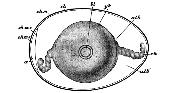
Fig. 185.—Semi-diagrammatic view of a fowl’s egg at the time of laying. a, air-space; alb, dense layer of albumin; alb´, more fluid albumin; bl, germinal disc; ch, balancers; sh, shell; sh.m, shell-membrane; sh.m 1, sh.m 2, its two layers separated to enclose air-space; yk, yolk. (After Marshall.) (× 1.)
The yolk is a golden-yellow fluid enclosed in a thin, elastic membrane and hence preserving a spherical shape. On its upper surface (but under the membrane) is a small circular patch (bl.) of paler colour. This patch, called the germinal disc, is about ⅛” in diameter; it contains the living matter from which the chick will be formed; the rest of the yolk, and the white, are simply a store of inert food which is used up by the growing chick.
The work of the balancers.—In order that the small living patch [Pg 286] of pale yolk, the germinal disc, may grow and develop into the chick it must be kept warm. When eggs are hatched in the natural manner, the heat is supplied by the body of the sitting hen, and the upper part of the egg is consequently kept warmer than the rest. It is important that the living part of the egg shall always lie nearest the hen’s body and thus be kept warm, and this is secured by an arrangement as effective as it is simple. The yolk is lightest in the neighbourhood of the germinal disc, and therefore always lies with this part uppermost. If the egg is slowly turned over, the yolk remains “right side up.” If it is turned over quickly, the yolk soon swings round into its original position. The twisted cords of white, the balancers, not only sling up the yolk and guard it from being thrown to one side by a sudden movement, but they also prevent it from rotating too quickly—and so possibly injuring the delicate body of the young chick—when it rights itself after the egg has been turned over.
The balancers are rendered necessary by the hen’s habit of turning her eggs two or three times a day. It is often supposed that the eggs are turned in order to keep them equally warm on all sides, but this is unnecessary. Most probably the egg is turned in order to alter slightly the young chick’s position from time to time, and allow its parts to grow naturally, unimpeded by the other contents of the egg.
1. A simple incubator.—Eggs are best incubated in the natural manner, that is, by the warmth of the hen’s body; but if a sitting hen cannot be obtained, an ordinary water-oven, such as is used in chemical laboratories, may be made to answer. It should be heated by a self-regulating burner, and kept at a temperature of about 40° C. The eggs should be turned two or three times a day, and the air of the oven should be kept moist by sprinkling water upon pieces of cloth, blotting [Pg 287] paper, or hay, kept with the eggs. Spring is the most favourable time of the year for making the observations, as eggs laid at other seasons are not always in a condition to produce chicks.
2. How to mark the eggs.—The most instructive changes take place during the first five days of incubation. If all the stages of the first five days are to form the subject of one lesson, an egg should be marked “5” with pencil, and then put into the incubator or under the hen five days before the lesson; a day later, an egg numbered “4” should be put in, and so on. The numbers will then indicate the length of incubation at the time of the lesson, and the eggs should be examined in order, from 1 to 5. If one egg is to be examined each day, five should be put in the incubator at the same time; no numbering will then be required.
3. How to examine the eggs.—Have ready a basin of water, heated slightly above the temperature of the hand (i.e. to about 40° C.), and dissolve table-salt in it in the proportion of a level teaspoonful of salt to a pint of water. The young chicks will keep alive longer in this solution than in ordinary water. Tap the shell in the middle of its broad end, and open the air-chamber (a, Fig. 185 ) completely. Then crack the shell in the middle of the length and, keeping the length of the egg horizontal, cut transversely round the middle of the shell with scissors in a vertical plane, until the halves are on the point of coming apart. Then lower the egg into the warm saline solution, pull the halves of the shell apart, and float out the contents. Examine the embryo carefully, making out as much as possible, and then snip round it with a pair of fine scissors to remove it from the yolk; float it into a watch-glass and cover it with weak alcohol (equal parts of water and spirits of wine). Examine it with a lens. After it has remained for a day in weak alcohol, put the embryo into strong alcohol in a small bottle (writing the age on a label), and preserve it.
Notice the gradual absorption of the white of the egg as development proceeds.
4. Chick after one day’s incubation.—Notice that the embryo is now to be distinguished as a streak crossing the germinal disc in a [Pg 288] direction at right angles to the long axis of the egg. Notice a rounded swelling at one end of the embryo; this is the head. Place the egg before you with the broad end to your left, and observe that the head of the embryo points away from you.
5. Chick after two days’ incubation.—Observe the increase in size of the embryo; make a note of its length. The head and neck of the embryo are now almost covered by a very thin transparent bag which has grown over it from the sides. This bag is called the amnion; it is filled with fluid, and protects the embryo from jars. Remove the amnion and notice the large head; it is now twisted so that its left side lies against the yolk, while the rest of the embryo still lies “face-down.” Observe the large eye on the right side of the head; the left eye cannot be seen without turning the head over. Notice the heart, a small red dot which by help of a lens can be seen to beat rapidly. Surrounding the embryo is a circular network of blood-vessels which bring food from the yolk to the heart, to be distributed to the various parts of the body. How large is the circular area of blood-vessels?
6. Chick after three days’ incubation.—The white of the egg is distinctly shrunken, and the network of blood-vessels is much larger than before. Remove the amnion and notice the marked increase in size of the embryo, especially of the head. The right side of the head and neck are still turned towards the shell. They are now quite free from the yolk, but the body of the embryo communicates with the yolk by a short, wide tube, the yolk-stalk. Try to see a small pit, a little above and behind the large eye. This is the beginning of the right ear. Measure the embryo and the width of the surrounding network of blood-vessels. Watch, through a lens, the beating of the heart.
7. Chick after four days’ incubation.—Carefully cut open the amnion to see the embryo better. Observe that the young chick is still more completely folded off from the yolk, and that the yolk-stalk is consequently narrower than before. The head is so strongly bent upon itself that the snout almost touches the tail. The body also has now turned over so as to lie with its left side on the yolk. Observe the two pairs of small buds which are the rudiments of the limbs. [Pg 289]
8. Chick after five days’ incubation.—Cut open the amnion, and notice the great increase in size of the embryo, and especially the enormous development of the head. The limbs now show signs of division into segments. Observe, under the hinder end of the body, a small, bladder-like outgrowth, the allantois, which in the later stages grows rapidly and spreads all round the inside of the shell. It is the breathing organ of the chick.
9. Effect of varnishing an egg.—Varnish an egg, and leave it under the hen with unvarnished eggs for the whole period of incubation (21 days). The varnished egg does not develop, because the varnish closes the pores of the shell and prevents the embryo from breathing.
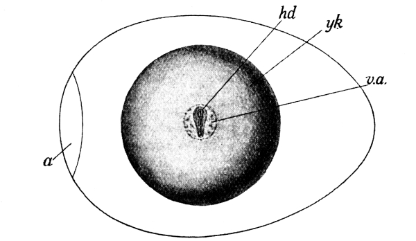
Fig. 186.—Hen’s Egg after 1 day’s incubation. a, air-chamber; hd, head of embryo; v.a., area in which yolk blood-vessels will appear later; yk, yolk. (× 1.)
The early development of the chick.—When the hen’s egg is exposed to a temperature of about 40°C., the germinal disc—the circular patch of living matter which lies, somewhat like a small inverted watch-glass, upon the upper surface of the yolk—grows larger, and its parts are gradually modified to form the various regions of the chick’s body. The white and yolk of the egg are used up during the process, and by the time they are absorbed, the young bird is in a [Pg 290] condition to break the shell and take up the activities of an independent life. To watch the orderly appearance and gradual development of the different systems of organs is most fascinating, and fortunately the observation of the main features presents no great difficulty if the foregoing instructions are followed.
The first signs of the chick are to be seen (Fig. 186), towards the end of the first day of incubation, in a streak which crosses the germinal disc in a direction at right angles to the long axis of the egg. One end of the streak is distinctly rounded, and this is the head end (hd.). In almost all cases, if the egg be placed so that its broad end is to the observer’s left, the head of the embryo will be directed away from him.
A day later, the embryo is markedly larger, and is partly covered by a double membrane called the amnion.[17] Surrounding the embryo is now (Fig. 188) a network of fine blood-vessels which [Pg 291] ramify over the yolk and carry small particles of yolk to feed the growing organs. At the end of the second day the network could be about covered by a sixpenny piece. On each side, its veins join to form a single small tube running to the heart (ht.), a tiny red dot which is situated behind the head and can be seen, by the help of a lens, to be beating rapidly. The head has grown more than the rest of the embryo, owing to the rapid development of the primitive brain, and is bent forwards. The head has turned over a little, so that its right side is directed towards the egg-shell; the rest of the embryo still lies “face-down.” The right eye (e.) can easily be seen as a dark spot on the side of the head.
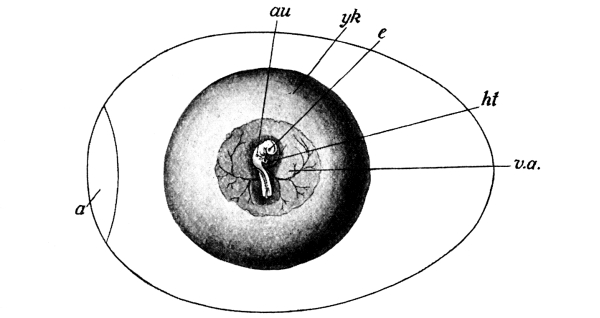
Fig. 188.—Hen’s Egg after 2 days’ incubation. The amnion has been removed. a, air-chamber; au, beginning of right ear; e, right eye; ht, heart; v.a., network of yolk blood-vessels; yk, yolk. (× 1.)
By the end of the third day (Fig. 189) the head and neck are raised distinctly above the yolk, and the tail is also more plainly marked off, so that the body is connected with the yolk only by a short wide tube, the yolk-stalk (yk.st., Fig. 187, B). The neck as [Pg 292] well as the head has now turned so as to lie with the right side directed towards the shell. The commencement of the ear is to be seen on each side as a small pit (au.) a little above and behind the large eye.
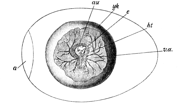
Fig. 189.—Hen’s Egg after 3 days’ incubation. The amnion
has been removed. References as in Fig. 188. (× 1.)
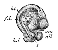
Fig. 190.—Chick Embryo after 4 days’
incubation. The amnion has been removed.
all, allantois; am, cut edge of amnion;
f.l., fore-limb; h.l., hind-limb;
ht, heart; t, tail. (After Duval.) (× 2.)
On the fourth day the embryo turns to lie entirely on its left side. It becomes more completely folded off from the yolk, and the connecting yolk-stalk is a narrower tube than before. The previously formed organs increase in size and complexity. The disproportionate size of the head, owing to the great development of the brain, is still more marked than before, and the head is so strongly bent upon itself that the snout almost touches the tail. One of the most noteworthy features of the fourth day is the appearance of the limbs. As yet these are merely a pair of small buds (fl., hl., Fig. 190), and no joints can be detected in them. [Pg 293]
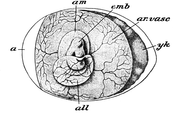
Fig. 191.—Hen’s Egg after 5 days’ incubation. a, air-chamber; all, allantois; am, amnion; ar. vasc., area of yolk blood-vessels; emb, embryo; yk, yolk. (After Duval.) (× 1.)
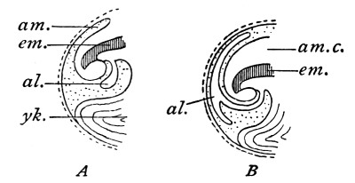
Fig. 192.—Diagrams illustrating the method of development of the allantois. al, allantois; am, amnion fold; am.c., amniotic cavity; em, embryo; yk, yolk. (After Foster and Balfour.)
By the end of the fifth day (Fig. 191) the head is enormous, and the limbs now show signs of being divided into definite segments. A thin bladder-like structure—the allantois (all.), which appeared on the fourth day—has grown out from the lower part of the body, behind the yolk-stalk. Its mode of origin is well shown in Fig. 192. It rapidly increases in size, and soon extends over the embryo (Figs. 192 B, and 191) and becomes closely applied to the shell-membrane. Air passes through the pores of the shell, and its oxygen is taken up by the blood which circulates in the vessels of the allantois. At the same time, waste carbon dioxide is able to escape from the blood [Pg 294] to the outer air. The allantois is therefore the breathing organ of the developing chick. If the egg is varnished and the shell thus rendered air-tight, the embryo dies of suffocation at an early stage.
The chief organs of the bird are now established, and the later development may be sketched more briefly. By the end of the ninth day (Fig. 193) the white of the egg is almost used up; the yolk, however, is still large, and is connected with the chick’s body by the narrow yolk-stalk. It thus appears that the white is not directly absorbed by the chick, but is first taken up by the yolk and afterwards passed on by the yolk blood-vessels which run to the heart. By this time, too, the allantois has spread at least halfway round the inside of the shell, that a supply of oxygen adequate to the increased needs of the animal may be obtained from the air. The chick has now a characteristic bird-like appearance; the beak has appeared; feathers have begun to sprout; the neck is long and slender; and the segments of the limbs, including the fingers and toes, are well defined.
About the fourteenth day the chick turns so as to lie lengthwise in the shell, with its head near the broad end. The yolk-sac dwindles in size, and at last, about the twentieth day, it is drawn into the interior of the body. Now the chick becomes restless, and—usually on the twenty-first day—thrusts its beak through the inner shell-membrane into the air-chamber at the broad end of the egg. For the first time it draws refreshing air into its lungs, and is stimulated to break the shell by a knob on its beak, and to creep out into the world. [Pg 295]
1. Hatching.—On the 20th or 21st day of incubation remove three or four eggs from the clutch under the hen, and keep them in a warm cosy place so that you may watch the process of hatching. Can you hear the chick tapping the inside of the shell? Could it have been taught to tap? Which part of the shell cracks first? When the shell has cracked, can you hear the chick chirping? Could it have been taught to chirp? Imitate the chirp and listen for a response. Keep the newly-hatched chicks in a warm, soft place. How soon do they recover from the exhaustion of hatching? Do the chicks show a liking for warm corners? Are they afraid of being touched gently?
2. Locomotion.—How soon do the chicks begin to run about? Do they stumble upon obstacles, or do they avoid or leap over them? Do they use their wings in running, or in jumping down a small step? Put a chick into a basket, and lower the basket quickly through the air, being careful not to let the chick fall out. Does it move its wings? How?
3. Feeding and drinking.—Lay a few grains of soaked wheat upon the ground before a chick which has not yet been fed. Does it peck at them instinctively? Tap a grain with the point of a pencil; does the chick now peck at it? Does it hit the grain at the first try? Does it ever strike at a grain which is out of reach? Watch a hen with her chicks; does she teach them to peck at food? How? Do the chicks know instinctively the difference between food and grains of sand, or have they to learn? Try if a chick can distinguish between a small worm and a bit of red worsted. Are young chicks afraid of large insects?
Take a chick, which has not yet drunk, to a small pool (a few drops) of water. Does it seem to know the use of the water? Induce it to peck at the water; does it now drink? How? [Pg 296]
4. The crouching-instinct.—Clap your hands suddenly and loudly near young chicks. How do the chicks behave? Does this behaviour render them less conspicuous?
5. Non-recognition of hen.—Having made the above observations on chicks hatched apart from the hen, put the chicks among their brothers and sisters with the parent hen. Do the new-comers respond, as readily as the others, to the clucking, etc., of the hen?
6. Maternal training and protection.—How many different meanings can you recognise in the different sounds of the hen’s voice? How does she call and protect the chicks when any danger threatens them?
7. The voice of the chick.—Notice the different sounds by which a chick expresses pleasure, alarm, distress, etc.
8. Preening and scratching.—At what age do chickens begin to preen their feathers, and to scratch the ground? Try to discover if these are instinctive activities, or whether they have to be learnt from the hen.
9. Change in plumage.—At what age are the down feathers replaced by the true plumage? At what age is it possible to distinguish the young cocks from the young hens?
10. External characters, etc., of adults.—Notice the flowing tail-feathers, hackles (the long feathers on the neck and loins), comb, and spurs of the cock-birds, and their absence or small size in the hens. What differences are there as regards voice? How does the hen announce that she has laid an egg? Which sex is the more pugnacious?
The recently hatched chick.—The newly-hatched chick is clothed with fine down-feathers (pp. 273 and 277). It is generally exhausted by its struggles to escape from the shell, but it soon recovers, and is able to run about freely on the second day after hatching. Young birds which, like the chick, are active immediately after hatching, are said to be precocious.
Instinct and education.—From the first, the chick performs [Pg 297] certain movements which are obviously instinctive, that is, which have not been acquired by any process of imitation or instruction. Even in the egg, it may be heard chirping soon after it has taken its first breath of air; and the complex activities of walking, running, jumping over obstacles, and, later, preening the feathers and scratching the ground—each of which involves the nicest adjustment of several muscles—are also instinctive. Certain other powers have to be learnt; the hen, for example, lifts and drops before the chick a particle of food which she wishes it to seize, and it soon learns to peck. At first its pecks generally fall a little short of the objects, but presently it becomes very adroit at catching food. “A chick a day or so old will catch a running fly at from the seventh to the twelfth shot.”[19] There does not appear to be much instinctive recognition of the difference between objects which are valuable as food and those which are the reverse. Such discrimination comes only by experience, and to a very young chick a piece of red worsted and a small worm are equally attractive before being seized. In the same way, the nature of water has to be learnt. Chicks peck at drops of water, but do not seem to know the use of water until their beaks are wetted, when they drink instinctively in the usual manner.
The use of the wings is also a matter of instinct, but it is acquired somewhat late, and is assisted by parental encouragement and example even in birds which are noted for their powers of flight. Fowls do not fly much, but chicks may be observed to use their wings as an aid in running or jumping. Prof. Lloyd Morgan[20] quotes an interesting experiment which shows well how deeply the reliance on flight is stamped in bird-nature. “If a chick a day or two old be placed in a basket, held firmly in the hand, and then lowered rapidly through the air, the fledgling will stretch out his little [Pg 298] immature wings in such an attitude as would make them break the fall were they fully developed; or will, if he be a little older, flap them with flight-like action, in either case showing an instinctive response.”
Hens and chickens.—Chickens which have been hatched in the natural manner are, from the first, under the control of the hen (Fig. 194), and her anxious care for their welfare has always been considered one of the most beautiful examples of the maternal instinct. In their defence her courage is unbounded; should she discover a succulent worm or other dainty morsel, her ordinary complacent cluck changes to a note of invitation, to which the brood at once responds; and at her danger-signal the chickens run to take refuge under her wings, or crouch motionless against the ground. The chirp of a chick also is capable of expressing several states of feeling, such as contentment, pleasure, alarm, and distress. These different notes are not only perfectly intelligible to the hen, but are familiar to anyone who has had experience of poultry.
In the face of these well-known instances of the perfect understanding between the hen and her chickens, it is somewhat surprising to find that chickens, hatched apart from the hen and then placed with the rest of the brood after a few days, at first pay very little attention to the hen, except for their instinctive tendency to nestle into warm places, and do not seem to understand her clucks. [Pg 299]
Adult fowls.—The down-feathers are soon shed and replaced by the true plumage, and the chicken gradually takes on the appearance of the adult. A marked difference is generally to be seen between the cocks and hens (Fig. 195) of any breed of poultry. The cock is usually provided with flowing tail-feathers, hackles (elongated feathers on the neck and loins), a prominent comb, and “spurs”—features which are either absent or much less perfectly developed in the hens. As great differences exist between the voices of the two sexes, the crow by which the pugnacious male challenges his rivals, and the cackle of the hen when she has laid an egg, being familiar examples.
EXERCISES ON CHAPTER XVI.
1. Point out the differences in appearance and position between the white and yolk of a fowl’s egg. What becomes of each during the development of the egg?
2. When does the heart of a chick begin to beat? Describe its appearance at this time, and explain what must be done to expose it to view.
3. Explain exactly where the chick is to be found in a fertile egg which has been incubated for three days. How big is a chick of that age? Of what colour is it? Does it give any signs of life? (1898)
4. What is the amnion? Where, in a developing chick, is it to be found, what is its appearance, when and how is it formed, and what is its use?
5. Explain the nature and use of the “balancers” of an egg.
6. Explain how a developing chick breathes.
7. What new organs are first seen distinctly on the fourth and fifth days respectively of incubation of a hen’s egg? Explain exactly how you would expose them to view, and describe their appearance.
8. What is the period of incubation of a duck’s egg? Observe and describe exactly how the hatching duckling breaks open the shell and escapes from it.
9. Observe and describe a duckling’s first attempt to swim. Is the action instinctive or not?
10. Make other observations upon ducklings, comparing them in as many respects as possible with chicks, and write careful accounts of the results you obtain.
11. How does a fowl’s egg which has been hatched for four days differ from a fresh-laid egg? Describe the new structures which have formed in it, so far as they can be made out by the unaided eye. (1904)
12. Where does the chick begin to form in the egg? Explain the arrangement which brings it as near as possible to the body of the sitting hen. (1905)
13. What is the use of the cloudy masses attached to opposite sides of the yolk of a fowl’s egg? (1906)
1. The song-thrush, mavis, or throstle.—Throstles are to be seen throughout the year. Take every opportunity of observing their habits, and make careful notes of these at the time. Especially attend to the following characters:
(a) General appearance.—What is the average size of the bird? What is the length of the tail? At what angle is the tail held? Does the bird move it in any special manner? What is the prevailing colour of the body? Notice the light-coloured and spotted breast. Compare the male and female as regards size, colouration, etc.
(b) Habits.—What situations do throstles chiefly inhabit? Do they keep near the edge of the wood, or have you often seen them inside thick woods? Does the bird hop, or walk, when on the ground? Upon what does it feed? Have you ever seen it cracking the shell of a snail? If you find a heap of snail-shells near a stone, watch, at a little distance, for the throstle coming to crack snail-shells on the stone. Does it also eat worms, or fruit? Describe a throstle’s beak. How would you describe the flight—as high or low, swift or slow, straight, in wide curves, or undulating? Does the bird perch on trees? How does it use its toes in perching?
(c) Song.—In what months does the throstle sing? Does it sing late at night and early in the morning? How does it compare with other birds [Pg 302] in this respect? In what positions have you seen the bird singing—when flying, perching, or on the ground? Try to write down the syllables which seem to you most like the throstle’s song, and notice how the phrases are repeated. Notice that throstles sing less during August than in autumn. Listen, in September or October, for young thrushes learning to sing, and contrast their efforts with the song of the adult birds.
(d) The nest.—At what time of the year do throstles pair and build their nests? In what situations have you found the nests? Write a description of the shape, size, and materials of the first throstle’s nest you find. Visit it frequently, try not to disturb the birds, and note the dates of laying of the eggs. How many eggs are laid? What are their size and colour? How are they arranged in the nest? Do the broad or the narrow ends point inwards? What are the advantages of this arrangement (i) for purposes of packing, (ii) when the eggs hatch? Do both birds sit on the eggs? If only one, which? What is the time of incubation?
(e) The young.—Are the young birds naked and helpless, or feathered, at hatching? How do the parent-birds feed them? What is the appearance of the fledglings? In what respect do they (i) resemble, (ii) differ from, the parents? How long do the birds remain in the nest after hatching? Write an account of any education you have observed them to receive from the parents.
{Make similar observations and notes on other birds, in addition to the special observations mentioned below, and learn to recognise their appearance, flight, song, nest, and eggs. A field-glass will be found of great assistance.}
2. The missel-thrush, or storm-cock.—Compare the missel-thrush with the song-thrush. Notice its larger size, the brighter colour of its spotted breast, and its somewhat less musical song. Listen for its song during, or immediately after, a storm, even in winter. In the nesting season observe the marked courage and pugnacity of the bird.
3. The blackbird.—Compare the blackbird with the thrushes in respect of appearance, food, habits, and song. Notice the difference in [Pg 303] the colouration of the male and female blackbird. Observe that the young bird has a spotted breast like that of a thrush, but that old blackbirds are not spotted. Have you noticed a blackbird show a preference for singing in any particular place? What observations would lead you to suppose that the blackbird is a near relative of the thrushes?
4. Fieldfares.—Distinguish these birds from thrushes. Notice that they arrive about October, and frequent fields in large parties.
The thrush family.—For many reasons the thrush family forms a convenient starting point in the study of the habits of common British birds. It includes some of our very finest songsters and therefore most popular birds; many of its members are abundant in most parts of the country, so that nearly every student has the opportunity of observing them at first hand; the birds are not specially shy; and, lastly, the family affords excellent material for the method of comparison and contrast, which, be it repeated, is the essence of all sound work in nature-study.
The song-thrush, which is also known as the throstle and mavis, is a shapely bird, easily recognisable by its grey, spotted breast (Fig. 196), which may be seen all the year round in wooded parts of the country. The male and female are very similar in appearance, and of almost equal size, measuring 8 or 9 inches from head to tail. The thrush generally feeds on the ground, hopping along on a pair of sturdy legs and feet, seizing and pulling out worms which incautiously show themselves at the mouths of their burrows, and catching insects and snails. A thrush usually has a favourite stone on which it cracks the shells of snails, and it is not uncommon to find, near the stone, a little heap of broken snail-shells—the remains of many a feast. In winter, when the summer diet can no longer be obtained, the thrush subsists largely upon the fruit of hawthorn, mistletoe, etc. Its beak, as is usual with birds, is distinctly adapted [Pg 304] to the capture of its food, being, in this case, narrow, round, and fairly long. The thrush is a strong and rather rapid flier.
Thrushes begin to sing at the first signs of spring, pairing and commencing the work of nest-building in February or early March. The throstle’s song is surpassed by very few birds; and many people consider it equal, if not superior, to the nightingale’s. The song begins before sunrise, and may be heard when almost all other birds have retired to rest. The thrush has a curious habit of repeating each strain two or three times, “lest,” as Browning says, “you should think he never could recapture the first fine careless rapture.” Many attempts have been made to imitate the song by words; Mr. R. Kearton[21] quotes with approval the following rendering by the famous Scottish naturalist Macgillivray:
[Pg 305] But, as Mr. Kearton remarks, “no human words can ever represent, especially in cold type, the passionate vehemence, the sprightliness, or the tender pleading of a thrush’s song.”
The nest of the thrush (Fig. 197) is a massive structure, deep and cup-shaped, and open at the top. It is built of small twigs and grass, and is plastered with mud or cow-dung until the inside is smooth and hard. It is usually placed in an evergreen bush or tree, but may also be found in cavities in tree-trunks or in holes in walls, or even in sheltered places on the ground. The eggs number from 4 to 6; they are about an inch long, are sky-blue in colour, and marked with black spots, which are most numerous near the thick end. The eggs are arranged with their pointed ends inwards; this method not only allows them to be packed more closely together and therefore to be more easily [Pg 306] covered by the sitting bird, but is also of convenience at the time of hatching, as the young birds usually emerge (p. 294) at the thick end. The eggs hatch after a fortnight’s incubation. The young thrushes are at first blind, almost naked, and are quite helpless. They make rapid progress, however, and are sufficiently fledged to leave the nest in about a fortnight after hatching. The fledgling throstle (Fig. 198) is lighter in colour than the adult, and much more distinctly spotted. In October the young birds may be heard learning to sing; their first efforts are hesitating and uncertain, but by tireless practice and careful imitation of the old birds they presently come into full possession of their delightful powers.
The missel, or mistletoe, thrush (Fig. 199) much resembles the song-thrush in appearance and manner of life, but is distinctly larger, measuring about 10½ inches from head to tail. It has a very pleasing song, which may be heard, even in the depth of winter, [Pg 307] when all other song-birds are mute. The missel thrush’s habit of singing vigorously during storms has led to its alternative name of “storm-cock.” The bird is noted for its pugnacity in the breeding season, and for the courage with which it defends its nest and young; any other birds approaching the nest are promptly attacked and driven to a distance. The nest is generally built in a tree, where a branch springs from the trunk. It is made of dried grass, moss, and wool, and the hen lays from 4 to 6 pale-green eggs, which are speckled with brown.
The blackbird (Fig. 200) may easily be distinguished from the [Pg 308] thrush by its colour—the male being black, with a yellow beak, and the female blackish brown, slightly mottled below. The blackbird is slightly larger than the thrush, but in shape and habits the two birds are very similar; they haunt the same woods, live upon similar food (i.e. worms, insects, snails and, in winter, fruit) and build their nests in similar situations. The blackbird’s song is, however, quite distinctive, and consists of mellow, flute like notes, without the repetition which is so characteristic of the throstle and missel-thrush. The blackbird is fond of perching, when singing, upon a bare branch which commands a good view of the surroundings. The nest (Fig. 201) is much like that of the song thrush, but the inside has a soft lining of fine grass instead of being hardened with mud. The 4, 5, or occasionally 6 eggs are bluish-green in colour, and are marked with blurred brown spots.
[Pg 309] Curiously enough, young blackbirds, like young thrushes, are very distinctly spotted. There can be no doubt that blackbirds and thrushes are descended from the same stock, and that their ancestors had spotted breasts. For some reason, the blackbird has lost its family colours, although, as is so often the case among animals, it is still compelled to bear the marks of its ancestry during its infancy. Young blackbirds also receive a musical education from their parents, and may often be heard practising their song in September or October.
The fieldfare, another near relative of the thrush, arrives in this country about the beginning of October, and stays with us for the winter, returning in spring to its nesting places among the pines and firs of Norway. The general colour of the bird is grey, with reddish-brown on the wings, and its breast is speckled in the thrush-manner. Fieldfares frequent fields in large parties, especially in the evenings, but are readily alarmed.
Other birds of the thrush family.—The redwing, wheat-ear, whin-chat, stone-chat, redstart, robin, nightingale, and hedge-sparrow are other members of the thrush family which can here be only referred to. They have many points of resemblance, one of the most interesting of which is that the young birds are invariably spotted. The bill is usually rather long, stout, and straight, the staple food being insects, worms, etc., though fruit is also eaten, especially in winter. The nests are typically cup-shaped, and the eggs greenish or blue, with or without spots. The young birds are quite helpless and almost naked when hatched, and can only open their mouths to be fed by the parents. Even after they have left the nest they need careful and continuous teaching by the parents before they can be made to understand that in future they will have to obtain their own food. Birds of this family, [Pg 310] on account of the variable character of their food, are not so markedly migratory as many others, although they generally move further southward in winter, as the supply of insects becomes scarce—their place being taken by birds which have spent the summer in more northerly regions. This habit is very obvious in the case of the fieldfare and redwing, which visit us for the winter and return to Norway and Sweden for the summer. The nightingale, on the other hand, arrives here from the south in April to build its nest and rear its young.
1. The swallow.—(a) General appearance.—Notice, first of all, the bird’s colouration, as this readily distinguishes the swallow from the martins. The whole of the upper surface is steel-blue; the only white is on the ventral surface. A black band stretches across the breast; the throat and forehead are pink. Observe the long, deeply forked tail, and the broad, short bill.
(b) Habits.—What are the earliest and latest dates on which you have seen swallows? Do the birds seem to prefer the neighbourhood of water? Have you ever seen them (i) perching on trees, (ii) on the ground? Why do swallows occasionally alight on the ground? Do they seem comfortable on the ground? Observe the small and weak feet; are these adapted to a terrestrial life? Do swallows feed on the ground? What do they eat? How are the insects caught? What is the advantage of the very wide gape? How would you describe the flight of the swallow?
(c) Voice.—Does the swallow sing? Describe its voice. Have you heard it when the bird was on the wing, or only when it was at rest? What kind of sound is uttered by the swallow when it is alarmed or angry?
(d) Nest.—In what situations have you found swallows’ nests? (Do not confuse the nest with that of the house-martin, which lays white, unspotted eggs.) How soon after their arrival do the birds begin to [Pg 311] build? What is the shape of the nest? Of what materials is it composed? How many eggs does it contain? What is their colour?
(e) Young.—Look for the newly-hatched young about the last week in June, and describe their appearance. How soon after hatching do they leave the nest? Do they at once feed themselves, or are they fed by the parents? Keep the nest under observation after the young birds have left it, and notice whether the old birds rear another brood the same summer.
In September look for the annual congregation of swallows which precedes their departure for Africa.
2. The house-martin.—Distinguish the house-martin from the swallow by the patch of white on the upper tail-coverts (Fig. 179), and by the feathered toes. Make notes of the dates of arrival and departure of the birds. Do house-martins occupy old nests or build fresh ones? What are the position, material, shape, and size of the nest? How does it differ from the swallow’s nest? Is the martin of sociable or solitary habits? Watch the birds building new nests or repairing old ones, and describe the process. Examine and count the pure-white eggs. What are the newly-hatched young like? How many broods have you known one pair of martins to rear in one season? Have you ever known martins to be turned out of their nests by sparrows? Watch for the autumn congregation.
3. The sand-martin.—Notice the small size of the bird, the mouse-colour of the upper parts, and the black feathers of the wings and tail. Note the dates of arrival and departure. Is the bird often seen near houses? Look for the nesting-holes in cliffs, banks, etc. Examine the holes, and notice that they lead into tunnels which slope slightly upwards. What is the use of the slope? If possible examine the nest at the end of the tunnel, and compare it and the eggs with those of the swallow and house-martin. Observe the peculiar jerky flight. Does the bird sing? Describe its voice.
4. The skylark.—Does the skylark frequent woods or open ground? Describe the appearance, size, and colouration of the bird, and try to see the long toes. Especially notice the long claw of the hind-toe. Have you ever seen the bird perching on trees or bushes? Are the feet [Pg 312] well adapted for perching? Does the skylark run or hop? In what month does it begin to sing? In March look for larks’ nests in hollows and ruts of fields. What are the materials of the nest? Describe the number and appearance of the eggs. Listen to the song, and try to say in what respects it differs from that of other birds. Does the bird sing only when flying, or also when on the ground? Describe the skylark’s flight, and study its method of soaring (p. 278). Observe that the birds collect into flocks in autumn. Do they leave the country for the winter? How do they spend the winter? Do they sing in winter? If possible examine the beak; for what kind of food do you think it best adapted?
5. The rook.—Notice the size, shape, and colouration of the bird. Do rooks live merely in pairs or in large communities? Do they build on the ground or in trees? Are the trees high or low? What is the largest number of rooks’ nests you have seen in one tree? Watch the birds repairing and building nests in February and March. Of what are the nests composed? Have you ever seen rooks stealing sticks from other rooks’ nests? Are the birds quarrelsome? Where do they feed? Do they hop or walk? Do they keep together when feeding? Do all the birds of one rookery feed at the same time, or do some remain in the trees? Why?
What is the shape of the beak? How is this associated with the food of the bird? Do the farmers in your neighbourhood consider rooks useful or the reverse? Why? What is the voice of the rook like? How does it vary to express warning, anger, etc. Describe any observations which lead you to suppose that a colony of rooks has a code of laws. Describe the flight of the rook; what other birds fly in a somewhat similar manner? In spring, look beneath the trees for the broken shells of eggs which may have fallen from the nests. What is their colour? Describe the appearance and habits of the young birds. Where do rooks spend the winter? Have you ever seen them visiting and inspecting their nests in winter?
6. Other birds of the crow family.—With the rook compare and contrast the crow, raven, jackdaw, magpie, and jay.
[Pg 313] The swallow family.—Swallows and martins are popular with all lovers of birds, for many reasons. Their arrival is welcomed as visible evidence of the approach of summer; their graceful and rapid flight delights the eye; their obvious liking for the neighbourhood of human dwellings wins our sympathy; and, lastly, they claim our gratitude by their incalculable services in keeping down insect pests.
The swallow (Fig. 202) may readily be distinguished from the martins by the absence of white from its upper parts, which are glossy and of a steel-blue colour. The forehead, chin, and throat are of a pink or chestnut colour; the ventral surface is white, with a black band crossing the neck. The tail is long, and very deeply forked. The bill is short and broad, and the gape stretches nearly to the eyes—a great advantage to a bird living on insects which are caught flying. The feet are very small and weak, but as the swallow spends most of its time in the air, and rarely alights on the ground except to collect materials for its nest, it does not need very sturdy hind-limbs, and the feet are chiefly used for perching.
Swallows arrive in this country about the middle of April, and about a month later proceed to build their nests in the chimneys of houses and on the rafters of barns and outhouses. The nest is basin-shaped and open at the top; it is chiefly composed of mud, which the birds collect on the ground, place in position, and allow to dry. Short straws are used to bind the mud together, and the nest is lined with soft grass and feathers. The hen lays from 4 to 7 eggs, which are a translucent white, with reddish-brown or grey markings. The first brood of young is usually hatched about the end of June, but a second or even a third [Pg 314] brood may be raised before the parents depart for the south. Gilbert White[22] gives the following charming account of the training of young swallows: “For a day or two they are fed on the chimney-top, and then are conducted to the dead, leafless bough of some tree, where, sitting in a row, they are attended with great assiduity, and may then be called perchers. In a day or two more they become flyers, but are still unable to take their own food; therefore they play about near the place where the dams are hawking for flies; and, when a mouthful is collected, at a certain signal given, the dam and the nestling advance, rising towards each other, and meeting at an angle; the young one all the while uttering such a little quick note of gratitude and complacency, that a person must have paid very little regard to the wonders of Nature that has not often remarked this feat.” When the young bird has learnt to feed itself “it at once associates with the first brood of house-martins; and with them congregates, clustering on sunny roofs, towers, and trees.... All the summer long is the swallow a most instructive pattern of unwearied industry and affection; for from morning to night, while there is a family to be supported, she spends the whole day in skimming close to the ground, and executing the most sudden turns and quick evolutions.... When a fly is taken a smart snap from her bill is heard, resembling the noise at the shutting of a watch case; but the motion of the mandibles is too quick for the eye.”
The swallow drinks whilst flying, sipping the water from the surface of pools. Its song is a delicate and pleasing warble, which is uttered both at rest and during flight. The voice becomes a squeak when the bird is alarmed or angry. Just before the autumn migration, swallows perch in crowds on roofs, hedges, and the branches of trees. They leave for Africa about the beginning of October. [Pg 315]
The house-martin may be distinguished from the swallow by the patch of white upon the upper tail-coverts, and by its feathered toes. Like the swallow, it arrives in this country about the middle of April. The birds soon begin to repair their old nests and to construct new ones under the eaves of houses. The nest is largely composed of clay or mud, which the birds collect from damp places in the road, etc., place in position against the wall, and allowed to harden. The completed nest is somewhat bag-shaped, having only a small hole at the top; it is lined with soft grass. The eggs are white, without spots. It is worthy of notice when a wild bird lays white eggs, they are hidden from sight by the shape or position of the nest, as in the case of the martins, or the nest is built in an inaccessible place. Exposed eggs are usually made more or less inconspicuous by being coloured or spotted. House-sparrows (Fig. 208) have often been known to expel the defenceless martins from their nests, and to lay their own eggs therein. The eggs of the sparrow may be recognised by their grey colour, and by the brown blotches with which they are marked.
The structure of the martin, like that of the swallow, is admirably adapted to a life in the air, and to a diet of flying insects—the bird having powerful wings, small and weak feet, and a soft and short but widely-opening beak. House-martins leave us about the beginning of October.
The sand-martin is distinctly smaller than either the swallow or house-martin, and it may be further distinguished from them by the “mouse-colour” of its upper parts. It usually arrives in this country about a fortnight in advance of its two relatives, and also departs before them, leaving in August or September. These birds live in colonies in tunnels which they excavate in banks or friable cliffs (Fig. 203)—generally in the near vicinity of water. The excavation may [Pg 316] be three feet in length; it slopes slightly upwards to the nest (which is placed at the end of the tunnel), in order that rain water may not collect in it. The eggs are pure white, and five or six in number. The birds are exclusively insectivorous; they do not sing, but utter a little twitter. The flight is very characteristic, consisting of “odd jerks and vacillations, not unlike the motions of a butterfly.”
The skylark.—At the first glance, it is obvious that the skylark (Fig. 204) is adapted for a life spent largely upon the ground. The large toes lie flat on the earth, and the long claw of the hind toe is plainly not very suitable for grasping the twigs of trees in the act of perching. In fact, the skylark only occasionally perches, on low [Pg 317] bushes. Walking or running on the ground, it lives upon grubs, caterpillars, flies, worms, etc., and also upon seeds, which its strong beak enables it to extract from the husks. The body is brown, the wings being streaked with black, and the dull-white throat and breast being marked with brown spots—a colouration which is a considerable protection. The nest is built in March, in a hollow or rut on the ground; it is lined with dry grass. The four or five eggs are greenish-grey, and spotted with brown. Whilst the hen sits upon her eggs (Fig. 205) the cock-bird either hunts for food or sings his delightful song as he soars in the air. The wonderful powers of soaring of this bird have already (p. 278) been referred to. When the lark drops to the ground he alights at a little distance from the nest, and then runs up to it through the grass. By this device he avoids revealing the position of his mate and young.
[Pg 318] Skylarks rarely sing in the depth of winter; in autumn they form large flocks which patrol the fields until spring in search for food—rendering incalculable services to mankind by the destruction of insect-larvae. During the coldest weather of winter the birds crouch under banks and hedges.
The rook.—The rook (Fig. 206)—often called the crow—is a rather large bird, measuring about 17 inches from head to tail. Its plumage is black, but as it becomes adult the feathers covering the face and nostrils are shed, leaving the skin of these parts bare. The bird has long and pointed wings, and is a strong flier. The bill is stout and almost straight. Rooks are no songsters, their harsh “caw” being destitute of musical qualities. When heard at a distance, however, the cry “becomes a confused noise or chiding; or rather a pleasing murmur, very engaging to the imagination, and not unlike the cry of a pack of hounds in hollow, echoing woods, or the rushing of the wind in tall trees, or the tumbling of the tide upon a pebbly shore.”[23]
One of the most interesting features of rook-ways is the habit of living in organised communities.[24] It is true that a primitive form of social life is found among swallows, martins, and (in winter) larks, reminding us of the [Pg 319] gregarious habits of the rabbit (Chapter XII.) among mammals; but a rook-society is rather to be compared with the pack-life of the wild dogs (p. 252), which depends for its integrity upon the observance of certain rules of conduct by its members. Naturalists have long known that such a code of laws is in force in every rookery, but what these laws are we do not fully understand. Here is a profitable and interesting field of observation for the student. It has sometimes been observed that certain birds are prevented by the rest from building until the nests already commenced are finished, and cases have been known of birds being expelled from the rookery, or even put to death, after a consultation or “trial,” presumably for some breach of law. On the other hand, it must be admitted that rooks are generally arrant thieves, not scrupling to rob their neighbours’ nests of building-materials to save themselves trouble. They are also very quarrelsome.
[Pg 320] The nests are built on high trees (Fig. 207); they are composed of sticks and turf, and are lined with moss and soft grass. As each of several adjacent trees often contains a large number of nests, a sort of rook-town called a “rookery” is formed. This is added to from year to year, as the number of the birds increases. During the summer the rookery forms the headquarters of the community, but about August or September, before the leaves are shed, the birds leave their nests, to spend the winter in thicker or evergreen woods, often at some distance. At intervals during the winter they revisit their spring nests. They come back to the rookery in February or March, and at once set about repairing old nests or building new ones. The male bird begins to feed the female even before the bluish-green eggs are laid, and is a pattern of domestic virtue throughout the period of incubation. For some time after hatching, the young birds are fed by the parents. Rooks feed on the ground in flocks, a few birds remaining behind in the trees as sentinels to give the alarm when danger threatens. The food consists very largely of the larvae of injurious insects, especially the grubs of the cockchafer beetle, and although the birds undoubtedly eat corn and other young crops, they do far more good than harm.
Other birds of the crow family.—The crow, raven, jackdaw, magpie, and jay are so similar in many respects to the rook and to each other that naturalists include them all in one group, which they call the crow family.
The passerine order.—The families of the birds hitherto considered in this chapter, with the flycatchers, wrens, chats, tits, finches, linnets, starlings, etc., are further grouped together to form what is called an order. The order which consists of these families derives its name passerine from the sparrow[25] (Fig. 208), perhaps the commonest of the birds included in it. It is probably in [Pg 321] this order that bird structure attains its most perfect development. Again, the birds of the crow family are generally considered to stand at the head of the order; if this view is correct, rooks must be regarded as occupying, among birds, a position which roughly corresponds to man’s place in the class of mammals.
Passerine birds are nearly all singers, and their toes are generally adapted for perching. They feed upon insects, seeds (especially the seeds of grasses), and soft fruit. Their beaks vary in form according to the nature of the food upon which they most depend, as is well shown by the soft, widely opening bills of the swallows, the strong but slender beaks of the thrushes, larks, and crows, and the short, hard beaks of such seed-eaters as the canary, sparrow, and other finches. Birds which are exclusively, or almost exclusively, insectivorous are compelled to migrate to warmer countries to obtain food during the winter. The eggs of passerine birds are usually coloured or spotted, and the young birds are hatched in a helpless and almost naked condition, being carefully tended and fed by both parents until they are able to fend for themselves. A few familiar birds of other orders will now be briefly considered.
1. The swift.—Distinguish the swift from the swallow by its larger size, its sooty black colour (except for the dull white of the throat), and its long, bowed wings. In what respect does the swift [Pg 322] resemble the swallow? Watch for the appearance of swifts about the end of April. Do the birds arrive singly, in pairs, or in flocks? Notice the rapid turns and twists of the flying bird. Does it as a rule fly near the ground, or at somewhat great heights? Have you ever noticed the bird perching, or settling on the ground? Have you seen it clinging to walls with its feet? Were these the walls of houses or barns, or of high towers? What actions would lead you to suppose that the nests are built in high towers? Are more swifts to be seen in the evening, or during the day? Can you explain this? Watch during the first half of August, and make a note of the day on which swifts were last seen.
2. The cuckoo.—What is the earliest date on which you have heard the cuckoo? Imitate the cry by putting your hollowed hands together and blowing between the thumbs. With practice you will be able to deceive the bird into thinking that another cuckoo is present, and it will approach near enough to be seen distinctly. Estimate the size of the bird, and write down a careful description of its form, colouration, and method of flight. Try to see that two of its toes point forwards and two backwards. Have you ever seen cuckoos mobbed by small birds?
Carefully examine as many nests of small birds as possible, and try to find one containing an egg slightly larger than the rest and perhaps differently coloured. This will probably be a cuckoo’s egg. Visit the nest at frequent intervals, both before and after the hatching of the eggs, and write descriptions of the eggs, the appearance and size of the young birds, what becomes of each, and the behaviour of the old birds. What is the latest date on which you have heard the cuckoo?
The swift.—Though the swift bears a strong general resemblance to the swallow, the two birds are not at all closely related, but belong to different orders. The swallow, like all other passerine birds (p. 320), has 12 tail-feathers, and its first toe is separately movable and directed backwards. The swift, on the other hand, has only 10 tail-quills, and all its toes point forwards. There are, however, more [Pg 323] obvious differences, which readily enable one bird to be distinguished from the other, even at a distance. The swift is, with the exception of a dull-white patch on the throat, of a sooty black hue over all the body, and in flight its long and powerful wings take the form of a bent bow (Fig. 209). The swallow flies in bold, sweeping curves, while the more rapid swift turns sharply in the air, in pursuit of the flying insects upon which it feeds, in a manner suggestive of the flight of a bat. While, again, the swallow flies much near the ground, the swift is usually seen at greater heights. The harsh scream of the swift is, moreover, in marked contrast with the swallow’s pleasing song.
Swifts do not willingly alight on the ground, even to collect materials for their nests, nor do they perch on trees or roofs, the arrangement of their toes being quite unsuitable for either action, and the length of the wings being such that, even with toes of the normal type, walking would be almost impossible. The nest, a somewhat rude structure of grass and feathers, is generally built in high towers or other tall buildings. The eggs are two in number; they are more conical in shape than those of the swallow, and, as the position of the nest renders them independent of protective colouration, are white. The hen-bird sits on them during the day, but generally leaves the nest and flies abroad in the evening to hunt for insects. Only one brood is reared, and the birds take their departure early in August, to spend the winter in South Africa or Madagascar.
[Pg 324] The cuckoo.—The cuckoo is placed by naturalists, not among the passerine birds, but in the order to which the swift belongs. The reasons for this cannot be fully considered here, but it may be mentioned that the cuckoo has only 10 tail-feathers, and that it differs from all passerine birds in having its fourth toe, as well as the first, directed backwards, the second and third pointing forwards (Fig. 210).
The cuckoo has a total length of about 14 inches. The upper surface of the body, and the throat, are grey; the rest of the lower surface is white, crossed with black bars. The wings are large and powerful, and the thighs are covered with long feathers. In general appearance, as well as in manner of flight, the cuckoo bears a great resemblance to a hawk, which may perhaps be the reason why small birds so often join forces and attack it. A keen-eyed observer will, however, at once distinguish the birds by the head and the bill, both of which are markedly longer in the cuckoo than in the hawk. Cuckoos arrive here in April, the males usually appearing first. The well-known call, to which the bird owes its name, is uttered by the male only; the voice of the female is quite different, and is often compared to the sound of bubbling water. The cuckoo feeds entirely upon insects; it is believed to be the only bird which eats the hairy caterpillar of the tiger-moth (p. 369).
Of all bird-habits, probably none has excited more interest than the manner of life of the young cuckoo. Its parents build no nest, but [Pg 325] depend entirely upon other birds for rearing their offspring. The female cuckoo lays her egg upon the ground and then carries it in her bill to a nest which contains similarly coloured eggs, if such a nest is to be found. If not, another convenient nest is selected. It is said that the male sometimes renders assistance by distracting the attention of the rightful owners in the meantime. In most cases the owners of the nest, apparently unaware of the trick, sit upon the strange egg with the rest, and in due course the young birds are hatched. The young cuckoo, whilst still blind and naked, wriggles itself under its foster brothers and hoists them over the edge of the nest. Far from resenting the crime, the duped parents now devote their energies to feeding the murderer, and continue their attentions until after it is fully fledged (about a fortnight after hatching) and perhaps several times their combined size.
The small size of the cuckoo’s egg—about one-fourth of what might be [Pg 326] expected from so large a bird—and its general similarity in colour to their own eggs, both aid in deceiving the victimised birds in the first instance, and the disproportionate size of their supposed offspring shortly after hatching apparently does not arouse their suspicions. It is believed that any particular female cuckoo lays only eggs of one type and deposits them, as far as possible, in nests of the mimicked species—the species, probably, by which she herself was reared. During the summer, therefore, she visits one such nest after another, until she has disposed of all her eggs. Further, it is supposed that her offspring inherit her tendency to lay eggs of the particular colour, and therefore to prey in their turn upon the species of their foster-parents. In this country, the meadow pipit, pied wagtail, robin, and reed-warbler are perhaps the most usual victims of the cuckoo’s parasitism, but the nests of other species (Fig. 211) are not uncommonly selected.
The old cuckoos depart about the end of July, apparently leaving the young ones to find their way south alone.
1. Tame ducks.—Closely observe ducks, both on land and when they are swimming. Where is the heaviest part of the body? Are the legs attached in front of, directly below, or behind the centre of gravity? Is the position of the legs an advantage or a disadvantage (i) in walking, (ii) in swimming? How does the foot differ from the feet of the birds previously mentioned? Between which toes is the web stretched? How are the toes held when the bird lifts its foot in walking? Does it walk gracefully? How does it swim? Are the legs moved together or alternately in swimming? Notice how the web folds up when the foot is moved forwards, and spreads out for the back-stroke. What [Pg 327] does the duck eat? How is the food obtained? Why are a duck’s feathers so little wetted by the water? Watch, and describe exactly, how the bird preens its feathers.
How can you distinguish the male (drake) from the female duck, (i) by appearance, (ii) by the voice? Do the colours of the drake’s plumage differ at different periods of the year? What relation have the changes of plumage to the moulting-season? What differences can you observe in the methods of moulting of ducks and fowls? Is there any time of the year when (i) ducks, (ii) fowls are unable to fly? Why? When and where do ducks lay their eggs? Describe the appearance of the eggs. Are young ducks helpless or precocious (p. 296), naked or clothed, at hatching? Can they feed themselves? Can they swim?
Examine a dead duck. Notice the thick covering of down-feathers next the skin. What is its use? Examine and draw a foot, and see how the web folds between the toes. Observe the soft, sensitive skin on the outside of the beak; what do you suppose is its use? Notice the shape of the beak. Open it to see the horny plates fringing its inner edge, and notice how these, with other plates on the thick, fleshy tongue, form a strainer.
The duck.—The birds of the duck order—which includes also the geese and swans—differ in many important respects from all our previous types, and it is interesting to observe how perfectly these differences are adapted to the manner of life. On land, the ungainly waddle, which is entailed by the insertion of the hind limbs so far back on the body (Fig. 212), shows that the duck is not in an entirely congenial element; in the water the bird is a model of graceful movement, perfectly balanced, progressing smoothly by alternate strokes of its webbed feet, and altering its course to any desired direction with the utmost ease. The duck feeds largely upon the small animals which abound in the water of ponds, and in the mud of the sides, and the beak is beautifully fitted for the duties it has to perform. It is [Pg 328] covered on the outside by a soft and highly sensitive skin, which enables the bird to detect with certainty the presence of its prey in the mud; the inner edge of the beak is provided with horny plates which, with similar plates situated on the edge of the thick, fleshy tongue, form a very efficient strainer, by means of which the useless water and thin mud can be forced out at the sides of the mouth and separated from the worms, etc., which were taken into the mouth at the same time. A thick coat of down-feathers, which lies next the skin and contains a great deal of entangled air, forms a non-conducting layer which prevents the undue escape of the heat of the body, and saves the bird from becoming chilled when in the water. Ducks are, moreover, careful to keep their feathers well oiled, and may often be seen preening themselves—applying the bill alternately to the oil-gland on the tail and to the feathers. The completeness with which water flows off a duck’s back is proverbial.
[Pg 329] During the greater part of the year the brilliant plumage of the drake forms a striking contrast to the sober brown and grey feathers of the female. Another point of difference is that in the male the four middle tail-feathers are curled upwards. From July to October, however, before the moult takes place, the two sexes are very similar in appearance. Birds of the duck order moult in a somewhat different manner from most other birds, in that all the quill-feathers are shed at once, instead of in pairs. Until the new feathers develop, flight is of course out of the question, and the birds remain as secluded as possible in the meantime.
The eggs are white and greasy-looking; they are laid in a rough, open-topped nest, lined with down, which is placed on the ground. The wild duck covers up the eggs when she leaves the nest. The young are active immediately after hatching. It has been noticed that the eggs of precocious birds are generally larger, in proportion to the size of the parent, than those of birds which are naked and helpless at hatching—the larger store of egg-food allowing a more complete development of the young bird in the shell. Further, the nests of precocious birds are, as a rule, less elaborately constructed.
1. The sparrow-hawk.—Watch for sparrow-hawks near farms. Notice the general resemblance of the bird to the cuckoo, but distinguish them by the short head and beak of the hawk. Observe the bluish-grey colouration of the upper parts. Describe the flight of the hawk and, if possible, its method of catching its prey.
2. The kestrel or windhover.—Distinguish the kestrel from the sparrow-hawk (i) by the reddish colour of its upper parts, and (ii) by its habit of hovering in mid-air. Have you ever known the kestrel to prey upon small birds? Upon what does it feed? [Pg 330]
The sparrow hawk and kestrel.—These two birds are the only British hawks which the average nature-student is likely to see during a country walk; and as one of them is given to preying on young game-birds, chickens, etc., while the other as generally confines itself to animals which are universally regarded as vermin, it is important to be able to distinguish them at sight.
The sparrow-hawk (Fig. 213) attains a length of 13 inches; the female is slightly larger than the male. The upper parts of the body are bluish-grey in colour; the lower parts are buffish white, and crossed with brown bars. The head is short and round; the bill is hooked and sharp, as in birds of prey generally, and the toes are armed with sharp claws. The bird is often to be seen near farms, lurking behind hedges and waiting for an opportunity of dashing upon chickens or other small birds and carrying them off.
The kestrel, which in size and shape much resembles the sparrow-hawk, is really a species of falcon (Fig. 179). It is unrivalled among common British birds in its power of remaining poised [Pg 331] in one position in mid-air by that rapid motion of the wings which is called hovering, a power which has earned for it the name of “windhover.” The kestrel, though in reality one of the farmer’s best friends, from its wholesale destruction of field-mice, voles, and many injurious insects, is often ignorantly confused with the sparrow-hawk. The reddish hue of the plumage of the kestrel’s upper parts, together with the hovering habit, ought to render such a mistake impossible to observant eyes, and to secure the bird from a persecution which may reasonably be directed against the sparrow-hawk.
EXERCISES ON CHAPTER XVII.
1. Extend the foregoing methods of study to the following passerine birds:—Starling, finches, wagtails, pipits, nuthatch, tits, warblers, wrens, and flycatchers, and make notes of the observations.
2. Try to discover reasons for grouping (a) woodpeckers, nightjars, and kingfishers with swifts and cuckoos; (b) pheasants and grouse with fowls.
3. Compare geese and swans with ducks, and make notes of as many points of resemblance and difference as possible.
4. Compare and contrast owls with hawks.
5. Arrange the above birds in lists according to (a) food, and the characters of the beak; (b) characters of feet and arrangement of toes; (c) nests (open-topped, covered, built in holes or tunnels); (d) colour and number of eggs; (e) condition of young at time of hatching.
6. How many birds do you know which (a) spend only the summer, (b) spend only the winter, (c) stay all the year, in this country? State, in each instance, upon what food the bird most depends.
1. Manner of life.—Where have you found frogs? Are they commonest in dry or in damp situations? At what time of the year have you seen them actually in water? How do frogs move about? Do they walk or hop? Chase a frog, and notice that its hops become shorter as it is pursued. Upon what does the frog feed? Have you ever seen frogs abroad in the depth of winter? Are insects common in winter? How do you suppose frogs spend the winter? What are the principal enemies of frogs?
2. External characters.—Catch a frog; is its body dry or moist? Does it feel cold or warm? How does it behave when caught? Does it soon become tame if well treated? Put a frog under an inverted pickle-bottle and observe it carefully. What is its size? What was the size of the smallest frog you have seen? At what time of the year can the smallest frogs be seen? What is the colour of the frog? Put one frog with dark-coloured leaves and soil, and another with light-coloured leaves, and try to find out if the skin of the first becomes lighter and that of the second darker in colour. Is it an advantage to the frog to be able to change slightly in colour? Why? What is the position of the frog when at rest? Notice the shortness of the body, the hump on the back, the relative lengths of the limbs, the 4 unwebbed fingers, the 5 webbed toes, and the absence of a tail. What is the advantage of the long hind-limbs? Mention other leaping animals whose hind-limbs are [Pg 333] longer than the fore-limbs. Is a short-bodied animal (i.e. one with its four limbs pretty close together) less likely to be injured by the fall, after a leap, than a long-bodied animal? Why? Put the frog into water, and watch it swim. What is the use of the webs between the toes? Examine the head. Notice the very wide stretch of the mouth, the small openings of the nostrils, the large prominent eyes, and the ear-drum—a dark-coloured disc on each side, a little below and behind the eye.
3. Breathing.—Watch the up-and-down movements of the floor of the mouth, by which the animal pumps air, through its nostrils, in and out of its mouth-cavity.
4. Method of feeding.—Put live insects, small worms, etc., under the upturned bottle enclosing the frog, and watch the animal’s method of feeding. Try to see how it uses its tongue. What is the advantage of the wide gape of the mouth? Can you get the frog to accept dead insects?
5. The inside of the mouth.—To kill a frog painlessly, soak about a teaspoonful of chloroform on cotton wool, and put it with the frog under a bottle or tumbler. After 15 or 20 minutes the frog will be quite dead.
(a).—Open the jaws widely, and examine the inside of the mouth. Notice how the rounded eyeballs project into the mouth-cavity when they are pressed from the outside. Pass your finger-end round the margins of the jaws, and feel the row of fine teeth borne by the upper jaw; the lower jaw is destitute of teeth. Feel also, in the roof of the mouth, two small patches of teeth (Vo., Fig. 216); these are carried by small bones called the vomers, and are therefore called the vomerine teeth. Just to the outside of the vomerine teeth and in front of the eyeball, notice on each side the internal opening of the nostril (Ch., Fig. 216); pass into one nostril a stiff bristle, and observe that it emerges at the external nostril. Pull forward the tongue (T, Fig. 216) and observe that it is attached at its front end; feel how sticky the tongue is. Behind the eyes, and at the angles of the jaws, notice the openings of the Eustachian tubes (E, Fig. 216); push a stiff bristle into one and observe, from the outside, that the end of the bristle presses the inner surface of the ear-drum. [Pg 334]
(b).—If the roof of the mouth has dried, moisten it with water, and place a very small cork-shaving or a tiny snip of paper on it, far back between the eyeballs. Notice that the shaving travels slowly down the throat. This experiment ought to be made soon after the frog has been killed.
6. The bones.—Feel, through the body-wall, the various parts of the skeleton, making out the skull, backbone, limb-bones and their manner of attachment, and breast-bone (protecting the heart). Observe the absence of ribs.
7. The skin.—Examine the skin of the dead frog, and notice that it is damp or even somewhat slimy, and that it differs from the skin of a mammal or bird in being naked, bearing neither hairs, feathers, nor scales. Pinch up the skin, and notice how very loosely it is attached to the underlying body-wall. Snip through the thin skin, turn it back from the body-wall, and see the network of blood-vessels upon its inner surface.
The habits of the frog.—The common grass frog (Fig. 214) is to be found in abundance, from early spring to October, in ditches, marshy land, and other damp places. As winter approaches, frogs generally bury themselves in the mud at the bottom of ponds, etc., and remain there in a state of torpor until the spring, when they emerge and the females lay their eggs. Frogs are said to be “cold-blooded” because their temperature never varies much from that of their surroundings. Birds and mammals, on the other hand, maintain an almost constant temperature,—a healthy man’s blood, for example, being just as warm in the depth of winter as it is on a hot summer day. So long as they are not actually frozen hard, therefore, frogs can endure the winter cold without much inconvenience. Although the frog is essentially a land animal, it is quite at home in the water, and swims gracefully and easily by the help of the webs which connect the long toes of its hind feet. On land, it progresses by long leaps, its limbs and body being [Pg 335] well adapted to this habit. As is usual in leaping animals, the hind legs and feet are markedly longer than the fore limbs; and the shortness of the body enables the two pairs of limbs to be brought together to break the shock of the fall.
Food.—The frog’s method of catching its prey is very interesting, and is graphically described by Dr. Hans Gadow[26] as follows:—“The food, which consists chiefly of insects, snails, and worms, must be moving to excite interest; then the frog, whose favourite position is half squatting, half supported by the arms, erects itself and, facing the insect, turns round upon its haunches, adjusts its position anew by a shifting of the legs, and betrays its mental agitation by a few rapid movements of the throat. All this time [Pg 336] the prey is watched intently until it moves; then there follows a jump, a flap of the tongue, and the insect is seen no more.” This flap of the tongue is well illustrated in Fig. 215. The frog’s tongue is free behind, but is attached, by its anterior end, close to the middle of the lower jaw,—an arrangement which enables it to be flicked out to its full length. Further, it is covered by a glutinous secretion, which sticks tenaciously to the prey.
The frog is fortunate also in the extremely wide gape of its mouth, which stretches, almost literally, from ear to ear. Once in the mouth, the captive is prevented from escaping by the teeth. Of these there are two sets; a row of fine teeth is present along the greater part of the margin of the upper jaw, and, in addition, two small patches of teeth—the vomerine teeth (Vo., Fig. 216)—occur on the roof of the mouth. Fairly large insects are promptly gulped down into the stomach; those which, owing to their minute size, escape being swallowed in the ordinary manner, are slowly but surely forced down the throat by the incessant lashing of thousands of tiny threads—too small to be seen except by high powers of the microscope—which are carried by the skin [Pg 337] of the roof of the mouth. The action of these invisible threads may easily be seen if a frog’s mouth be opened widely and a small cork-shaving be placed near the top of the throat (Expt. 60, 5, b). The shaving steadily travels backwards and is soon lost to sight.
It should be clearly understood that frogs and toads are of incalculable value in keeping down insect pests, and deserve systematic protection for this if for no other reason.
How a frog breathes.—When a resting frog is watched, the floor of the mouth is seen to be raised and lowered alternately. It is a common belief among children that these movements are a sign that the animal is “getting ready to spit.” Frogs do not spit, however, and the action is simply a part of the breathing process, which is performed in the following manner. The mouth being closed, the nostrils are opened, and, by alternate up-and-down movements of the floor of the mouth, the air present in the mouth-cavity is completely replaced by fresh air. The nostrils are then closed, and the slit-like glottis (gl., Fig. 163) which leads to the lungs (l. lng., r. lng.) is opened. The foul air is forced out of the lungs and mixed with the pure air in the mouth-cavity. Then, immediately, the floor of the mouth is raised—pumping the mixed air into the lungs—and the glottis is closed. In the lungs an exchange takes place between the oxygen of the refreshed air, and the surplus carbon dioxide in the blood which is circulating in the capillaries (p. 242) of the walls of the lungs. In the meantime the nostrils have again been opened, and the first stages of the process are already being repeated.
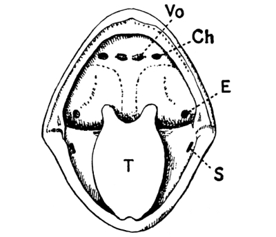
Fig. 216.—Inside of mouth of Edible Frog. Ch, internal opening of nostril; E, opening of Eustachian tube; S, opening leading into vocal sac; T, tongue; Vo, vomerine teeth. (× 1.)
[Pg 338] A considerable part of a frog’s breathing is carried on, in these first stages, through the thin membrane forming the roof of the mouth-cavity. This is richly supplied with blood capillaries, and is therefore admirably adapted for the exchange of gases which constitutes respiration. Moreover, the skin covering the general surface of the body has also a very abundant blood-supply, and forms yet a third respiratory organ; so that it is practically impossible to drown a frog in ordinary water—in water, that is, which contains dissolved air. This power of breathing through the skin is of great importance to a frog during the winter sleep at the bottom of a pond.
The frog’s skin.—The frog’s skin is kept moist by a slimy fluid which is continually being discharged from small glands in its substance. The moisture not only facilitates skin-breathing as described above, but its evaporation keeps the body cool even in hot weather—a matter of vital importance to an animal to which a temperature of 40° C. or so is fatal. A third most interesting property of the frog’s skin is its power of changing somewhat in colour to match the colour of its surroundings. The change depends upon an alteration in form and size of certain small brown specks imbedded in the thickness of the skin. When the animal finds itself in dark-coloured surroundings these specks enlarge, and the skin as a whole takes on a darker hue. On a light background the reverse change takes place.
A case of evolution.—The advantage which accrues to a frog from being thus rendered less conspicuous to enemies and prey alike is obvious, and there is no difficulty in picturing to ourselves the probable manner in which the advantage was developed. Widely different races of animals have colour-specks in the skin, and we may assume that frogs and their ancestors have possessed such spots for countless generations. Now suppose that, ages ago, a frog happened to be born [Pg 339] with the power of altering very slightly the size of his colour-specks. If this power rendered him less conspicuous, in however slight a degree, than his neighbour frogs he would, other things being equal, be more likely than they to escape from enemies, grow to maturity, and in due course have sons and daughters. All his offspring, and they might number thousands, would tend to inherit, to varying extents, the power of changing colour. In some—perhaps in most—the power would be practically absent; in others it might be equal to that of the parent; while in a few instances it would probably be greater. In any case, those frogs of the second generation which had the greatest power of changing their colour would be the most likely to survive the keen struggle for existence and therefore to leave offspring. The survival of the “fittest” frogs of each generation, and the transmission to the next generation, in ever-increasing intensity, of their favouring accomplishment, would naturally result at last in the production of a race of animals in which, as in the frogs of to-day, the power of changing colour is universal.
The laying of the eggs.—About the end of March the frogs resort in great numbers to shallow ponds and ditches, pair with much croaking, and the females lay their eggs or “spawn.” Both sexes croak, and the male of the edible frog, though not of the common grass frog, is able to make more noise in virtue of a pair of vocal sacs (Fig. 217), which he can inflate with air from the mouth (Fig. 216), and which act as resonators. After spawning, the frogs leave the water, abandoning the eggs to their fate, and resume their ordinary terrestrial life, until approaching winter prompts them to hide in the mud and go to sleep. [Pg 340]
1. A simple aquarium.—A simple aquarium, in which the development of the frog from the egg may be watched, is easily made. Obtain a fairly large basin, and cover the bottom with sand, mud, and stones from a pond. Arrange these so that the bottom shall shelve from the surface of the water at one side to a depth of 3” or 4” at the other. Put in some stones covered with green slime, which will almost certainly be found in the pond, plant a few water-weeds, and allow the water to clear.
2. Frog spawn.—Having prepared the aquarium, obtain, towards the end of March (or earlier in a mild spring) a handful of frog spawn from a pond or ditch. It forms a mass of jelly in which the true eggs—small balls about ¹/₁₀” in diameter—are imbedded. Put this in the aquarium and examine it carefully every day, making the observations described below. If possible, obtain a pair of spawning frogs, and place them in a bucket with a little water, so that the earliest stages also may be studied.
3. The eggs before hatching.—Observe the globe of jelly which surrounds each egg. Try to pick it up between your finger and thumb. Do you think it is of any use in protecting the eggs from being eaten by fishes, birds, etc.? Remove the jelly as completely as possible from one egg, put the egg in a watch-glass with a little water, and examine it carefully with a strong lens. To which of the stages shown in Fig. 218 does it correspond? If possible, treat a newly-laid egg in the same manner and examine it every hour or so through the day. Make notes of the time elapsing between the various stages shown in Fig. 218.
4. Hatching.—Notice that the developing eggs change from the spherical to the ovoid form. What is the shape when the embryo begins to move? Notice the appearance of a neck and a tail. At this stage the embryo makes its way out of the jelly, or hatches. The jelly may now be thrown away, as it is of no further use. How does the embryo behave immediately after hatching? Put one in a watch-glass and with a lens try [Pg 341] to see the sucker by which it attaches itself to water-weeds, etc.
5. Tadpolehood.—Examine the tadpoles at frequent intervals by the help of a lens, and write careful accounts of the changes which take place in them. Notice particularly the fine threads—the external gills—which grow out from the sides of the neck. How many are there? After a time they shrivel up and are replaced by other gills which cannot be seen. Write a description of a tadpole at the time when it begins to feed. How large is it?
Count the tadpoles in the aquarium at intervals of a few days, carefully removing all dead ones. Supply fresh green slime from the pond from time to time. Have you any reason to suppose that tadpoles are cannibals?
How soon do the tadpoles come to the surface to breathe air? Describe the appearance of one when it begins to do this. Has it any legs? How many can you see? In what order do the legs grow out?
Notice now the dwindling of the tail. (It does not drop off, as is so often stated.) The tadpole has become a frog. What percentage of the original eggs have developed into frogs?
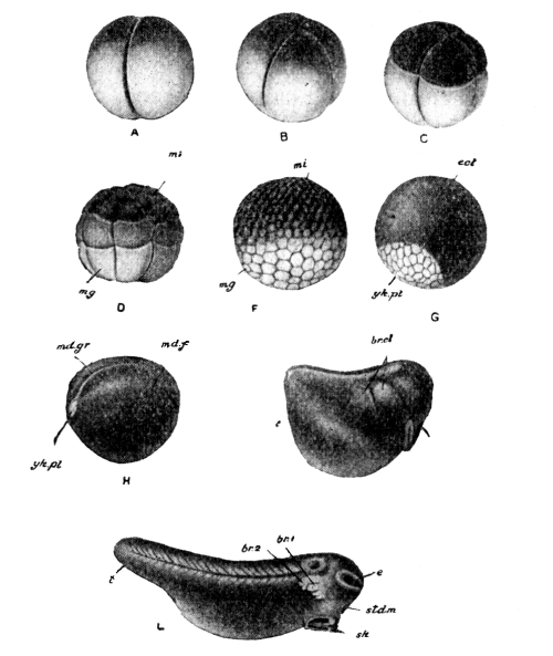
Fig. 218.—The early development of the Frog. mi, small black cells; mg, large white yolk-cells; ect, black cells overspreading yolk-cells; yk.pl, yolk-plug; md.gr, groove of commencing nervous system; md.f, right margin of groove; br.cl, depressions marking position of future gill-slits; stdm, pit which will become mouth; t, tail; br.1, br.2, external gills; e, eye; sk, sucker. (× 5.)
The early development of the frog’s egg.—A frog’s egg is a little spherical mass about one-tenth of an inch in diameter. When laid, it is covered by a thin gelatinous layer which soon swells up in the water to form a transparent globe of about half-an-inch diameter, in the centre of which the true egg can be seen. This jelly is extremely slippery and difficult to grasp, and is consequently an efficient protection against the attacks of birds, fishes, insect-larvae, etc., and even of parasitic fungi and other small organisms. The jelly also acts as a float. At the time of laying, each egg consists of a black and a white portion. In the lower, or white, part there is a store of food, on which the little embryo subsists [Pg 342] until it acquires a mouth and begins to fend for itself. The development begins in the upper, or black, part of the egg, and may easily be watched with a lens. And to be appreciated properly, the changes should be watched, and not merely read about. The student should get, if possible, some freshly-laid frog’s eggs, and remove the gelatinous investment from one or two. It will require care to do this without injuring the eggs. They should now be put, with a little fresh water, into a watch-glass, and carefully examined at intervals of half an hour or so. Soon a little pit makes its appearance in the middle of the black half, and gradually extends until it becomes a groove, which [Pg 343] little by little reaches quite round the egg (Fig. 218, A). In the meantime another groove begins to form at right angles to the first, and, in its turn, grows down round the egg (B). If we compare the whole egg to the earth, and the middle of the black half to the North Pole, these two grooves may be said to be along meridians at right angles to each other. The third groove (C) may be considered as along a parallel of latitude, but it is somewhat to the north of the Equator. These first three grooves deepen until the whole egg is cut up into eight separate pieces. The sight of the apparently lifeless speck dividing itself up in this regular and orderly manner, “while you wait,” is an intellectual treat which should not be missed. The cleavage of the egg goes on rapidly, but in a less regular manner now, until the whole is cut up into a hollow sphere of segments (F), black and small (mi) in the northern hemisphere, whitish and larger (mg) in the south.
It is worth while to pause here to consider how these early changes are assisted by the peculiar condition of the egg at the time of laying. The southern hemisphere of the egg is laden with a store of food. The food is dead, and acts as a mechanical hindrance to the activity of the living matter. In the northern half of the egg but little food is present to impede its activity, and it is plainly important that this half shall receive as much warmth as possible from the uncertain sunshine of early spring. Two circumstances ensure this. In the first place, the food-laden region is the heaviest part of the egg, so that the latter—buoyed up as it is by the jelly—tends to float with its most “alive” part upwards. Secondly, this upper part is coloured black,—a great advantage, since black objects absorb heat readily. Both these peculiarities therefore favour the more rapid development of the “northern” hemisphere. Hence the third cleft is to the north of the egg’s equator, instead of being halfway between the upper and lower [Pg 344] poles, and hence, too, at the close of segmentation the northern segments are smaller and more numerous than the southern. In the case of the hen’s egg (Chapter XVI.), the amount of stored food contained in the egg (the yolk) is so enormous that segmentation is confined entirely to a small patch on the upper surface. Another result of the relatively small amount of stored food in the frog’s egg is that the tadpole is compelled to turn out and earn its own living at a stage when the chick’s inherited fortune is still considerable.
The tadpole.—The spherical mass soon becomes ovoid, and is divided into head and trunk by a neck-constriction (J). An occasional wriggle shows that the creature is alive. Shortly afterwards a tail grows out from the hinder end of the trunk, giving the animal something of the appearance of a fish (L). In this stage the tadpole makes its way out of the jelly, and thus hatches, about a fortnight after the laying of the eggs. The little creature is quite helpless; it has no mouth (the egg food is not yet exhausted, however), and its attempts at swimming are still feeble and uncertain. In this defenceless condition (Fig. 219, 1) it will be seen to attach itself to the water-weed of the aquarium by means of a sucker (Fig. 218, L, sk) on the underside of its head. It is in a very favourable position for examination, and by help of a lens, two—soon there are three—pairs of fine, thread-like outgrowths can be distinguished on the sides of the neck (Fig. 219, 2 and 2a). These are the external gills. In a few days the mouth breaks through, and the animal begins to nibble at the vegetation with little horny jaws, and soon swims about the aquarium with confidence.
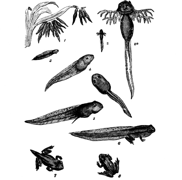
Fig. 219.—The Frog. Stages in the life-history, from the newly-hatched Tadpoles (1) to the young Frog (8). (× 1.) 2a is a magnified view of 2.
The gills are the organs by which the young tadpole breathes, and each little gill-thread is seen, when viewed with a low power of the microscope, to contain tiny loops of blood-vessels. Every time the tadpole uses his jaws, wags his tail, or, in short, does anything at [Pg 345] all, he uses up some oxygen and produces some carbon dioxide. The water of the aquarium contains dissolved oxygen—green water-weeds are kept in aquaria for the express purpose of liberating oxygen (p. 51)—and this oxygen makes its way through the excessively thin membrane which divides the blood of the gills from the water, to be swept away by the current of the blood to the various parts of the body. The carbon [Pg 346] dioxide produced is brought to the gills by the blood stream, and passes through the membrane into the water, where it is utilised by the water-weed as food (p. 51). Animal and plant are thus mutually serviceable.
A series of slits soon opens through the sides of the neck, and along their margins are formed folds which are usually called the internal gills. The external gills dwindle and shrivel up as the internal gills are being formed, and at the same time a flap of skin grows backwards from each side of the head and covers over the slits so that they cannot be seen. Presently the two flaps fuse at their edges—except at one point on the left side, where a spout is left—and so enclose a chamber. The water which enters the tadpole’s mouth pours through the gill-slits, into the chamber, and out through the spout. As it swills over the folds of the internal gills there is an exchange of carbon dioxide for oxygen in the manner just described.
The tadpole in the meantime is growing strong and active, and the tail has grown out to form a powerful organ, the sinuous motion of which propels the animal with relatively great speed through the water.
From the point of view of the biologist, perhaps the most interesting feature of this stage of tadpolehood is the almost entire correspondence of the structure with that of a fish, although the adult frog is not in any sense a fish. This curious state of things is explained by supposing that frogs have descended from fish-like ancestors, and that every frog, in the course of its development, is under the necessity of repeating, in a more or less modified manner, the chief stages of its ancestral history. As Marshall happily expressed it,[27] a frog during its development climbs up its own genealogical tree.
[Pg 347] The metamorphosis.—Just as the external gills are replaced by internal gills, so these, in their turn, are replaced by lungs, and advanced tadpoles frequently come to the surface of the water to breathe air. Limbs have now grown out from the sides of the body, and the webbed hind feet considerably assist the tail in swimming. Changes take place in nearly all the internal organs, fitting the animal for its life on land; and these changes are so extensive that there is necessarily a short period when the creature is neither tadpole nor frog, and is incapable of feeding. The tail, however, which would be useless to the terrestrial, leaping frog, is gradually absorbed, and forms a store of nutriment during the transformation.
The gills shrivel up, and the slits close; the outer layer of the skin (including the horny jaws) is thrown off, the hind limbs lengthen, and the animal leaves the water—a frog.
EXERCISES ON CHAPTER XVIII.
1. Make observations upon toads. In what respects do they differ in appearance from frogs? How do they move about?
2. Are the skins of toads dry or moist? In what situations have you found toads? How do they protect themselves from the heat of the summer sun?
3. At what time of the year, and in what places, do toads lay their eggs? Compare the voice with that of a frog. Do toads inflate any part of the body when they sing? Compare the vocal sacs with those of frogs.
4. Look for toad-spawn in the spring. It forms long, gelatinous ropes in which the eggs are embedded. How large are the eggs? In what respects do they differ from frogs’ eggs?
5. Keep toads’ eggs in an aquarium, and carefully compare all the stages of development with those of the frog.
6. Count how many caterpillars you can persuade a toad to eat “at one sitting.” What would be one result of the extermination of toads and frogs? [Pg 348]
7. Describe carefully what happens when a frog leaps. Point out the special arrangements which enable a frog to leap safely. (1897)
8. Describe the ordinary process of feeding of a frog, and show how it is assisted by the peculiar structure of the frog’s mouth and tongue. (1895)
9. Explain the special use of the wide gape of the frog. Where are the teeth of the frog situated? (1898)
10. Mention some remarkable features of the mouth of a frog, and try to show that they are adapted to meet special needs. (1901)
11. Describe the process of filling the lungs with air, as observed in the frog and the rabbit. (1896)
12. What changes take place in a tadpole during the first week after hatching? Illustrate your answer by drawings. (1898)
13. Describe the appearance of one of the gills of a very young tadpole as seen by a low magnifying glass. (1901)
14. Describe the appearance, size, and mode of life of a tadpole about a fortnight after hatching. On what does it feed? Describe its mouth carefully. (1897)
15. Compare a fresh-hatched tadpole with one in which the hind limbs have recently appeared. (1898)
16. Trace the history of a tadpole from hatching to the time when the tail begins to grow less. What does it feed upon during this time? (1898)
17. Relate the life-history of the frog, from the time of hatching to the end of the first summer. (1897)
18. Give some account of the habits of a young tadpole a few days after hatching, especially with respect to locomotion and feeding. Make a drawing of such a tadpole in side view, three times the natural length. (1904)
19. Describe the hind leg of a frog, and explain the ways in which it is used. (1905)
20. Show how a full-grown frog is enabled to live either in air or in water. (1906)
1. Habits.—In what places have you seen cockroaches? Are they often to be seen during the day, or do they, in general, come forth only at night? What is the colour of the body? Put a live cockroach under a tumbler, and watch its movements. In what position is the head held? Notice the long feelers; how are they used? Look at the lower part of the head, and try to see the palps, which resemble small feelers. How many legs has the cockroach? Watch the rhythmical movement of the hinder part of the body. Soak a small piece of bread in milk or water, and put it under the tumbler; watch the cockroach feed. How does it use the palps? Notice that the jaws move from side to side. How does the insect clean itself?
2. External characters.—Put a cockroach into a test-tube, and dip the tube into boiling water; this kills the insect instantly. Notice the thin “shell” which covers the outside of the animal. Make out that the body consists of (a) head, carrying the eyes, feelers, and jaws; (b) thorax, carrying the wings and legs; and (c) abdomen. Examine them in turn.
(a) The head.—Notice the vertical position, the black, kidney-shaped eyes, the feelers, the “upper” lip (labrum), and the small, blackish mandibles at the sides of the labrum. Pass your knife-point close behind the labrum and into the mouth, and see the “lower” lip (labium), which lies behind the knife, i.e. behind the [Pg 350] mouth. Cut off the head and carefully strip off the labium. Put it on a sheet of paper and examine it with a lens, comparing it with Fig. 222 (Mx. 2). Examine the same head from behind with a lens and see the pair of first maxillae. Notice how they are attached to the head, and then remove one and compare it with Fig. 222 (Mx. 1). Work one of the mandibles backwards and forwards with the point of a pin, and then remove it and examine it with a lens to see the toothed inner margin.
(b) The thorax.—Notice the overlapping fore-wings (wing-covers). Pull them aside with forceps; notice that they are narrow and rather stiff, make out their points of attachment at the corners of the second segment of the thorax, and then cut them off with scissors. Pull out the delicate hind-wings in the same way, carefully noticing their fanlike method of folding, and their points of attachment to the corners of the third segment of the thorax. Stretch one out, cover it with a piece of glass to flatten it, and then draw it, marking the lines of folding. (Notice that some cockroaches of the common species found in kitchens are destitute of wings, and have only very small wing-covers. These are females.) After removal of the wing-covers and wings, the three segments of the thorax are very distinct. Notice that each segment bears a pair of legs. Take off one of the legs and draw it. How many joints has it? What is the use of its bristles?
(c) The abdomen.—Observe the line which runs along the dorsal middle line of the abdomen; this marks the position of the heart. Examine the method of telescoping of the segments of the abdomen, and the soft membrane which connects the dorsal and ventral plates of each segment. Notice that the abdomen bears neither wings nor legs. Observe the pair of short palp-like bodies (cer., Fig. 223) at the end of the abdomen, and between them, in the male, a pair of more slender styles. In the female observe the boat-shaped “brood-chamber” (Fig. 223, st. 7), on the ventral surface.
(d) The spiracles.—In the thin membrane between the dorsal and ventral plates, at the junction of two abdominal segments, look for a small hole (spiracle) leading into the interior of the body. How many [Pg 351] abdominal spiracles can you find on each side? Observe the two larger spiracles on each side of the thorax, between the first and second, and the second and third legs.
What is an insect.—The word insect is so commonly applied to animals having no claim whatever to the title, that it is advisable to point out at once some of the features which distinguish insects from other animals with which they are often confused. Insects, spiders, crustaceans, centipedes, and their near relatives, all have jointed bodies and legs, which are covered by a continuous suit of armour of a substance called chitin. In some cases, as in lobsters and crabs, this is for the most part hardened by mineral matter to form a stout shell, being soft and flexible only at the joints where ease of movement is required. In other cases the layer of chitin may remain thin and delicate, and all gradations between the two extremes may be found. Though soft where movement of one part on another takes place, the chitin is always firm enough, elsewhere, not only to form a protection for internal organs, but also to afford attachment to the muscles which move the body and limbs. It is therefore a skeleton, but as it is on the outside of the body it is called an exoskeleton, to distinguish it from the internal skeleton (p. 224) of the vertebrata. Animals such as insects, spiders, centipedes, and crustaceans, which have jointed bodies and legs, and are covered by a chitinous exoskeleton, are called arthropods. Insects may be at once distinguished from all other arthropods by the single pair of feelers and the six legs. Many of them, but not all, possess wings—structures which are found in no other arthropods. Insects are always air-breathers when adult.
The cockroach.—The cockroach is not a general favourite, but it displays so well the essential features of insect structure that it affords an excellent introduction to the study of the more popular [Pg 352] members of the class, which in many respects are highly specialised. It is also easily obtained, and of fairly large size.
The body of the cockroach (Fig. 220) is very distinctly divided into three regions: (1) the head (Fig. 221) which carries the feelers, the eyes (ey.) and the jaws (man., max.¹, and max.²); (2) the thorax, separated from the head by the slender neck, and bearing the legs and the wings; and (3) the abdomen, which bears neither jaws nor limbs.
The head (Fig. 221) hangs nearly vertically from the neck. The large, compound eyes (ey.) are somewhat kidney-shaped; the long, jointed feelers are set in sockets just below the eyes. The front of the head is smooth and rounded; hinged on its lower edge is a flap which hangs down in front of the mouth, and is called the labrum or upper lip. Just behind the labrum, and attached to the side and front plates of the head, is the first pair of jaws, the mandibles (man., Fig. 221; Mn., Fig. 222); they work from [Pg 353] side to side, and their inner edges, which bite against each other, are strongly toothed. The second pair of jaws, called the first pair of maxillae (max.¹, Fig. 221; Mx. 1, Fig. 222) is situated behind the mandibles. Each first maxilla consists of a two-jointed base and two branches; the inner branch bites against the inner branch of the first maxilla of the other side, while the outer and longer branch forms the maxillary palp, which acts as a small feeler. The two second maxillae (the third pair of jaws) are broadly like the first maxillae, but are partially fused to form the labium or lower lip (Fig. 222, Mx. 2), which hangs down behind the mouth. The outer branches of the second maxillae are called the labial palps (Lab. Pa.). It is well to acquire clear notions of the arrangement of the jaws in the cockroach, in order to understand the great modification which the mouth-parts of many other insects have undergone. It is important to notice that the jaws of all arthropods work from side to side, not vertically as do the jaws of vertebrates.
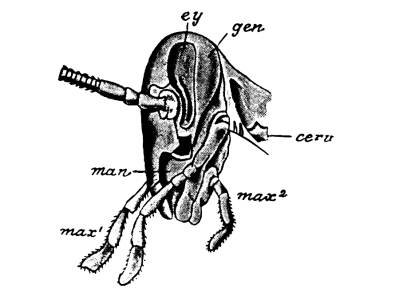
Fig. 221.—Cockroach. The head and its appendages seen from the left side. cerv., one of the neck-plates; ey., eye; gen., side plate of head; man., mandible; max.¹, first pair of maxillae; max.², second pair of maxillae (labium). (× 5.)
The thorax consists of three segments, each of which bears a pair of legs, upon which the weight of the resting or walking insect is supported. It has been found, by instantaneous photography, that in a walking insect the weight is carried at any instant by the first and third legs of one side and the second leg of the other side. At the next step the body is carried by the remaining three legs. In the American (Fig. 220) and German cockroaches both sexes possess two [Pg 354] pairs of wings, which are fixed at the front angles of the second and third segments of the thorax. The hind-wings, which alone are used in flight, are folded up fanwise when not in use, and are covered by the smaller fore-wings, which are generally called the wing-covers. In the common cockroach of this country, only the male has well developed wing-covers and wings. In the female the wing-covers remain small, while the wings themselves have disappeared.
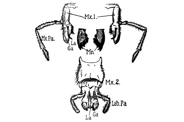
Fig. 222.—Jaws of the Cockroach. Mn., mandibles; Mx. 1, first pair of maxillae; Mx. Pa., maxillary palp (outer branch); La., Ga., inner branch; Mx. 2, second pair of maxillae (labium); Lab. Pa., labial palp (outer branch); La., Ga., inner branch. (× 8.)
The abdomen consists of ten segments, although only eight can be clearly seen without dissection. The “telescopic” arrangement of the segments is well shown in Fig. 223. Where one segment joins the next, the chitin remains thin and flexible. In each segment the chitin forms a dorsal (p. 217) and a ventral plate, which are joined together at the [Pg 355] sides by a flexible membrane. The chitinous covering of the upper surface of the abdomen is so transparent that the heart, a median tube, may be seen through it.
How an insect breathes.—If the sides of the abdominal segments be carefully examined, a small aperture will be seen perforating the thin layer of chitin between the dorsal and ventral plates, at the point where each segment joins the next; and, in the thorax, larger but similar openings will be found on each side between the first and second and the second and third legs. These holes are called spiracles (Fig. 223, spir.); they lead into a complex system of air-tubes which ramify throughout the whole system and supply the organs with oxygen. The tubes are prevented from collapsing by a spiral lining thread of chitin. This peculiar method of respiration, which is characteristic of insects, should be carefully contrasted with the manner of breathing in a rabbit or a man (p. 242) and in a tadpole (p. 344). It ought to be borne in mind, however, that the essence of respiration is the same in all living things, and consists in a replacement of excess carbon dioxide by fresh oxygen (p. 242); the difference lies merely in the manner of effecting the exchange. In the rabbit, the blood, carrying the excess of carbon dioxide, is brought to the air (in the lungs); in the tadpole it is exposed, in the gills, to the dissolved air of the surrounding water; while in the cockroach the air is carried directly to the tissues needing fresh oxygen. The air of the tubes is renewed by a rhythmic action of the abdomen, which can readily be observed in the living insect.
The internal organs.—Figs. 163 and 223, which represent a general dissection of a frog and of a cockroach respectively, exhibit in the most striking manner the essential differences in the structure of vertebrates and arthropods. The fact that the skeleton of the frog is wholly internal and that of the cockroach wholly external, has [Pg 356] already been mentioned. It will be seen, further, that while in the frog the central nervous system (the brain and spinal cord) lies entirely dorsal to the digestive canal, in the cockroach the great nerve chain (Fig. 223, n, n,) is mainly ventral—the only dorsal part of the central nervous system being the so called brain (brn.), which is connected with the ventral chain by a ring surrounding the gullet (gul.). The heart of the frog is ventral to the digestive canal; that of the cockroach is the most dorsally placed organ of the body.
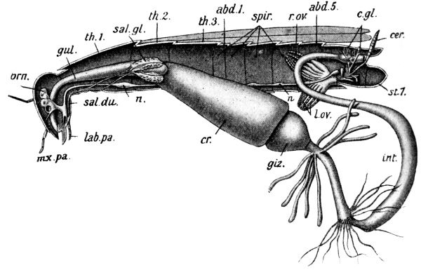
Fig. 223.—Cockroach; general dissection of female from the left side. abd. 1, first, and abd. 5, fifth abdominal segments; brn., “brain”; cer., cercus; c.gl., glands which form the egg-case; cr., crop; f., feeler; giz., gizzard; gul., gullet; int., intestine; lab. pa., labial palp; mx. pa., maxillary palp; n, n, nerve chain; r. ov., right, and l. ov., left ovary; sal. du., salivary duct; sal. gl., salivary gland; spir., spiracles; st. 7, brood-chamber; th. 1, first, th. 2, second, and th. 3, third thoracic segments. (× 2.)
Habits and life-history.—Cockroaches infest kitchens and pantries; they are of social habits, and hide together in crevices during the day, but come forth at night to feed. They are not at all [Pg 357] fastidious as to diet, but are especially fond of starchy foods, which they are able to digest (p. 234) by means of the fluid formed in their large salivary glands (Fig. 223, sal. gl.). The eggs are laid sixteen at a time in a little oblong case, which the female carries about in a boat-shaped receptacle (st. 7, Fig. 223) at the end of her abdomen, until she finds a suitable place in which to deposit it. When the little cockroaches hatch, they are quite white, but except for the absence of wings they closely resemble the parents in form, and run about and feed freely from the first. The chitinous exoskeleton is shed from time to time as the animal increases in size, a new coat, at first soft and wrinkled, but rapidly stretching and hardening, having previously formed beneath the old one. Shortly before the last moult, the wing-covers and wings begin to grow out from the angles of the second and third thoracic segments. In this manner the young animal gradually takes on the form and dimensions of the adult.
The position of the cockroach in the insect class.—The cockroach is familiarly known as the “black-beetle,” but the name is a very misleading one, because the true beetles differ from cockroaches not only in structure, but also in life history. Cockroaches are grouped with earwigs, grasshoppers, crickets, and locusts in the order Orthoptera,[28] a term alluding to the fanlike folding of the hind-wings.
1. The habits of Dytiscus.—Search a pond for water beetles. Put them in a wide-mouthed bottle with some of the weeds to be found in the pond, and take them home and observe their habits. Among the larger beetles—especially from ponds with a clear surface (not covered with [Pg 358] duckweed, etc.)—will probably be seen some with a yellow band round the edge of the upper surface. These are Dytisci. Notice the ovoid, smooth body, the breadth of the hind legs, and—in the male—the cup on each fore leg. Try to see how the legs of each pair are used. On what does the animal feed? Take a small piece of meat in a pair of forceps and hold it near the beetle’s head. If the animal does not notice it, stroke one of the feelers with the meat; how does the beetle behave? What is the use of the feelers? Notice how the beetle rises to the surface immediately it stops paddling. Which end of the body sticks out of the water? Why does the beetle need to come to the surface? In April look for larvae (Fig. 224, A), and describe their appearance and habits.
2. External characters.—Kill a Dytiscus by dropping it for a moment into boiling water. Examine it first from the dorsal surface, noting the almost unbroken oval outline, and the firmness of the armour. Notice that the inner edges of the two wing-covers fit closely together in the middle line. How does the beetle compare in this respect with the cockroach? Observe the small triangular plate between the anterior ends of the wing-covers; open out the covers and wings to find of which segment of the thorax it is a part. Examine the wing-covers and wings closely; notice that the wings are folded transversely as well as longitudinally. Spread and pin out one wing-cover and wing of one side, and draw a dorsal view of the animal. Dissect out the jaws and compare them with the jaws of the cockroach (Fig. 222). Examine and draw one leg of each pair.
The bordered little diver.—In nearly every English pond there at times occurs a beetle—an inch or more in length—which is known to naturalists as Dytiscus marginalis. The name is somewhat cumbrous, but it would be difficult to find a more appropriate one; for “bordered little diver”—the plain English of the scientific term—indicates at once the peculiarities which soonest strike the observer: the creature’s skill in diving, and the yellow band which runs round the edge of the olive-green body. The outline of the beetle is an almost [Pg 359] unbroken oval, and it is worth noticing that either such a boat-shape (Fig. 224, B), or the cigar-shape of the torpedo is adopted by almost all actively swimming aquatic animals. The resistance of the water is further lessened by the smoothness of the beetle’s armour, which forms a hard shell enclosing the body. As in all insects, the body is divided into head, thorax, and abdomen. The eyes are large, and are so arranged that the animal can at the same time see objects both above and below—a great advantage to a creature living so active a life. The feelers are very sensitive organs of touch, and possibly of smell also. The jaws consist of one pair of mandibles and two pairs of maxillae, and are shaped much like those of the cockroach; they are powerful, and render Dytiscus a very formidable enemy to the more peaceable inhabitants of the pond.
The thorax consists of the usual three segments, but only the first and a small triangular area of the second are to be seen until the wing-covers are pulled aside. These last differ from the wing-covers of the cockroach not only in being much harder and stronger, but also in their inner edges meeting accurately along the middle line of the body. The delicate, filmy wings, which alone are of use in flight, are folded transversely as well as longitudinally. On the lower side of the thorax are three pairs of legs; they are very interesting and are worth examining in some detail. In the male, the first leg on each side is furnished with little circular areas which were at one time believed to [Pg 360] be suckers. It is now known, however, that they give off a sticky substance which adheres firmly to any object clasped by the legs. The middle pair of legs seems to be used chiefly for steering. In both sexes the third or last pair of legs is modified to form a pair of sculls. Ordinary land-beetles can move their legs in a vertical direction, as well as in a horizontal one; but the hind legs of water-beetles are jointed to the thorax in such a manner that they can only move backwards and forwards, not up and down. The resemblance of the hind leg to an oar does not stop here, however. On one side there is a fringe of stiff hairs, forming the blade of the oar. The joint carrying the hairs is so arranged that the beetle can “feather” its oar—by turning the edge of the blade to the water—at each stroke.
Occasionally, usually after sunset, the beetle quits his watery home, and “wheels his droning flight” in search of pastures new. His flights are, however, only temporary and merely from one pond to another. Although Dytiscus thus normally lives in water, very cursory observation only is needed to see that he cannot exist without a regular supply of fresh air. He no sooner stops paddling than his body rises naturally to the surface, and as the tail is lighter than the head, it rises out of the water. The wing-covers are now raised a little, so that the space between them and the wings is put into communication with the outside air. The impure gas contained in this space is soon replaced by a bubble of fresh pure air, the wing-cases are lowered, and the “little diver” plunges once more into the depths. The water is prevented by hairs from getting into the air-space below the wing-cases, and the true wings are thus kept always dry. The spiracles (p. 355) are in communication with the air-space, so that the animal is enabled to remain below the surface for a relatively long time. [Pg 361]
The life-history of Dytiscus.—In March or April the female Dytiscus lays her eggs in slits which she cuts in the submerged stems of pond-weeds, and the eggs hatch in about three weeks. The creature which emerges from the egg is of active habits, but is not at all like the parent in appearance. A young animal which leads an independent and self-supporting life, and differs markedly in structure from the adult, is called a larva. Thus a tadpole is a larval frog, and a caterpillar is the larva of a butterfly or moth. The larva of Dytiscus when of full size is about 2 inches in length. Like other larvae of its family (Fig. 224, A), it has six slender legs, which serve both for swimming and for crawling over the bottom of the pond, and its head is provided with a pair of sickle-shaped mandibles, with which it seizes its prey. Each mandible is grooved on its inner side, the groove being converted into a tube by a membrane which covers it in. The savage larva sucks the blood of its victim until literally nothing is left but the shrivelled husk. At the end of the Dytiscus larva’s tail are two appendages which are fringed with hair. When the creature wishes to breathe it comes to the surface, and the tip of its tail protrudes out of the water. As each of the appendages just mentioned is pierced with a hole which leads into one of the two main air-tubes of the body, an interchange of vitiated for pure air readily takes place.
When the larva is about six weeks old, it leaves the pond and buries itself in the soil on the banks. Its exoskeleton is shed, and a thin, transparent layer of chitin—the “pupa-skin”—takes its place. In this condition the animal sinks into a state of torpor, and apparently becomes as motionless as a mummy. In this resting stage it is called a pupa. The pupal stage is necessary for the completion of the great changes—commenced some time previously—which must take place [Pg 362] before the larva can acquire the structure of the adult. The pupal stage lasts two or three weeks, and when at last the creature emerges from its cell it is a beetle like its parents.
In consisting of three well-marked stages, the life-history of a beetle thus differs essentially from that of a cockroach. All beetles agree with Dytiscus in this respect, though in manner of life almost every conceivable variation is found. The beetle-order of insects receives its scientific name—Coleoptera[30]—from the sheathing character of the strong and closely-fitting wing-covers. The wings themselves are large, and folded in a somewhat complex manner. The mouth parts greatly resemble those of the cockroach.
1. Cabbage-white butterflies.—(a) The eggs.—In May or September, search the leaves of cabbages, turnips, and other crucifers (p. 95) for the tiny eggs of cabbage-white butterflies. Do the eggs occur singly or in clusters? Are they found on the upper or the lower surface of the leaf? Cut off a piece of leaf which carries eggs, put it under a tumbler, and examine it every day until the larvae (caterpillars) emerge from the eggs.
(b) The larva.—Have ready a “breeding-cage,” i.e. a box measuring, say, 18” × 8” × 6”, one large face of which is of perforated zinc or fine wire gauze, and the other of glass; the box should be without bottom, so that it can be placed over a food-plant. Put the larvae, with leaves of the plant on which they were found, under the box, and observe them carefully. Replace all rotted or soiled leaves by fresh ones. It is best to keep the food-plant in a bottle of wet sand inside the case; the leaves then remain fresh for several days.
Describe the appearance of the caterpillar; notice its general worm-like form and absence of wings. In the head, observe the [Pg 363] small feelers, and watch the action of the mandibles in feeding. Behind the head come the three segments of the thorax; notice that each bears a pair of short, jointed legs. How many segments can you see in the abdomen? Which of the abdominal segments bear legs or feet? How do these differ from the thoracic legs? Look for the spiracles (p. 355) at the sides of the body. Which segments have spiracles? Kill a full-fed caterpillar by immersing it in methylated spirit, or by putting it into a small box with a few drops of ether (a most inflammable liquid) or chloroform. When it is dead, examine it more closely. Try to make out the mouth-parts clearly; they are best seen from the back of the head. The labium (p. 353) is here represented by a conical body called the spinneret, out of which come the silken threads used to protect the pupa.
(c) The pupa.—When the larva is full-fed, it seeks out a sheltered place, and fixes itself in position by means of silk threads which issue from the spinneret; the larval skin is shed and replaced by a pupal skin, and the animal remains quiescent until the change from larva to butterfly is complete. Examine the manner of attachment of the pupa to its support. Kill a pupa by immersion in methylated spirit, or by dipping it for a moment in boiling water; carefully strip off the skin, and make a drawing of the animal. Especially notice the arrangement of the wings and legs.
(d) The imago or winged insect.—When the internal organs of the butterfly are completed, the pupal skin splits, and the perfect insect comes out. Look for pupae between September and March, or in July, and try to see the transformation. Notice the method of flight of the butterfly, and the position in which the wings are held when it settles. At what time of the day, and in what kind of weather, have you seen cabbage-whites flying? Kill a butterfly by putting it into a bottle containing crushed laurel leaves, or a few lumps of potassium cyanide (a deadly poison) wrapped in blotting paper, and examine it closely. [Pg 364]
(i) The head.—Notice the large, compound eyes, the knobbed feelers, the long and coiled proboscis, and the labial palps (which look like tusks).
(ii) The thorax.—The true form of the thorax and abdomen is concealed by the hairs which clothe the body. Wet the body with methylated spirit to make the hairs lie down. Notice that the first and third segments of the thorax are very small, while the middle segment, which carries the fore-wings, is large. Observe that the fore-wings are not modified into wing-covers, but are, generally speaking, much like the hind-wings. Determine the sex of the specimen by means of Fig. 225. Notice that, when you touch a wing, a little white dust comes off on your finger; the dust consists of very small scales. Do both surfaces of the wings bear scales? With a rather stiff camel-hair brush, brush the scales from the wings of one side. What markings have been removed, and what new markings have been made clear, by the removal of the scales? How many legs has the butterfly?
(iii) The abdomen.—Count the segments.
2. The tiger moth.—In early summer examine lettuce, strawberry, and nettle leaves for “woolly bears”—the hairy caterpillars of the tiger moth. Keep these in the breeding-cage with the food-plants, and observe and describe their appearance and habits. How do they behave when alarmed? Watch them spin their cocoons (pupa cases) about the end of June, and describe the process. Examine the pupa, and state in what respects it differs from the caterpillar. About the end of July watch for the emergence of the perfect moth.
Notice the position of the wings of a resting moth. Are they held like those of a butterfly? Kill and examine the moth, and compare it further with a butterfly. Especially notice
(a) That the feelers of the tiger moth are not knobbed at the ends, but are either thread-like (in the female) or comb-like (in the male); and
(b) That a bristle at the base of the hind-wing hooks on a catch in the fore-wing of the same side.
3. The vapourer moth.—About the end of June examine rose trees, fruit trees, willows, oaks, etc., for caterpillars of the vapourer moth. They may be recognised (Fig. 228) by the reddish warts and the tufts of hair which stand out from various parts of the body. Notice that the caterpillars fall into two groups according to size, being, [Pg 365] when full-grown, about 2 in. and 1¼ in. long respectively. Keep the larvae in a breeding-cage until they pupate and change into moths; notice that the moths (the females) which come from the large caterpillars have no wings; while the male moths, which are derived from the small caterpillars, have well-developed wings.
A typical butterfly.—The great beauty of butterflies and moths, and the ease with which the stages of their wonderful life-history can be followed, have made these insects favourite objects of study among naturalists of all ages.
Among the commonest of butterflies are the well-known cabbage-whites, the caterpillars of which work so much havoc upon crops of cruciferous plants (p. 95). The eggs are laid in May and September upon the leaves, and soon hatch out into small larvae called caterpillars, which feed voraciously and grow rapidly. The larval skin is shed from time to time as it becomes too small. The caterpillar (Fig. 225, A) is somewhat worm-like in appearance, but insect characters may easily be recognised in it. The head is small and shiny. It carries six eye-spots, but the large compound eyes of the adult are not yet visible. A pair of short feelers is present; and the mouth-parts are obviously comparable with those of the cockroach, although the labium (p. 353) has become converted into a spinneret, which gives out the silk threads used for the protection of the pupa. The mandibles, which are used in gnawing leaves, are stout and toothed; the first maxillae are rather small. Behind the head come the three segments of the thorax, each of which bears a pair of short jointed legs; wings have not, as yet, developed. The abdomen really consists of ten segments, although only nine can be seen without dissection. Segments 3 to 6 of the abdomen bear short, unjointed legs called pro-legs or cushion feet, and the last segment bears a pair of appendages called the anal feet. Spiracles are to be seen on the [Pg 366] first eight abdominal segments. In the larva of the green-veined cabbage-white, each spiracle is reddish and surrounded by a yellow border.
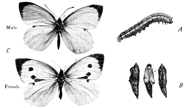
Fig. 225.—Cabbage White Butterfly. A, larva;
B, pupa; C, perfect insect.
(All × ⅔.) (B and C from photographs by Mr. A. Flatters)
When the caterpillar has attained its full size, it stops feeding and seeks out a sheltered place—often a chink in a wall. Silken threads are given off by the spinnerets until a little heap of silk is formed into which the hooked end of the abdomen is fixed. Then a girdle of silk is made, passed round the thicker fore-part of the body, and so attached to the wall that the animal is supported in an upright position. The larval skin now splits and is peeled off, and the pupa, or chrysalis, stage (Fig. 225, B) is entered upon. All the external parts of the butterfly are complete at the time of pupation, but profound changes are still necessary in the internal organs; it is to allow these changes to take place in tranquillity that the resting, or pupal, stage is interposed between larval and adult life.
At last the new organs are ready for their work, the pupal skin cracks, and the perfect insect (Fig. 225, C) emerges. The head now [Pg 367] carries a pair of large compound eyes, and two slender feelers with knobbed ends. The jaws also have been remodelled in accordance with the completely different manner of life upon which the insect is now entering. The mandibles are now only doubtfully recognisable; the first pair of maxillae are elongated and grooved, and are closely applied to each other to form a long tube called the proboscis (Fig. 226, Mx. 1), which, when not in use for sucking up the sweet juices of flowers, is kept coiled up beneath the head; the palps of the labium (Lab. Pa.) project like tusks on the sides of the head. The thorax is provided with two pairs of broad wings, which are covered with minute overlapping scales—forming a delicate “bloom” which is readily detached by rough handling. In many butterflies and moths the scales are gorgeously coloured and arranged in symmetrical patterns. The name Lepidoptera,[32] which is applied to this order of insects, was suggested by the scaly covering of the wings. When the butterfly is at rest the wings are either fully expanded horizontally, or are held vertically over the back, the upper surfaces of the fore wings being in contact. The first segment of the thorax, which bears the first of the three pairs of legs, is greatly reduced; the third segment also is but small.
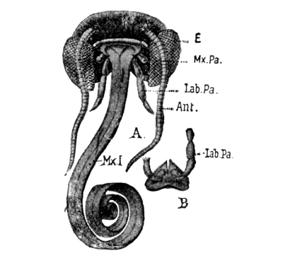
Fig. 226.—A, Head of a Lepidopterous insect; B, the labium; Ant., feeler; E, eye; Lab. Pa., labial palp; Mx. 1, proboscis; Mx. Pa., maxillary palp (magnified).
In short, the whole organisation of a butterfly is definitely adapted to the special duties which belong to this period of its life. The [Pg 368] growing stage is over; the sole object of life is now to seek a mate and—in the case of the female—to lay the eggs in a place where the future larvae may find plenty of food. Ease of locomotion and conspicuousness are secured by the broad and brilliantly-coloured wings; the peculiar manner of flight is considerable protection against the attacks of birds; large eyes aid in the recognition of the mate; and a concentrated and easily-digested food is supplied by the nectar of flowers, and made accessible by the long, sucking proboscis—the service of cross-pollination (p. 92) often being unconsciously rendered in return for the sweet draught.
The common species of cabbage-white butterflies spend the winter as pupae; the perfect insects emerge in April, lay their eggs, and then die. The caterpillars pupate and a second generation of butterflies appears, their offspring reaching the pupa stage about the end of autumn.
Moths.—Moths pass through a life-history which is identical, in its broad features, with that of butterflies. The larva which hatches from the egg is a caterpillar, whose life is spent in feeding and growing. At the same time the external features of the adult are gradually taking form under the skin. When at last the full larval size is attained, and a resting stage is necessary for the perfection of the internal organs, the caterpillar’s skin splits, and is shed, and the animal becomes a pupa or chrysalis. A moth-pupa is generally somewhat egg-shaped (Fig. 227), whereas the pupa of a butterfly is usually conical, though there are many exceptions.
The winged moth, which at length emerges from the pupal skin, differs from a butterfly in certain obvious respects. Its body is usually broad and thick; its feelers are either comb-like or thread like, not knobbed at the ends; the two wings of one side are in most cases secured together at the base by one or more bristles on the hind-wing hooking over a catch on the fore-wing; in rest, the wings usually slope and are [Pg 369] not fully extended. Whereas butterflies usually fly only in the sunshine, moths often fly by night, and the flowers which night-flying moths frequent for nectar are as a rule white and strongly-scented, and close during the day. In finding their mates, moths seem to depend largely upon the sense of smell, which is probably lodged in the feelers.
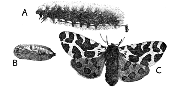
Fig. 227.—Stages of Tiger Moth. A, Caterpillar, from left side; B, pupa (removed from cocoon), ventral view; C, perfect insect (female). (From a photograph by Mr. A. Flatters.) (× ⁵/₇.)
The life-history of a typical moth is well exemplified by the Tiger Moth (Fig. 227), which is easily reared in captivity. The larva—often called the “woolly bear,” from its thick covering of hair—may be found in early summer on the leaves of lettuce, strawberry, nettle, and other plants. It pupates about the end of June, working its hair, together with silk spun by the spinneret, into a cocoon, in which the resting stage is passed. About a month later the perfect moth emerges; its fore-wings are beautifully mottled with cream colour and chocolate brown; the hind-wings are red, with metallic violet spots. The feelers of the male are comb-like, and are probably very sensitive organs of smell, by means of which he seeks out his mate. The female’s feelers are thread-like.
In the Vapourer Moth (Fig. 228), whose “looping” flight may often be observed even in the streets of towns during the day, the two [Pg 370] sexes are remarkably different from each other. The male (C) alone can fly; the female (D) is wingless, and is confined for the whole of her short adult life to the place where she emerged from the cocoon. Here she lays her eggs and then dies. Neither she nor her mate is capable of feeding.
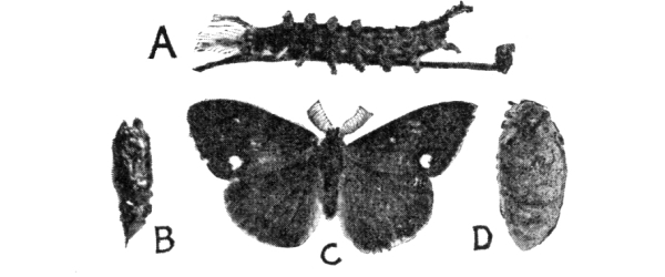
Fig. 228.—Stages of Vapourer Moth. A, Larva (male); B, male pupa; C, male moth; D, female moth. (From a photograph by Mr. A. Flatters.) (× ⅚.)
It would be difficult to find a more striking example of the fact that the one duty of the adult moth is reproduction. The female vapourer is even debarred from the privilege of choosing favourable places for her eggs; but a compensation for this disadvantage lies in the agility of the larvae (A), which are able to migrate without difficulty to another plant whenever food becomes scarce. As Prof. Miall remarks:[33] “Whatever the larva can do for itself, the female can safely leave undone; but what the larva cannot do, by reason of sluggishness or restricted diet, the parent must provide for. Hence activity and intelligence in the one lead to degeneration in the other.... Wings are to insects what spores are to ferns, plumed seeds to dandelions, and hooked seeds to burrs—a ready means of dispersal.”
Other insects.—Space does not permit of more than a reference to other insects, and the work of this chapter is to be regarded as the merest introduction to the study of this fascinating class of animals. The chief remaining orders are the Neuroptera, including May flies, dragon flies, and caddis flies; the Hemiptera, among which are included the various “bugs,” water boatmen, plant lice, etc.; the Diptera (two-winged flies), such as the house-fly, gnat, harlequin fly, daddy-long-legs, etc.; and the Hymenoptera, including bees, wasps, ants, gall flies (p. 146), and ichneumons. Many [Pg 371] of these insects have aquatic larvae which may be found in ponds; and their life-histories should be studied in aquaria and careful notes made of the transformations.[34]
EXERCISES ON CHAPTER XIX.
1. Examine the following animals, and find out (a) which are arthropods, (b) which are insects: tortoise, spider, grasshopper, lobster, earwig, centipede. Give reasons for your conclusions.
2. Keep a grasshopper under a tumbler with a small sod of grass. Observe its habits, and find out how it “chirps.” Compare its structure with that of the cockroach.
3. Compare a cockchafer with a water-beetle. In what order of insects would you place the cockchafer, and why?
4. Compare other water-beetles with Dytiscus, and try to trace their life-history.
5. Observe the habits and examine the structure of the water-boatman. What reasons can you find for excluding it from the beetle-order?
6. Examine a daddy-long-legs, and try to find the two stumps which are all that remain of the hind-wings.
7. Look for “blood worms” (larvae of the harlequin fly) in horse-troughs and sluggish streams in summer; keep them in a saucer of water with a few dead leaves. Observe their habits and describe the appearance of the pupa. What kind of insect emerges from the pupa?
8. Keep caddis-worms in an aquarium and describe their habits.
9. Examine the leaves of stinging nettles for caterpillars in June, and try to rear butterflies from them. Carefully notice from which kind of caterpillar each butterfly is derived.
10. Look for caterpillars of the Privet Hawk Moth on privets and lilacs on August and September evenings. Keep some, with earth and twigs of the food plant, in a covered flower-pot, and observe their method of pupation.
11. Compare the colouration of the wings of butterflies and moths with that of the plants they most frequent, and describe any cases of protective colouration which you find.
1. The crayfish and lobster.—(a) Habits.—Readers in limestone districts will probably be able to find crayfishes in the streams, and the habits of the animals in their natural surroundings should be observed and described. Other readers will be able to obtain live specimens from dealers.[35]
Place the animal on the table or desk. Notice that the body consists of an anterior, unsegmented portion, the cephalothorax (corresponding to the head plus the thorax of an insect), and a posterior, jointed abdomen. Watch the movements of the stalked eyes, the feelers and legs; allow the claws of the largest pair of legs (the “pincers”) to grasp a pencil. Put the crayfish in a white dish, with an inch or two of water, and by means of a pipette discharge a few drops of water containing some suspended colouring matter, such as carmine or indigo, near the point of attachment of the last walking leg. Describe the movements of the coloured water. When the animal is startled, notice the sudden vertical flexure of the abdomen, by means of which the body is pulled backward in the water. Feed the crayfish with meat or worms, and try to see the action of the jaws.
(b) External characters.—Kill the crayfish instantaneously by [Pg 373] dropping it into boiling water. Compare the animal with a cockroach (p. 349). Examine more closely the stalked eyes, the two pairs of feelers, the four pairs of walking legs and the single pair of large pincers, and the fusion of the head and thorax to form the cephalothorax. Count the segments of the abdomen.
(c) The appendages.—Examine the ventral surface of the abdomen, and notice that each segment, except the last, bears a pair of small jointed organs; these are called swimmerets. Remove one of the pair carried by the last segment but two, examine and draw it; make out that it consists of a stalk and two branches. Notice that the branches of the swimmerets of the last segment but one are expanded, and form, with the last segment, the tail fin.
Study the appendages of the cephalothorax from behind forwards. They consist of (i) four pairs of legs used for walking, and a larger pair of pincers; (ii) three pairs of foot-jaws or maxillipedes; (iii) two pairs of maxillae; (iv) one pair of mandibles; (v) two pairs of feelers.
(d) The gills.—With strong scissors cut off the side part of the exoskeleton of the cephalothorax, and notice the plume-like gills in the gill-chamber thus laid open. Move the adjoining legs, and see that some of the gills also move.
2. The crab.—Obtain a crab and compare it, point by point, with the crayfish, or lobster. Notice the great width of the shell of the cephalothorax; much of this width is due to the gill-covers, which stand out from the sides of the true body. Look for the opening of the gill-chamber at the base of the pincers. Make out the stalked eyes (in sockets), the two pairs of feelers, and the five pairs of legs. Notice how the animal runs; in what respects does the method differ from the gait of the crayfish? Examine the ventral surface of a dead specimen, and notice that the abdomen is tucked under the cephalothorax. Stretch out the abdomen; compare it with that of the crayfish, and count the segments. Notice the absence of the tail fin; it is not required, as the crab does not swim. Extend the abdomen, and make drawings of the animal (i) from above, and (ii) from below, and label the parts. Carefully remove the foot-jaws and the true jaws, and compare them with [Pg 374] the corresponding appendages of the crayfish. With strong scissors cut off one of the gill-covers, and examine the gills. Observe that in the crab the gills are not in any case attached to appendages; they all spring from the sides of the body-wall.
Tabulate the respects in which you have found the crayfish and crab (i) to agree with, (ii) to differ from, insects.
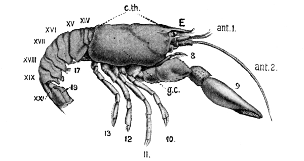
Fig. 229.—Crayfish, seen from the right side. The appendages are numbered in Arabic and the abdominal segments in Roman numerals. ant. 1, first, and ant. 2, second, feelers of the right side; c.th., cephalothorax; E, eye; g.c., gill-cover. (× ⅔.)
The student who has worked through Chapter XIX. will at once recognise in a crayfish, or a lobster, an animal possessing many features in common with the insects, and will find it interesting to try to discover for himself why it is not called an insect, but is placed by naturalists in a different class.
The crayfish.—The crayfish (Fig. 229), or the lobster (which agrees closely in structure with the crayfish), is obviously an arthropod (p. 351), for it is covered by an exoskeleton of chitin, and has hollow jointed limbs and a segmented body. In these respects it agrees with the insects. On the other hand, it plainly possesses at least five pairs of legs, and has two pairs of feelers [Pg 375] (ant. 1 and ant. 2, Fig. 229), whereas no insect has more than three pairs of legs (when adult), or more than one pair of feelers.
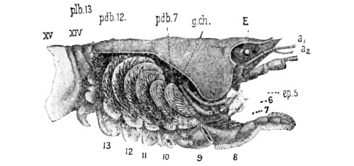
Fig. 230.—Crayfish; the right gill-chamber and gills as seen after removal of the right gill-cover. a₁, a₂, feelers; 6-13, sixth to thirteenth appendages; xiv, xv, first and second abdominal segments; E, eye; ep. 5, scoop on second maxilla; g.ch., gill-chamber; pdb. 7, gill attached to seventh appendage; pdb. 12, gill attached to twelfth appendage; plb. 13, gill on body-wall above thirteenth appendage. (Slightly reduced.)
These differences are apparent at the first glance, and closer examination reveals even greater contrasts. The three primary divisions of the body into head, thorax, and abdomen, which are so characteristic of insects, are not obvious in the crayfish; for the head and thorax are here fused into one mass, the cephalothorax[36] (Fig. 229, c.th.), which is covered by a shield called the carapace. Moreover, in the crayfish every segment except the last bears a pair of appendages, which vary in form in different regions of the body according to their duties, but which can all be shown, by careful comparison, to be modifications of one primitive form, which is Y-shaped and consists of a basal stalk and two branches. This form of appendage is well seen in the swimmerets (17, Fig. 229) of the abdominal region; further forwards the appendages become walking legs (9-13, Fig. 229); next come three pairs which combine the characters of legs and jaws, and are called maxillipedes (8, Fig. 229); then are the true jaws—two pairs of maxillae, and one pair of biting and crushing mandibles; and lastly, in front of the jaws, two pairs of feelers (ant. 2 and ant. 1). It is believed that the first pair of feelers corresponds to the single pair of feelers of the [Pg 376] cockroach; that the jaws correspond to the jaws; that the maxillipedes of the crayfish are the equivalents of the walking legs of the cockroach; while the remaining appendages of the crayfish have no representatives in the insect.
Lastly, the crayfish differs essentially from the insect in its method of respiration (p. 355), for it is an aquatic animal and breathes dissolved oxygen. It therefore possesses neither lungs nor spiracles but gills (Fig. 230). These are situated at the sides of the true body-wall, in gill-chambers formed by the downgrowth of the sides of the carapace. The gills are delicate plumes, containing fine blood-vessels, so that an exchange of gases readily takes place between the blood and the surrounding water. On each side, a scoop (Fig. 230, ep. 5) on the second maxilla is continually baling water out of the front of the gill-chamber, fresh water flowing in from behind to take its place. Certain of the gills are attached to the legs, so that the motion of the legs also is some assistance to respiration.
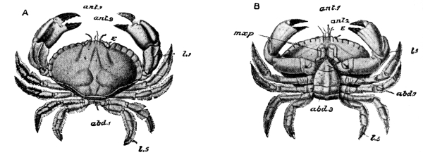
Fig. 231.—Crab; A, from above, B, from below, ant. 1, first, and ant. 2, second feelers; abd. 3, third, and abd. 7, seventh abdominal segments; E, eye; l. 1, pincers; l. 5, last walking leg; mxp, third maxillipede. (× ⅙.)
The crab (Fig. 231) is markedly different in shape from the crayfish or lobster, but is nevertheless easily seen to be built on essentially the same lines of structure. It also consists of twenty segments, of which the first thirteen are fused to form a [Pg 377] cephalothorax; and the appendages of this region are quite comparable with those of the crayfish. The great width of the shell is largely due to the gill-covers, which stand out from the sides of the body much further than do those of the crayfish. As a result the crab finds it easy to run side-first. The crab is essentially a walking, not a swimming, animal, and the abdomen—upon which the crayfish and lobster so much depend in swimming—is in the crab reduced in size and kept tucked under the cephalothorax.
Crustaceans.—These facts are sufficient to show that the title “insect” cannot with any propriety be given to either the crayfish or the crab, unless, indeed, the term is to be applied to all arthropods indiscriminately. The crayfish, lobsters, shrimps, prawns, crabs, barnacles, and many less-familiar animals, are placed in the crustacean class of arthropods. Crustaceans have usually a distinct head, thorax, and abdomen; but some of the thoracic segments may be fused with the head to form a cephalothorax (p. 375). Like other arthropods, the animals are covered with an armour of chitin, and in many cases this is so hardened, except at the joints, by mineral matter that it becomes a rigid shell or crust. The head bears two pairs of feelers in addition to the jaws; and the segments of the thorax and abdomen are provided with appendages which are variously modified as jaw-feet, legs, swimmerets, etc. Typically, the animals breathe by gills and are aquatic, but forms are known which are able to live comfortably on land if the gills are kept moist. One of the most interesting examples of this is found in the common wood-louse (Fig. 232), which lurks under stones and logs in damp and dark situations, and breathes by plate-like gills on the abdominal segments. [Pg 378]
Other arthropods.—Other familiar arthropods, which are neither insects nor crustaceans, are spiders and centipedes. The spiders belong to the arachnid class, and the centipedes to the myriapod class of arthropods.
1. The fresh-water mussel.—(a) Habits.—Study the habits of the living animal (Fig. 233) in the aquarium. Notice the muscular foot which is protruded between the halves (valves) of the shell, and by means of which the mussel slowly makes its way along the sandy bottom. With a pipette, discharge some water, coloured with carmine or indigo, close to the more pointed end of the shell. Where is the coloured water taken in, and where is it expelled? Notice that the shell closes when the animal is handled.
(b) The regions of the shell.—The rounded end is anterior, the more pointed end posterior, the straight hinge-line dorsal, and the gape ventral. Notice the knob-like umbo on each side near the anterior end of the hinge; it is the oldest part of the valve. Make out the concentric lines of growth, showing the successive positions of the margin. Draw the shell from the right side and from above.
(c) General structure.—Kill the mussel by putting it for a few minutes into hot, but not boiling, water. Notice that the valves of the shell now gape apart somewhat. Hold up the animal and see, lining the shell, a thin soft membrane, the mantle. Carefully separate the mantle from one valve and notice, near each end, a thick white pillar which passes from one valve to the other. These pillars are the closing muscles. Pass the blade of a knife between the mantle and valve, and cut through each closing muscle quite near to the valve. Turn the valve back and remove it from the other valve by cutting through the elastic ligament (at the hinge) with strong scissors. Clean the inside of the separated valve and examine it; notice (i) the line of attachment of the mantle; (ii) the impressions of the closing [Pg 379] muscles; (iii) the lines of shifting of the closing muscles—triangular depressions stretching from the umbo to the muscles.
Examine the animal as it lies in the other valve. Make out: (i) The right and left lobes of the mantle which line the valves and (in the natural position of the mussel) hang down from the sides of the body; (ii) the two plate-like gills on each side, lying between the mantle and the foot; (iii) the median foot; (iv) the reddish, triangular palps surrounding the mouth—an aperture at the anterior end of the foot. Push a stout pin into the mouth and upwards into the gullet. Make a drawing showing the relative positions of the parts.
2. The garden snail.—(a) Habits.—Where have you seen snails? At what time of the year are they most active? Upon what do they feed? Place a snail upon a lettuce leaf and carefully watch its method of eating. How does it move about? Put a live snail upon a sheet of glass, and look through the glass to see the wave-like action of the flat sole—the foot—by means of which it moves.
Have you ever found snails in winter? How much of the animal was visible? How is the mouth of the shell closed in winter?
(b) General appearance.—Make a drawing of the animal from the left side and another from the right side, and notice the differences between the two. Is the shell placed over the middle of the animal, or does it lie to one side? How many turns has the spiral of the shell? In a fully expanded snail observe the fleshy “collar” round the margin of the shell; it is the edge of the mantle. Notice the rounded head, the two pairs of tentacles, the eye-spot at the tip of each of the larger and upper tentacles, the mouth, and, near the base of the shell on the right side, the respiratory pore, which opens into the lung. Touch various parts of the body in turn to see if they are irritable; how are the tentacles retracted? Can the animal close and open its respiratory pore at will?
3. An air-breathing pond-snail.—Obtain several fresh-water snails and study their habits in glass aquaria. The commonest fresh-water snails are species of Limnaea; identify some of these by [Pg 380] comparison with Fig. 234, A, and try to make out the parts already seen in the garden snail. Watch the action of the mouth as the animal feeds on the green scum which often collects on the sides of aquaria. Notice how it creeps over the glass or along the surface-film of the water. When the snail comes to the surface it replaces the air of its lung by fresh air, and a bubble may often be seen escaping from the respiratory opening under the lip of the shell. Look for the spawn (egg-masses) of this snail on leaves or on the sides of the aquarium, and examine the eggs frequently with a hand lens.
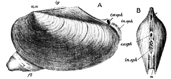
Fig. 233.—Fresh-water Mussel. A, from left side; B, from behind. ft, foot; in. sph., inhalant siphon; ex. sph., exhalant siphon; lg, ligament; m, mantle; um, umbo. (× ⅔.) (After Howes.)
Molluscs.—Molluscs are soft-bodied animals, in most cases protected by a hard, external shell, but they differ essentially from crustaceans and all other arthropods in not being segmented, and in not possessing jointed limbs. Familiar and instructive examples of two classes of the group are found in the fresh-water mussel and the garden snail.
The fresh-water mussel (Fig. 233) is to be found in streams, along the bed of which it ploughs its way by means of a muscular [Pg 381] foot (ft.). The body is enclosed in a brown shell, which consists of halves called valves, hinged together along the straight, dorsal edge by an elastic ligament (lg.). The action of the ligament is to separate the valves slightly unless they are forcibly held together by the contraction of closing muscles which run from one valve to the other. Hence the shell of a dead mussel always gapes open. The rounded end of the shell is anterior, the more pointed end posterior. The oldest part of each valve is the umbo (um., Fig. 233), a knob just in front of the ligament; and concentric lines surrounding the umbo mark successive positions of the margin of the valve as the animal increased in size. The valves are formed by the activity of the mantle lobes (m.)—a pair of delicate membranes which hang down from the sides of the body. The foot is a median prolongation of the body itself, and on each side a pair of plate-like gills lies between the foot and the mantle lobe of that side. “Thus the whole animal has been compared to a book, the back being represented by the hinge, the covers by the valves, the fly-leaves by the mantle lobes, the first two and the last two pages by the gills, and the remainder of the leaves by the foot.”[38]
When the living mussel is undisturbed, the mantle folds project slightly at the hinder end of the shell, their edges being so placed in contact that they form two short tubes. A current of water flows in at the lower of these (in. sph., Fig. 233), carrying to the mouth a supply of food-particles, and to the gills and mantle a store of dissolved oxygen; while an outgoing current leaves by the upper tube (ex. sph.).
The garden snail (Fig. 234, B) seems at first sight to have but little resemblance to a mussel; but it also is a mollusc—consisting of a soft, unsegmented body, which is produced ventrally into a foot, and is protected by a shell formed by the activity of a mantle fold of the body. In this case, [Pg 382] however, the shell is one piece, and is spirally coiled. The snail has also a distinct head, which bears two pairs of tentacles; at the tip of each of the longer and upper tentacles (t) is an eye (e). The animal crawls about by wave-like contractions of the muscular foot; it feeds upon vegetation, which it rasps into small particles by means of a toothed tongue, and then swallows. The snail is entirely adapted to a terrestrial life, and breathes air—the mantle fold under the shell enclosing a lung chamber with blood-vessels in its walls, which opens to the exterior by a respiratory pore (p.o., Fig. 234) on the right side. The snail spends the winter, in a state of torpor, under logs or stones, the body being entirely retracted into the shell, the mouth of which is closed by a plate of hardened slime.
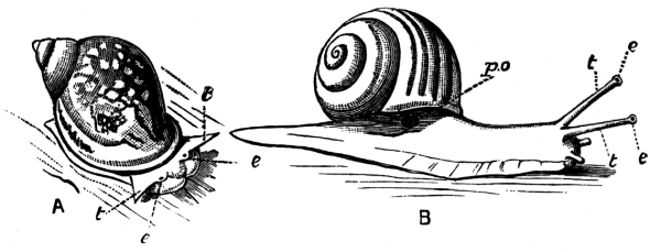
Fig. 234.—A, A Fresh-water Snail (Limnaea); B, Garden Snail. e, e, eyes; p.o., respiratory pore; t, t, tentacles, (× 1.)
Slugs (Fig. 235) are of very similar structure, but in them the shell has almost disappeared, even the trace which remains being concealed by the mantle fold.
Among the commonest of fresh-water snails are various species of Limnaea (Fig. 234, A). They may be found abundantly in ponds, [Pg 383] and are often kept in aquaria, where they perform a useful service by devouring the minute plants which are apt to accumulate to an undesirable extent and form green scum on the sides. These snails, like the garden snail, breathe air, and often come to the surface to take a fresh supply into their lung chambers. Some other water snails, however, breathe dissolved oxygen by means of gills beneath the shell.
1. External characters.—Dig up several earthworms from the soil and examine them. What is the length of the largest and of the smallest specimens? What is the thickness of the body? Watch the worms crawling about, and describe the method of locomotion. Has the animal any legs? Do the length and thickness of any one worm vary during its movements? Is the variation connected with locomotion? How? Can you distinguish between the fore (anterior) and hind (posterior) ends, and between the upper (dorsal) and lower (ventral) surfaces? How do they differ?
Kill a large worm by immersion in methylated spirit and examine it more closely. Notice the segmented character of the body, and estimate the number of segments. Which part of the body has the largest segments? Observe the swollen appearance of segments 32 to 37; this region is called the clitellum. Notice the mouth (overhung by a short lobe) in the first segment, and the vent in the last segment. On the ventral surface of segment 14 try to see a pair of small pores; these pores are the openings through which the eggs are discharged from the body. Pull the worm gently between your fingers, and notice the bristly feel; in which direction of motion is this most apparent? Examine the ventral surface with a strong lens in a good light, and try to see four double rows of very small bristles.
2. Habits.—Examine the surface of the ground of a garden or lawn at night by help of a lantern or lamp, being careful to tread [Pg 384] lightly, and observe the actions of any worms you see. Are more worms to be seen at night than during the day? Do the worms seem disturbed by the light of the lantern? Try to grasp one; is it easily caught? Why not? Where does the worm retreat? If you can grasp a worm before it has time to withdraw completely into its burrow, observe the difficulty of drawing it out without tearing it. Try if the worms are disturbed by a loud shout (be careful not to blow upon them when shouting), or by a heavy stamp of your foot.
Carefully lay open several burrows and notice:
(a) Whether the mouths of the burrows are plugged in any way, and, if leaves are used for this purpose, whether the leaves have been dragged into the hole (i) by the broad ends, (ii) by the narrow ends, or (iii) by the sides; do you find any signs of intelligence in the method adopted?
(b) The length and width of the burrow and the character of its lining.
(c) The end of the burrow; is it enlarged?
Examine worm castings. Mark out a square yard of surface and collect, dry, and weigh all the castings found on this area in a certain time, say a month. Such observations should be made at different periods of the year, and over different kinds of soil, and comparisons made. Estimate the weight brought up per acre by worms during a year. Are the particles composing the castings very fine, fine, or coarse?
Place worms in glass-covered flower-pots with earth of different degrees of firmness, and observe the methods of burrowing in loose and in firmly compacted earth respectively. Place pieces of leaf of carrot, onion, and cabbage on the surface, and at night observe how the worms grasp the pieces and drag them into the burrows.
Earthworms.—Few people except naturalists have any idea of the vast number of earthworms living in the surface soil of this and most other countries, or of the importance of the work which they do.
The common earthworm (Fig. 236) is much simpler in structure than any of the animals previously considered in this book. It is roughly [Pg 385] cylindrical in shape, though somewhat flattened on the ventral surface. It is divided into about 150 segments, which are marked on the exterior by grooves running round the body. The mouth is an opening in the first segment (Fig. 236, 1) and is overhung by a short fleshy lobe. The worm crawls along by alternate elongation and shortening of its body, being aided by short bristles which are directed backwards and act as pivots; limbs are entirely absent. The animal is very sensitive to touch, even to the vibrations of the ground; but it is stone-deaf, only just capable of distinguishing between light and darkness, and has very little sense of smell.
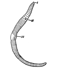
Fig. 236.—Earthworm,
seen from right side.
1, 15, 33, first, fifteenth,
and thirty-third segments.
(× ½.)
During the day, the earthworm generally remains in its burrow in the soil, with its head just inside the entrance. Its method of forming the burrow depends upon the texture of the ground. In loose soil the earth is simply pushed aside, but where the material is too compact for this, the animal actually eats its way through. The burrow is lined with soft earth or little stones, and is plugged at the mouth with leaves or other convenient objects. The animal was found by Darwin to display distinct intelligence in its manner of drawing leaves into the mouth of its burrow, seizing them in most cases by their narrow ends, so that they could be pulled in with as little difficulty as possible. Objects are generally grasped between the lobe, which overhangs the mouth, and the lower part of the first segment, the hold being maintained by a sucking action. The inner end of the burrow is enlarged to allow the worm room to turn round. At night, the fore part of the body is protruded in search of food, the tail being generally retained in the [Pg 386] burrow, ready for instantaneous retreat in case of alarm.
The earthworm feeds upon leaves—which are first softened by a fluid discharged over them, and then sucked into pieces small enough to be swallowed, for the animal has no jaws—and upon the half-decayed organic matter which is always present in ordinary soil. The soil itself is swallowed in large quantities; the nutritious portion is extracted, and the undigested matter deposited upon the surface of the ground, near the mouth of the burrow, in the form of castings. As a result of numerous experiments, Darwin estimated the weight of castings thus thrown up by earthworms on an acre of land as 15 tons annually. The following passage is worthy of very careful attention: “When we behold a wide, turf-covered expanse, we should remember that its smoothness, on which so much of its beauty depends, is mainly due to all the inequalities having been slowly levelled by worms. It is a marvellous reflection that the whole of the superficial mould over any such expanse has passed, and will again pass, every few years through the bodies of worms. The plough is one of the most ancient and most valuable of man’s inventions; but long before he existed the land was in fact regularly ploughed, and still continues to be thus ploughed by earthworms. It may be doubted whether there are many other animals which have played so important a part in the history of the world as have these lowly organised creatures.”[39]
The earthworm lays its eggs in a small cocoon formed by the hardening of a viscid material which is discharged by a swollen part of the body called the clitellum, extending from the 32nd to the 37th segments. After the formation of the cocoon the worm moves backwards, and the eggs leave the body by small pores on the ventral surface of segment 14, [Pg 387] as this region passes the cocoon. A small amount of food-material is also enclosed in the cocoon, and forms a store of nutriment for the young worms during their early development.
EXERCISES ON CHAPTER XX.
1. Compare the legs of a cockroach with those of a crayfish and a vertebrate. (1900)
2. Describe the respiratory organs of the crayfish. How are they continually supplied with fresh water? (1898)
3. Examine a spider. How many legs has it? Of what divisions does its body consist? Why do you consider that a spider is not an insect?
4. Make observations, and write descriptions, of the habits of spiders, paying special attention to the methods of construction of the webs, the manner of catching prey in different cases, and the care of the young by the parents.
5. Compare a centipede with an insect, pointing out the features of resemblance and difference. (1897)
6. How does a pond-mussel open and close its shell? (1900)
7. Compare an oyster with a fresh-water mussel, and try to find points of resemblance and difference, making careful notes and sketches of these. How many closing muscles has the oyster? Are its gills plate-like? Why do you consider the oyster a mollusc?
8. Where is the lung of a snail situated? How do we know that it is really a lung? (1897)
9. How does an earthworm resemble and differ from a caterpillar? (1900)
10. Explain the action of an earthworm in the formation of vegetable mould. (1897)
11. Classify the animal described below:
A land animal with a long narrow soft body and no legs; two pairs of tentacles on the head; a breathing hole on one side? (1904)
12. Give the characters by which insects can be distinguished from crustaceans. (1905)
An animal or a plant must be studied from several points of view before its manner of life can be understood in any real sense. It must, for example, be regarded, first, as a complicated piece of machinery, every part of which is beautifully fitted for the performance of a special duty; it must also be considered as an individual, having likes or dislikes—or at least tendencies—which are to some extent peculiar to itself; finally, it must be considered in its relation to other animals and plants, and to its surroundings. Field-work is especially concerned with the last of these methods of study—the observation of living things under natural conditions,—and this ought constantly to be borne in mind. To make nature-study a pretext for uprooting locally-rare plants and robbing birds’ nests is indefensible. The commonest plants are usually the most instructive, and afford ample material for the beginner’s experimental work; while the pleasure of finding and describing (perhaps photographing) a bird’s nest, and of keeping the eggs and young under observation, is something unknown to the mere collector of eggs. The life of both plant and animal is sacred in the eyes of every nature-student worthy of the name. At the same time, no [Pg 389] sentimentality ought to prevent the destruction of undoubtedly noxious insects and weeds. In general, however, specimens should be killed only for the purpose of a leisurely examination of structure, which would otherwise be impossible, or to make needed additions to the teaching-collection of a school museum.
Generally speaking, collecting is of very doubtful value, except to experts. Insects and common plants may, however, be collected without scruple by the beginner, though it is worth remembering that the most perfect specimens of butterflies and moths are those reared in captivity from the eggs, larvae, or pupae.
The student is strongly recommended to make a sketch map of some small area to which he has easy access, and to record upon this the positions of features of special interest. Such a map may, in the first instance, be copied on an enlarged scale from an ordnance map of the neighbourhood, which may be obtained at the local free library. It will be advisable to duplicate the drawing by means of one of the many appliances for such work, and to keep one copy each for trees, flowering plants, birds’ nests, etc.—the position of each object, or of a well-defined group, being carefully marked by a small number (Fig. 237), and the reference, with the date, being filled in on the margin. The varying character of the ground—sandy, marshy, clayey, etc.—should be indicated by diagrammatic shading or colouring. By following this method the student will more clearly realise that different plants are dependent upon different conditions of soil, drainage, etc.; and that, e.g., plants at home on a bleak moor, in a hedge, in marshy land or in water respectively, are characteristically modified so that they can make the best of their special conditions of life. In this manner he will, almost unconsciously, gain wider views of the relationships which exist between the facts learned from his more [Pg 390] detailed observations. It would be a distinct gain to biological science if field clubs also would adopt some such plan, each member undertaking to fill in upon his map information of the animal and plant life of the area allotted to him. The co-operation of various clubs, and the systematic arrangement of information thus obtained, would result in a store of knowledge of the highest value.
Many animals are so shy that they can only be approached with difficulty. In such cases it is especially necessary for the observer to move quietly and silently, and, when a favourable position is reached, to remain as motionless as possible, preferably with his back to the sun. If a field-glass can be obtained it will be of great assistance, but some practice will probably be required before it can be used easily.
It is well to start each ramble with some definite object of study in view—either trees, grasses, flowers, fruits, birds, caterpillars, or pond life, for example, and to be provided with pill-boxes, bottles, etc., according to circumstances. A notebook, pencil, pocket knife, and hand lens should, however, always be carried, and observations should be recorded on the spot.
Pond life.—Practically the only method of gaining a real knowledge of aquatic organisms is by the help of the aquarium. Specimens may be obtained by means of a net, or of a small wide-mouthed bottle tied to a stick. A pickle bottle is convenient for carrying home the material collected. The conditions of a natural pond should be imitated as closely as possible in the aquarium. At first, some little difficulty will probably be found in obtaining the necessary balance between the animals and plants of the aquarium. When this has been reached, it will only rarely be necessary to change the water, provided that dead or sickly specimens are promptly removed. [Pg 391]
In the organisation of school journeys so much depends upon local conditions of various kinds, so much must of necessity be left to the initiative of the teacher, that it is manifestly impossible here to do more than enunciate certain general principles. In the first place, it should be borne in mind constantly that the primary object of the school journey is the cultivation of habits of thoughtful observation; and that the chief danger to be guarded against is that out-of-focus condition into which the mind, like the eye, inevitably falls when it is concerned with too many things at once. To obviate this danger the teacher should go over the route in advance, noting carefully the features, physical and otherwise, which afford material for observation and investigation by the class. The order in which these features may be best studied should be decided upon, and a scheme of several visits, each to be concerned with one special subject of study, can then be drawn up. Such a preliminary survey should suggest a plan by which every member of the class may be allotted a definite task—to find something or do something, or to solve some problem on the spot.
These principles may be best illustrated by a special example, but it will be obvious that the same ideas, with modifications in detail, may be applied in any district. The sketch-map (Fig. 237) illustrates a walk through Healey Dell, near Rochdale, Lancashire. The rocks which are exposed at various places along the route belong to the Carboniferous formation, and are composed of shale, coal, or millstone grit. [Pg 392]
The object of the first journey will be in most cases to familiarise the class with the “lie of the land” and the most obvious features of the scenery. As a preparation, lessons should be given on the points of the compass and the various methods of finding the direction of the north. The simplest of these is by the use of the compass: it being remembered that the needle points about 16° to the west of true north. A second method depends on the fact that at noon the sun is in the south and that therefore (because the hour hand of a watch makes two revolutions in the twenty-four hours) the north and south line approximately bisects the angle between twelve and the hour hand, if the latter is pointed to the sun when the watch is horizontal. Incidentally, the method of finding the pole-star might be also explained to the class. Further, each pupil should be encouraged to find out how many steps he takes, on the average, in pacing a given measured distance. If the general direction of the walk is north and south, as in the example, it will be found best to begin the first journey at the south end (in this case from Shawclough Station), since to most children it is easier to conceive of a journey northward than in any other specified direction. Throughout the ramble constant reference should be made to the direction of the route and the relative positions of well-marked features of the landscape. The distances between certain points should be estimated, and, whenever possible, measured (by pacing), and notes made by the class. For example, from Shawclough to Ending the distances and directions are roughly: ¼-mile W.N.W., ⅓-mile N.N.W., ¼-mile E.N.E. In the first journey also the class should be made to notice where the ground slopes most and where least (the direction and angle of slope should be estimated in a few cases), and the names of neighbouring woods, farms, etc., should be learnt. The direction of flow of the river and the various bends in its course should also be noted, and reference made to the route by which, after joining those of other rivers, its waters ultimately reach the sea. Afterwards, the pupils should write an account of the journey and, in the higher classes, should be encouraged to draw a sketch-map, however crude, from memory. [Pg 393]
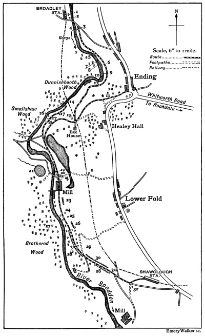
Fig. 237.—Sketch-map of Healey Dell, Rochdale.
1, birches; 2, willows; 3, beech; 4, potholes; 5, two waterfalls; 6, shallows, and vertical concave bank; 7, “Fairies’ Chapel”; 8, stratification of rocks; 9, mud deposits; 10, waterfall; 11, docks; 12, sloping trees; 17, weir; 14, railway viaduct; 15, horse chestnut; 16, aqueduct; 17, sycamore; 18, elm; 19, elm; 20, beech; 21, pond life; 22, well; 23, shale; 24, sycamore; 25, oak, bearing leaves in winter; 26, flagstones; 27, beeches, with wood-pigeons’ and magpies’ nests; 28, hawthorn hedge; 29, solitary oak; 30, gutter, with Pellia; 31, blackberry bush; 32, stunted oak.
Before the second journey each pupil should be provided with a blank sketch-map of the route. This may, in the first instance, be copied or traced from the six-inch Ordnance Map, and then duplicated in large numbers. Only the route and river, and a few of the more conspicuous landmarks, should be indicated on the maps as given to the class: details should be filled in, on the spot, by the pupils. The object of the second journey may conveniently be the study of the river and its work, and for this purpose it will be advisable to follow the stream in the direction of its flow. Variations in the speed of the current, and in the width of the stream and the hardness of the rocks or banks between which the water flows, should be noted, and the relations between cause and effect elicited by questioning. The hardness of the rocks at 4 has prevented the channel from being widened to a greater extent by the water, and accounts for the rapidity of the flow. A glass of water collected here is found to contain much suspended gravel. The considerable loss of weight of bodies in water is noteworthy, as explaining the great size of the stones which may be [Pg 395] transported by rivers. The scouring action of such stones is shown in the fine “potholes” at 4 and below the waterfall (Fig. 238) at 10, and has resulted also in the quaint stone portico of the “Fairies’ Chapel” under the right (west) bank of the river at 7. Again, the difference in the rates of erosion of hard and soft rocks has had much to do with forming the waterfalls at 5 and 10. Where the stream is wider and the flow slower (as at 6 and 9, and below 10), may be noticed sand and mud [Pg 396] deposits; and where the stream makes a bend it is found that the slowest flow and the maximum deposit are on the convex bank; while the concave bank is worn almost vertical (as at 6 and other places) by the swifter rush of the water, and may be undercut to such an extent as to cause the bank to give way. In this manner a river is constantly changing its course. The weir at 13, and the old water-wheel still to be seen in the ruined mill below, suggest remarks on the motive power of water, and on the circumstances which may cause the old industries of a district to be superseded by new ones. Along the rest of the route the bed of the river is less steep and its banks exhibit less variation, but they still afford plenty of material for study. The railway viaduct at 14 and an aqueduct at 16 suggest at least a casual reference to the derivation of the terms. Before the pupils are asked to write a “composition” on the ramble, a revision lesson on the features noticed should be given, and the accuracy of the entries on the sketch-maps checked by comparison with an enlarged map drawn by the teacher, or with a large “parish plan” of the Ordnance Survey, on the scale of 25.3 inches to the mile.
It will be well to devote two or more journeys to the study of the trees along the route. One journey should be taken in the spring, before the leaves are out, and another in the summer, when the foliage is well developed. It is far better to study three kinds of trees in some detail than to risk confusion at the beginning by attending to a dozen. In Healey Dell the commonest trees are beech, oak, and sycamore, and these serve admirably as an introduction to tree lore. If the first tree-journey be taken in the summer, the leaves of some three abundant species should be compared and contrasted, and each pupil should secure good specimens, to be drawn and preserved [Pg 397] afterwards. The presence of a little bud in the “axil” (the upper angle between leaf and twig) of most of the leaves should be pointed out by the teacher; and since the arrangement of the buds (and therefore of the subsequent branches of the twig) thus depends on the positions of the leaves, this last point is of considerable interest. In the sycamore, the leaves are in pairs at right angles to each other; in the beech and oak they are single and alternate, but much more crowded together in the oak than in the beech. The bark of the three trees is equally distinctive, and with the method of branching (obscured when the foliage is thick) serves to identify the trees from a distance in the winter. In winter and spring the interest of a tree is centred in its buds, and there are few things which more richly repay study. In spring, attention should also be given to the flowers—generally arranged in catkins—of common trees. Separate sketch-maps should be used as records of the positions of the more notable trees or plantations along the route. Any tendency to vandalism on the part of the pupils by tearing off branches should, of course, be sternly repressed; especially interesting twigs should, on occasion, be cut off by the teacher only, for later study.
There is much diversity of opinion as to the way in which flowers may be best studied in a limited number of school journeys. In most cases it will perhaps be impracticable to attempt more than teaching the names and calling attention to the habitat of the commonest. This, though a necessary introduction to the subject, tends to degenerate into a mere exercise of the memory, and in itself possesses little educational value. It should be supplemented by a detailed examination of a typical flower—say a buttercup—and by the explanation of the work of each part. Once the pupil has understood that the single duty of a flower is the production of healthy seeds, [Pg 398] and has been led to notice how, by the aid of ingenious devices, the up-to-date plants have learnt to call in the aid of insects, while the more conservative families still rely on the aid of the wind, he will be eager to discover for himself “how the thing works.” With young children it is folly to attempt any but the very broadest principles of classification of flowers; but quite young children can appreciate the advance from flowers without petals, through flowers with separate petals, to those with petals united to form a tube (thus restricting the nectar more and more to “useful” insects); and so understand the advantage which a primrose has over a buttercup, and a buttercup over an oak flower.
At least one journey should be given, in the autumn, to the study of the dispersion of fruits and seeds. The pupils should provide themselves with empty match-boxes or chip pill-boxes. In this ramble the class may with advantage be divided into four groups. Group A will collect examples of fruits and seeds which are dispersed by the wind; Group B, of fruits which by means of hooks or otherwise become attached to the hides of grazing animals, and are carried far from the place where they grow; Group C will collect fruits which tempt animals to eat them for the sake of sweet pulp (in these cases the pupils should find out a how the fruit is made conspicuous, b how the seeds themselves are protected from being injured by the animals); while Group D will search for specimens of plants which sow their own seeds.
There still remains abundant material for study in this walk, and mention only can be made of the sticklebacks, frog-spawn, snails, caddis-worms, dragon fly larvae, blood-worms—of the “things creeping innumerable, both small and great beasts”—which have been found in the [Pg 399] river, ponds, and wells along our route, and have been used to stock the aquarium; of the rabbits and birds; of the nests of ants and wild bees and wasps; of a certain blackberry bush (31) rich in interesting leaves; and of a thousand and one other things which, under the guidance of a judicious leader, may well be the means of teaching children to see what they look at and to think about what they see. For this is the first and last object of the school journey.
PLANT LIFE.
General.—As in December (p. 422).
Plants usually in flower.—Shepherd’s purse, daisy, snowdrop, and a few others.
ANIMAL LIFE.
General.—As in December (p. 422).
Mammals.—Bats (p. 255) reappear at the end of the month.
Birds.—Missel thrush (p. 306) sings.
Insects.—Winter pupae of butterflies and moths (Chap. XIX.) may be found. [Pg 401]
PLANT LIFE.
General.—As in December.
Plants usually in flower.—Shepherd’s purse, daisy, snowdrop, hazel, and a few others. Hedges are now clipped; study the effect which this treatment has upon subsequent growth.
Corn.— Barley and oats are sown.
Liverworts.—Spore-cases of Pellia (p. 200) become visible.
ANIMAL LIFE.
General.—As in December.
Mammals.—Bats reappear.
Birds.—Thrushes and blackbirds pair and begin to build; study differences in song (pp. 304 and 308). Rooks repair and build nests (p. 318).
Frogs. (p. 332) reappear.
Insects.—Pupae of cabbage-white butterflies and other Lepidoptera may be found. [Pg 403]
PLANT LIFE.
General Work for Spring Months.—Study the germination of seeds and the early stages of growth of the new plants (Chap. I.); the structure and methods of unfolding of buds (Chaps. IV. and VIII.); the movements of young twining stems; “bleeding” of stems, and paths of food-currents (Chap. V.); and examine and collect spring flowers (Chaps. VI., VII., and VIII.).
Plants usually in flower—Shepherd’s purse, marsh marigold, wild plum, daisy, dandelion, daffodil, snowdrop, hazel, alder, willow, poplar, elm, and others.
Horsetail.—Fertile haulms (p. 195) appear.
Corn.—Barley and oat sowing continued.
ANIMAL LIFE.
General Work for Spring Months.—Study the development of frogs and toads (Chap. XVIII.) and insects (Chap. XIX.); the development and education of the chick (Chap. XVI.); the nesting-habits of birds (Chap. XVII.); the education and play of lambs and other young mammals (Chap. XIV.); the habits of molluscs (Chap. XX.); and the work of earthworms (Chap. XX.). Stock aquaria.
Mammals.—Lambs are born.
Birds.—Thrushes, blackbirds, skylarks, rooks, and other birds lay eggs. Fieldfares and redwings begin to depart. Frogs and Toads lay their eggs.
Insects.—Dytiscus (p. 361) lays eggs. Pupae of cabbage-white butterflies and other Lepidoptera may be found. [Pg 405]
PLANT LIFE.
Plants usually in flower.—Wallflower, shepherd’s purse, buttercup, anemone, marsh marigold, common vetch, plum, pear, strawberry, primrose, cowslip, daisy, dandelion, speedwell, red deadnettle, daffodil, wild hyacinth, wild tulip, rushes, arum, annual meadow grass, oak, birch, alder, willow, poplar, elm, ash, and others.
Trees which unfold their leaf-buds.—Larch, horse chestnut, beech, elm, sycamore.
Elm fruits (p. 154) are abundant; study the method of dispersal (p. 173).
Corn.—Barley and oat sowing continued.
ANIMAL LIFE.
Birds.—Sand-martins, cuckoos, swallows, house-martins, nightingales, swifts, and other birds arrive. Kestrels and sparrow-hawks nest. Fieldfares and redwings depart.
Frogs and Toads.—Tadpoles hatch.
Insects.—Cabbage-white butterflies (p. 362) and other Lepidoptera emerge from pupae. Caterpillars are plentiful. Moths may be taken on willow flowers at beginning of month. Larvae of Dytiscus (p. 361) and other aquatic insects may be found in ponds.
Molluscs.—Garden snails (p. 379) reappear. [Pg 407]
PLANT LIFE.
Plants usually in flower.—Wallflower, shepherd’s purse, buttercup, anemone, marsh marigold, laburnum, common vetch, red clover, white clover, broom, cherry, apple, pear, strawberry, hawthorn, primrose, cowslip, daisy, dandelion, speedwell, red deadnettle, white deadnettle, lily of the valley, wild hyacinth, star of Bethlehem, wild tulip, rushes, sedges, arum, sweet-scented vernal grass, slender foxtail, meadow foxtail, annual meadow grass, perennial rye grass, oak, beech, birch, willow, elm, horse-chestnut, ash, sycamore, and others.
Most forest trees are now in leaf.
Liverworts.—Spore-cases of Pellia (p. 200) open.
ANIMAL LIFE.
Birds.—Swallows build nests. Most birds are incubating eggs (Chap. XVI.).
Frogs and Toads.—Tadpoles may be found in ponds.
Insects.—Moths and butterflies are common. Cabbage-whites lay eggs. Caterpillars of tiger-moth (p. 364) and other Lepidoptera may be found. Aquatic insect-larvae in ponds. [Pg 409]
PLANT LIFE.
General Work for Summer Months.—Study the forms and duties of leaves (Chap. III.); and the thickening of stems (Chap. V.). Examine, identify, and collect grasses (Chap. VII.) and summer flowers (Chap. VI.), and make observations upon cross-pollination of flowers by insects (Chap. VI.). Study the development and structure of fruits (Chap. IX.); and the life-history of ferns (Chap. X.). Compare and contrast mushrooms and toadstools (Chap. XI.).
Plants usually in flower.—Wallflower, shepherd’s purse, buttercup, anemone, meadow vetchling, common vetch, red clover, white clover, broom, wild rose, hawthorn, poison hemlock, cow parsnip, carrot, daisy, dandelion, speedwell, mullein, snapdragon, thyme, sage, red deadnettle, white deadnettle, lily of the valley, wild hyacinth, star of Bethlehem, rushes, sedges, sweet-scented vernal grass, slender foxtail, meadow foxtail, Timothy grass, Yorkshire fog, wild oat, annual meadow grass, smooth-stalked meadow grass, rough-stalked meadow grass, meadow fescue, sheep’s fescue, perennial rye grass, couch grass, lime, sycamore, and others.
Lime leaves unfold.
Fruits of strawberry, willow, and other plants are ripe.
Haymaking begins.
ANIMAL LIFE.
[Pg 411] Mammals.—Examine young wild rabbits (Chap. XII.).
Birds.—Most birds are hatching eggs. First brood of swallows hatched at end of month. Drake’s plumage becomes similar to duck’s (p. 329).
Frogs and Toads.—Tadpoles reach full size.
Insects.—All stages of Lepidoptera very abundant. Caterpillars of tiger-moth (p. 364) pupate at end of month. Vapourer caterpillars (p. 364) may be found at end of month. Larvae of aquatic insects in ponds; pupae of Dytiscus (p. 361) in soil on banks. [Pg 412]
PLANT LIFE.
Plants in flower.—Shepherd’s purse, candytuft, buttercup, meadow vetchling, red clover, white clover, wild rose, blackberry, poison hemlock, water hemlock, cow parsnip, carrot, hedge parsley, fool’s parsley, water dropwort, daisy, dandelion, thistle, foxglove, speedwell, snapdragon, mullein, musk, mint, thyme, sage, red deadnettle, white deadnettle, rushes, slender foxtail, Timothy grass, Yorkshire fog, wild oat, annual meadow grass, smooth-stalked meadow grass, rough-stalked meadow grass, meadow fescue, sheep’s fescue, couch grass, Spanish chestnut, lime, and others.
Fruits of gooseberry, cherry, raspberry, strawberry, and other plants are ripe.
Ferns.—Spores of male-fern, bracken, and hart’s tongue are ripe (Chap. X.).
Fungi.—Mushrooms (p. 203) and toadstools may be found.
ANIMAL LIFE.
Mammals.—Lambs are weaned.
Birds.—Many birds are hatching second broods. Old cuckoos begin to depart at end of month. Poultry moult. Young wild ducks may be seen in water meadows.
Frogs and Toads.—Young animals leave ponds.
Insects.—Cabbage-white caterpillars pupate. Tiger moths (p. 369) emerge from pupae at end of month. Caterpillars of vapourer moth (p. 364) may be found. [Pg 413]
PLANT LIFE.
Plants in flower.—Shepherd’s purse, candytuft, buttercup, meadow vetchling, red clover, white clover, wild rose, blackberry, water hemlock, cow parsnip, carrot, hedge parsley, fool’s parsley, water dropwort, daisy, dandelion, thistle, foxglove, speedwell, snapdragon, mullein, musk, mint, thyme, sage, red deadnettle, white deadnettle, rushes, slender foxtail, Timothy grass, Yorkshire fog, wild oat, annual meadow grass, couch grass, and others.
Leaves of horse chestnut begin to fall.
Fruits of cherry, plum, raspberry, and other plants are ripe.
Corn.—Wheat, oats, and barley harvests begin.
Ferns.—Spores of male-fern, bracken, and hart’s tongue are ripe.
Fungi.—Mushrooms and toadstools may be found.
ANIMAL LIFE.
Birds.—Swifts and old cuckoos depart. Many rooks go into winter quarters. Nightingales begin to depart. Sand-martins congregate. Song-birds are comparatively silent.
Insects.—All stages of vapourer moth may be found. [Pg 415]
PLANT LIFE.
General Work for Autumn Months.—Study the storage of food in twigs, underground stems, bulbs, etc. (Chaps. IV. and V.); collect good specimens of leaves showing autumn colours, observe the phenomena of leaf-fall and the formation of vegetable mould, and notice the order in which forest trees become leafless (Chaps. IV. and VIII.). Study the development and structure of fruits, and the methods of dispersal of seeds (Chap. IX.). Make collections of dry fruits (Chap. IX.), and of the “seed” of useful and injurious grasses (Chap. VII.).
Plants usually in flower.—Shepherd’s purse, buttercup, meadow vetchling, red clover, white clover, blackberry, hedge parsley, water dropwort, daisy, dandelion, thistle, foxglove, speedwell, snapdragon, musk, mint, red deadnettle, white deadnettle, slender foxtail, annual meadow grass, and others.
Ash and horse chestnut leaves fall.
Fruits of apple, pear, plum, blackberry, and other plants are ripe.
Corn.—Wheat-sowing begins.
Fungi.— Mushrooms and toadstools may be found.
ANIMAL LIFE.
General Work for Autumn Months.—Study the various methods by which animals prepare for the winter: e.g. migration of birds, hibernation of bats, frogs, insects, etc.; change of colour or thickness of coat, storage of food, etc. [Pg 417]
Birds.—Swallows and house-martins congregate. Sand-martins and nightingales depart. Rooks go into winter quarters. Young song-birds may be heard learning to sing.
Insects.—Moths may be taken on ivy blossoms, etc. Eggs of cabbage-white butterflies may be found. Caterpillars are mostly full-fed and ready to pupate. Pupae of vapourer moth may be found. [Pg 418]
PLANT LIFE.
Plants usually in flower.—Shepherd’s purse, white clover, daisy, dandelion, snapdragon, red deadnettle, white deadnettle, slender foxtail, and others.
Most forest-trees shed their leaves.
Fruits of apple, pear, plum, and other plants are ripe.
Corn.—Wheat-sowing continued.
ANIMAL LIFE.
Birds.—Young song-birds may be heard learning to sing. Fieldfares and redwings arrive. Swallows and house-martins depart. Drakes reassume their distinctive plumage.
Frogs hibernate (p. 334).
Insects.—October is the best month for pupa hunting. [Pg 419]
PLANT LIFE.
Plants usually in flower.—Shepherd’s purse, daisy, white deadnettle, and others.
Most forest-trees are now leafless.
Fruits of hawthorn, rose, holly, mistletoe, etc., are ripe.
Corn.—Wheat-sowing continued.
ANIMAL LIFE.
Mammals.—Bats hibernate (p. 257).
Birds.—Larks patrol fields in flocks (p. 318).
Insects.—Pupae may be found. [Pg 421]
PLANT LIFE.
General Work for Winter Months.—Arrange collections of flowers, grasses, leaves, etc. Study the methods of branching, and the bark, of trees, and make drawings of typical examples (Chap. VIII.). Examine bulbs and corms, (Chap. V.) and grow them in water-glasses. Trace the water-conducting strands in the flower-stalks of snowdrop, narcissus, etc. (Chap. V.).
Flowers of daisy, white deadnettle, and a few others may be found.
Fruits of mistletoe, holly, etc., are ripe.
ANIMAL LIFE.
General Work for Winter Months.—Prepare skeletons, etc., and study the structure and manner of life of rabbits, poultry, and pigeons (Chaps. XII., XIII., and XV.). In snowy weather, examine and draw the footprints of domestic and other animals (Chap. XIV.). Place grain, crumbs, suet, and other food for birds, and identify those which come to feed (Chap. XVII.).
Birds.—Missel-thrush sings. Sparrow-hawks may be seen near farms.
Insects.—Pupae may be found. [Pg 423]
The National Froebel Union.
(Questions 1 and 2 and any two others to be answered.)
1. Describe the flower provided, and draw it in longitudinal section. Explain how cross-pollination is ensured in this flower; give drawings to illustrate.
2. Give and plan out the subject-matter for one or more Lessons, to be given to children of seven or eight years of age, on the Dispersal of Fruits and Seeds by Animals.
3. Describe the structure of an Acorn and of a Wheat grain, and contrast these two seeds. Give enlarged drawings of each.
4. What is the work of the root of a plant, and how is this work carried out? What different forms of roots are to be found? Give examples and make a rough sketch of each.
5. Describe the bud in winter, and its method of unfolding in spring, of any two of the following trees:—Oak, Beech, Sycamore. Make sketches to illustrate.
6. Make as complete a list as you can of Flowering Plants which grow in ponds; state how these plants have adapted themselves to their habitat, and say at what time of the year each of them flowers. What plants would you expect to find growing round the margin of the pond?
(Questions 1 and 2 must be answered, and any three others.)
1. Draw up Notes of Lessons on one of the following subjects:—“A Mole,” “Domestic Fowls,” “A Cat.” State the age of the children, and the method you would pursue.
2. What is a ruminant? Give as many groups of ruminating animals as you can and the habits of one. [Pg 425]
3. Give the life-history of one of the following, with illustrations:—a Bee, a Caddis-fly, a Spider, a Butterfly.
4. Give the life and habits of the Squirrel.
5. How does a Starling differ from other Birds?
6. Draw a common Snail and a Slug. Give a short account of their life-histories.
7. Give instances of protective colouring amongst (1) Insects, (2) Birds, (3) Mammals, in this country.
Board of Education.
11. Give an example in each case of a plant with—(a) Plumed fruits or seeds. (b) Winged fruits or seeds. (c) Climbing stem covered with hooks. (d) Flowers which come out before the leaves. (e) Flowers in which the stamens are united to form a tube.
12. Show in the case of any two British wild plants the special means they possess for survival in the struggle for existence.
13. Name five of the earliest flowering wild plants in your neighbourhood, in the order in which they flower, and mention the chief characteristics of the flower in each case.
14. Describe the life-history of a fern so far as it can be observed by the naked eye and with the aid of a pocket lens.
15. Describe, with the help of drawings, the work of a bee in its mode both of collecting pollen and honey and of fertilising flowers.
16. Give a short account of the structure of a bird’s wing. How are the wings made use of during flight?
17. Give an account of some simple experiments you would employ to demonstrate the phenomena of respiration in animals and plants. [Pg 426]
Board of Education.
(You should answer six questions.)
1. Describe with the aid of a drawing the various structures seen by means of a pocket lens in a section across the middle region of a grain of wheat.
2. How would you measure the rate of transpiration of water from a small plant or a leafy stem?
3. How can it be shown that the root responds to external influences of moisture, light and gravity?
4. Give a brief account of the function of the green leaf in the nutrition of plants.
5. Compare by drawings the leaves of broad bean and garden pea. Then discuss the means by which the two plants obtain mechanical support.
6. When the bulb of a thermometer is placed in a jar of soaked and germinating seeds, what temperature change is observed? Explain the cause of this.
7. Describe experiments which show in what respects the air is affected in composition by passing through the lungs.
8. Describe with the help of drawings the structure of the flowers of the hazel or willow and show how they are adapted for cross-pollination.
9. What is meant by root pressure, and how would you demonstrate it? Illustrate your answer by drawings.
10. What are the conditions of the soil which make it a suitable medium for healthy root-action and vigorous plant growth? Conversely under what conditions of soil would the plant fail to thrive or die? [Pg 427]
Board of Education
(South Kensington).
(You are permitted to answer only eight questions.)
1. Write what you can of the habits of the common House Fly and of the common Clothes Moth; draw figures of their appearance at different stages of the life-history.
2. Where and when do you find Frog’s eggs? Of what use is the jelly with which they are surrounded?
3. How does the Tadpole swim, and how does the Frog swim? How does the Frog jump, and how does it catch a fly?
4. Contrast the characters of the mouth (including teeth if present) in the Frog, Bird, Cat, Rabbit and Sheep.
5. Describe the characteristic modes of locomotion in the Bird, Dog, Rabbit and Bat, and point out any peculiarities of the skeleton which are related to these habits.
6. Describe the heart of the Sheep, and account so far as you can for any differences you can point out between the various chambers.
7. In what way are bees useful to flowers? Explain in any one example you choose what happens when a bee visits the flower.
8. Describe the roots of a pea or bean. What importance do you attach to the different parts you mention?
9. What is starch? How would you show whether or not it was present in a leaf? What conditions are necessary in order that the leaf may produce it?
10. Describe the fruit of either the Sycamore or the Poppy, and explain the uses of the different parts in dispersing the seed.
11. Describe how you would proceed in arranging an experiment to enable you to study the germination of a seed. Give a brief account of the process of germination of any seed you may select.
12. Describe and sketch the specimen provided, and explain, as far as you can, the use of the different parts. [Pg 428]
GLASGOW: PRINTED AT THE UNIVERSITY PRESS
BY ROBERT MACLEHOSE AND CO. LTD.
A HEALTH READER
BY
C. E. SHELLY, M.A., M.D., M.R.C.P.
CONSULTING MEDICAL OFFICER, HAILEYBURY COLLEGE
AND
E. STENHOUSE, B. Sc.
ASSOCIATE OF THE ROYAL COLLEGE OF SCIENCE, LONDON
Globe 8vo.
SOME PRESS OPINIONS.
BOOK I.
Schoolmaster.—“All the facts are presented in the same lucid and simple style, so that very young readers can understand them. The book will carry health and happiness with it, so that we hope it will find its way into many a school for boys as well as for girls.”
Head Teacher.—“Full of good advice that should be within reach of all.”
Lancet.—“The book is divided into 30 lessons: the subject matter of each is well arranged and simply expressed.... Can only do good to those who read it.”
Educational News.—“It is appropriately simple in diction, carefully arranged, and quite sufficiently full of information for the young people whom it is meant to serve as a Class Reader.... A special word of praise is due to the photographic illustrations.”
Teacher.—“We have looked carefully through the pages of this little book, and believe it will be exceedingly helpful to teachers. It is simply written. Each lesson is practical, concise, and interesting.”
Teachers’ Aid.—“The physiology necessary to a proper appreciation of the laws of health is delightful in the simplicity of its treatment, while the simple way of pressing home the truths of hygiene in a language suited to the capacity of young children is considerably enhanced by the novel and original illustrations which consist of photographs specially taken for the purpose. To teachers taking the subject in school we can most cordially recommend this reader.”
Child Life.—“From the scientific point of view the matter is very valuable, and such as every boy and girl should know before they leave school.”
BOOK II.
Schoolmaster.—“This is a capital little book, attractively written and well illustrated.” Child Life.—“The book is an accurate and easy reader, provided with suitable illustrations.”
Bookseller.—“Illustrations are provided in profusion, and the book should certainly prove most useful and attractive for all those who have to teach or study the important matters with which it deals.”
Head Teacher.—“Is clearly and well written by practical men who understand the capacities of children.”
Lancet.—“Their book will be of real service to those teachers who are empowered to impart such instruction. The pictures are well planned to catch a child’s attention.”
MACMILLAN AND CO., LTD., LONDON.
MACMILLAN AND CO.’S
Books for Elementary Schools.
MACMILLAN’S “OFFICIAL” COPY BOOKS.
Nos. I.-XIII. 2d. each.
MACMILLAN’S “OFFICIAL” WRITING CHARTS.
Size, 32 by 21 in., on Cloth, Rollers, and Varnished.
MACMILLAN’S SPELLING FOR PROMOTION.
By R. F. Macdonald.
| Junior (Parts I. and II.) | 2d. each. |
| Intermediate. | 4d. |
| Senior. | 4d. |
MACMILLAN’S STORY READERS.
By Evelyn Sharp.
Introductory—For Infant Classes.
With Coloured illustrations. 8d.
| Book I. for Stage I. | 10d. |
| Book II. for Stage II. | 1s. |
THE BOYS’ BOOK OF POETRY.
THE GIRLS’ BOOK OF POETRY.
THE “GLOBE” POETRY BOOKS.
THE “GLOBE” POETRY READER.
For Advanced Classes. 1s. 4d.
MACMILLAN’S DEPARTMENTAL POETRY BOOKS.
Edited by S. C. B. Edgar, M.A.
Paper Covers.
| Infants, | 2d. |
| Junior I., | 4d. |
| Junior II., | 5d. |
| Senior I., | 5d. |
| Senior II., | 5d. |
MACMILLAN’S NEW GLOBE READERS.
| Primer I., | 4d. |
| Primer II., | 5d. |
| Infant Reader I., | 6d. |
| Infant Reader II., | 8d. |
| Book I., | 10d. |
| Book II., | 1s. |
| Book III., | 1s. 2d. |
| Book IV., | 1s. 4d. |
| Book V., | 1s. 6d. |
| Book VI., | 1s. 8d. |
MACMILLAN’S NEW LITERARY READERS.
Reading Sheets (17 sheets on Manilla. Size, 37×28) 12s.
| Primer I. | 4d. |
| Primer II. | 5d. |
| Infant Reader | 6d. |
| Introductory | 8d. |
| Book I. | 9d. |
| Book II. | 10d. |
| Book III. | 1s. |
| Book IV. | 1s. 4d. |
| Book V. | 1s. 6d. |
| Book VI. | 1s. 6d. |
MACMILLAN’S HISTORY READERS.
| Book I. Simple Stories | 9d. |
| Book II. Simple Stories | 10d. |
| Book III. Stories and Tales | 1s. |
| Book IV. 1066 to 1485 | 1s. 4d. |
| Book V. The Tudor Period | 1s. 6d. |
| Book VI. The Stuart Period | 1s. 8d. |
| Book VII. The Hanoverian Period | 1s. 9d. |
MACMILLAN’S NEW HISTORY READERS
ON THE CONCENTRIC PLAN.
| Primary. | 1s. |
| Junior. | 1s. 6d. |
| Intermediate. | 1s. 6d. |
| Senior. | 2s. |
| Summaries: | |
| Junior and Intermediate. | 3d. each. |
| Senior. | 4d. |
Scottish Edition.
| Primary. | 1s. |
| Junior. | 1s. 6d. |
| Intermediate. | 1s. 6d. |
| Senior. | 2s. |
| Summaries: | |
| Junior and Intermediate. | 3d. each. |
| Senior. | 4d. |
MACMILLAN’S GEOGRAPHY READERS.
| Book I. | 10d. |
| Book II. | 1s. |
| Book III. England | 1s. 4d. |
| Book IV. The British Empire | 1s. 4d. |
| Book V. Europe | 1s. 6d. |
| Book VI. The British Colonies, etc. | 1s. 6d. |
| Book VII. The United States, etc. | 1s. 9d. |
MACMILLAN’S NEW GEOGRAPHY READERS.
| Asia. | 1s. 6d. |
| America. | 1s. 6d. |
| Africa and Australasia. | 1s. 6d. |
MACMILLAN’S SHORT GEOGRAPHY OF THE WORLD.
By George F. Bosworth, F.R.G.S. 1s. 6d.
A SUMMARY OF GEOGRAPHY.
By George F. Bosworth, F.R.G.S.
With Maps. In Three Parts.
Sewed, 4d. each
MACMILLAN & CO., LTD.,
St. Martin’s Street,
London, w.c.
MACMILLAN AND CO.’S PUBLICATIONS.
MACMILLAN’S OROGRAPHICAL MAP OF EUROPE.
Designed by B. B. Dickinson, M.A., and A. W. Andrews, M.A.
Cloth, mounted on Rollers, and Varnished. 15s. Unmounted. 11s.
Mounted on Weekes’ Rollers with cords, unvarnished. 21s.
Notes to same. 1s.
MACMILLAN’S GLOBE GEOGRAPHY READERS.
By V. T. Murché.
| Introductory. | 1s. |
| Junior. | 1s. 6d. |
| Intermediate (England). | 1s. 9d. |
| Intermediate (Our Island Home). | 2s. |
| Senior. | 2s. 6d. |
MURCHÉ’S OBJECT LESSONS FOR INFANTS.
Two Vols. 2s. 6d. each.
MURCHÉ’S OBJECT LESSONS IN ELEMENTARY SCIENCE.
Vol. II.—Stages III., IV. 3s.
MURCHÉ’S OBJECT LESSONS IN
ELEMENTARY SCIENCE.
New Edition.
| Stage I. | 2s. |
| Stage II. | 2s. |
| Stage III. | 2s. |
| Stage IV. | 2s. |
| Stage V. | 2s. |
| Stage VI. | 2s. |
| Stage VII. | 2s. |
MURCHÉ’S SCIENCE READERS.
| Book I. | 1s. |
| Book II. | 1s. |
| Book III. | 1s. 4d. |
| Book IV. | 1s. 4d. |
| Book V. | 1s. 6d. |
| Book VI. | 1s. 6d. |
| Book VII. | 1s. 9d. |
MURCHÉ’S TEACHERS’ MANUAL OF OBJECT LESSONS
IN DOMESTIC ECONOMY.
| Vol. I.—Stages I., II. | 2s. 6d. |
| Vol. II.—Stages III., IV. | 3s. |
MURCHÉ’S DOMESTIC SCIENCE READERS.
| Book I. | 1s. |
| Book II. | 1s. |
| Book III. | 1s. 4d. |
| Book IV. | 1s. 4d. |
| Book V. | 1s. 6d. |
| Book VI. | 1s. 6d. |
| Book VII. | 1s. 9d. |
MURCHÉ’S TEACHERS’ MANUAL OF
OBJECT LESSONS IN GEOGRAPHY.
MURCHÉ’S TEACHERS’ MANUAL OF
OBJECT LESSONS IN
ELEMENTARY SCIENCE AND GEOGRAPHY.
A Complete Scheme.
| Vol. I.—Stage I. | 1s. 6d. |
| Vol. II.—Stage II. | 1s. 6d. |
| Vol. III.—Stage III. | 1s. 6d. |
MURCHÉ’S READERS IN ELEMENTARY SCIENCE
AND GEOGRAPHY.
| Book I. | 1s. |
| Book II. | 1s. |
| Book III. | 1s. 4d. |
A HEALTH READER.
By C. E. Shelly, M.A., M.D.,
and
E. Stenhouse, B.Sc.
| Book I. | 1s. |
| Book II. | 1s. 4d. |
| [Book III. In preparation. | |
MURCHÉ’S OBJECT LESSONS IN
NATURE KNOWLEDGE.
| Junior. | 1s. 6d. |
| Intermediate. | 2s. |
| Senior. | 2s. 6d. |
MURCHÉ’S NATURE KNOWLEDGE READERS
(RURAL READERS).
| Junior. | 1s. |
| Intermediate. | 1s. 3d. |
| Senior. | 1s. 9d. |
BUCHANAN AND GREGORY’S
JUNIOR COUNTRY READERS.
| Book I. True Animal Stories. | 1s. |
| Book II. More True Animal Stories. | 1s. 2d. |
| Book III. Talks on Country Life. | 1s. 4d. |
BUCHANAN’S SENIOR COUNTRY READERS.
| Book I., | 1s. 6d.; |
| Book II., | 1s. 6d.; |
| Book III., | 2s. |
BUCHANAN AND GREGORY’S LESSONS ON COUNTRY LIFE. 3s. 6d.
MACMILLAN’S WALL PICTURES OF FARM ANIMALS.
| Unmounted, varnished or unvarnished, | 3s. each; |
| mounted on cards, bound edges, and hanger, varnished, | 3s. 6d. each; |
| in oak frame, varnished, | 5s. each. |
| 1, | Thoroughbred Horse. |
| 2, | Shire Horse. |
| 3, | Shorthorn Cow. |
| 4, | Ayrshire Cow. |
| 5, | Lincoln and Southdown Sheep. |
| 6, | Large White and Berkshire Pigs. |
MACMILLAN’S MENTAL ARITHMETIC.
With Answers.
| Stages I., II. | 6d. |
| Stages III., IV. | 6d. |
| Stages V., VI. | 6d. |
| Stages I. to VI. separately. | |
| Without Answers. | 2d. each. |
THE RATIONAL ARITHMETIC.
By George Ricks, B.Sc.
| Teachers’ Books for First, Second, Third, Fourth, | |
| Fifth, Sixth, and Seventh Years’ Courses. | 8d. each. |
| Scholars’ Books for First, Second, Third, Fourth, | |
| Fifth, Sixth, and Seventh Years’ Courses. | 3d. each. |
MACMILLAN’S RATIONAL TEST CARDS
IN ARITHMETIC.
Third, Fourth, Fifth, Sixth, and Seventh Years.
64 Cards in each. 1s. net per packet.
MACMILLAN’S PICTURE ARITHMETIC.
| Book I. | 3d. |
| Book II. | 3d. |
| Book III. | 3d. |
| Answers I.-III. | 4d. |
MACMILLAN’S ARITHMETIC FOR
IRISH ELEMENTARY SCHOOLS.
By W. H. Adair.
| Parts I. and II. | 1½d. each. |
| III. and IV. | 2d. each. |
| V., VI. and VII. | 3d. each. |
METRIC ARITHMETIC.
With Copious Examples and Answers.
By Richard Wilson, B.A. (Lond.).
Globe 8vo. Sewed. 6d.
ARITHMETIC FOR THE STANDARDS.
Scheme A.
By Rev. J. B. Lock, M.A.,
and
Geo. Collar, B.A. B.Sc.
| Part I. | 2d. |
| Part II. | 2d. |
| Part III. | 2d. |
| Part IV. | 2d. |
| Part V. | 3d. |
| Part VI. | 4d. |
| Part VII. | 6d. |
| Answers to Parts I., II., III., IV., | 3d. each; |
| Parts V., VI., VII., | 4d. each. |
ARITHMETICAL TEST CARDS.
Scheme A.
| Stage II., 60 Cards and 2 sets of Answers, | 1s. 6d. per packet. |
| Stage VI., 48 Cards, | 1s. 6d. per packet. |
| Stage VII., 40 Cards, | 1s. 6d. per packet. |
ARITHMETIC FOR PROMOTION.
Scheme B.
By Rev. J. B. Lock, M.A.,
and
R. F. Macdonald.
| Part I. | 3d. |
| Part II. | 3d. |
| Part III. | 3d. |
| Part IV. | 3d. |
| Part V. | 3d. |
| Part VI. | 4d. |
| Part VII. | 6d. |
| Answers to Parts I., II., III., IV., | 3d. each; |
| Parts V., VI., VII., | 4d. each. |
ARITHMETICAL TEST CARDS FOR
ARITHMETIC FOR PROMOTION.
Scheme B.
By R. F. Macdonald.
Stages III. to VII., 1s. 6d. each.
MACMILLAN’S MONTHLY TEST BOOKS IN
ARITHMETIC.
Scheme B.
By R. F. Macdonald.
| Part I. | 3d. |
| Part II. | 3d. |
| Part III. | 3d. |
| Part IV. | 3d. |
| Part V. | 3d. |
| Part VI. | 3d. |
| Part VII. | 3d. |
| Answers, | 4d. each. |
ALGEBRA FOR ELEMENTARY SCHOOLS.
By H. S. Hall and R. J. Wood.
| Parts I., II., and III., | 6d. each |
| Cloth, | 8d. each. |
| Answers, | 4d. each. |
MENSURATION FOR BEGINNERS.
With the Rudiments of Geometrical Drawing.
By F. H. Stevens, M.A. 1s. 6d.
ENGLISH GRAMMAR FOR
ELEMENTARY SCHOOLS.
By J. C. Nesfield, M.A.
| Book I. | 3d. |
| Book II. | 4d. |
| Book III. | 5d. |
| Book IV. | 6d. |
WORD-BUILDING AND COMPOSITION.
By Robert S. Wood.
| Book I. | 2d. |
| Book II. | 2d. |
| Book III. | 3d. |
| Book IV. | 3d. |
| Complete in 1 Vol. Cloth | 1s. |
| Book V. | 6d. |
| Book VI. | 6d. |
| Book VII. | 1s. |
| Complete in 1 Vol. Cloth | 2s. |
THE TEACHERS’ MANUAL OF
COMPOSITION.
By Robert S. Wood.
Vol. I. Junior.—For Infants and Stages I. and II. 1s. 6d.
SIX LARGE CHARTS IN COLOURS FOR THE
CLASS TEACHING OF COMPOSITION.
Price 3s. each, Mounted and Varnished.
The Six on Roller to turn over, 13s.
(E. J. Arnold and Son, Publishers, Leeds.)
A TEXT-BOOK OF NEEDLEWORK,
KNITTING, AND CUTTING
OUT,
WITH METHODS OF TEACHING.
By Elizabeth Rosevear, Training College, Stockwell.
With Illustrations.
Crown 8vo. 4s. 6d.
NEEDLEWORK, KNITTING, AND CUTTING OUT
FOR OLDER GIRLS.
By Elizabeth Rosevear.
Globe 8vo.
| Stage V. | 8d. |
| Stages VI., VII., and Ex-VII. | 1s. |
VARIED OCCUPATIONS IN WEAVING,
AND CANE AND STRAW WORK.
By Louisa Walker.
With Illustrations.
Globe 8vo. 3s. 6d.
VARIED OCCUPATIONS IN STRING WORK.
Comprising Knotting, Netting, Looping,
Plaiting, and Macrame.
By Louisa Walker.
Illustrated.
Globe 8vo. 3s. 6d.
THE TEACHERS’ WORK BOOK;
Syllabus of Instruction and Examination;
Progress and Mark Book; and
Head Teachers’ Report Book.
Arranged by A. Flavell
and
G. H. Robinson.
Fcap. folio. 1s. 6d.
THE INFANT SCHOOL TEACHERS’ WORK BOOK:
Syllabus of Instruction and Examination;
Progress and Mark Book; and
Head Teachers’ Report Book.
Arranged by A. Flavell
and
G. H. Robinson.
Fcap. folio. 1s. 6d.
MACMILLAN’S ATTENDANCE REGISTER.
For the Latest Requirements of the Code. 1s. 6d.
MACMILLAN’S REGISTER OF ADMISSION,
PROGRESS, AND WITHDRAWAL.
8s. 6d.
SUMMARY OF ATTENDANCES FOR FIVE YEARS.
Arranged for a Mixed or an Infants’ Department. 3s. 6d.
Arranged for a Boys’ or Girls’ Department. 3s. 6d.
Arranged for an Infants’ Department. 3s. 6d.
MACMILLAN’S “THREE TERM”
CLASS RECORD BOOK.
4to. 1s.
MACMILLAN’S CLASS TIME TABLE.
Arranged by A. Flavell
and
G. H. Robinson. 6d.
MACMILLAN’S SCHOOL TIME TABLE.
Arranged by A. Flavell
and
G. H. Robinson.
Mounted, 2s.
Paper,1s.
THE CLASS TEACHERS’ JOURNAL.
Arranged by G. H. Robinson
and
H. M. Cohen.
Fcap. folio. 1s. 9d.
MACMILLAN’S “OFFICIAL” DRAWING BOOKS.
No. I.-XX. Price 3d. each.
BLACKBOARD DRAWING.
By M. Swannell.
Royal 4to. 4s. 6d.
THE FIRST ELEMENTS OF SCIENCE.
Arranged as Observation Lessons,
and correlated with Drawing.
By George Ricks, B.Sc.
Illustrated by Arthur Wilkinson.
In 3 parts. Med. 4to. 1s. 6d. each.
MACMILLAN’S ART STUDIES OF ANIMALS,
PLANTS,
COMMON OBJECTS AND
CONVENTIONAL FORMS.
8vo. Sewed. 6d.
A GRADUATED COURSE OF DRAWING
FOR INFANTS.
By Constance H. Fowler.
Royal 4to. 4s. 6d.
MACMILLAN’S BRUSHWORK COPY-BOOKS.
A Graduated Scheme of Lessons
Designed and Arranged by
A. R. Cartwright
and
F. C. Proctor.
Nos. 1 to 4.4d. each.
Nos. 5 and 6. 5d. each.
MACMILLAN’S BRUSHWORK CARDS.
Being advanced Drawing Copies of Animals and Birds
illustrating expression of Mass and Form
with the Brush.
Selected and arranged by F. C. Proctor.
Series A.: Animals.
Series B.: Birds.2s. each.
MACMILLAN’S FREE BRUSH
DESIGN DRAWING CARDS.
By Francis W. Wallis.
| In Three Sets. | |
| Junior | 2s. |
| Intermediate | 2s. |
| Senior | 2s. |
NATURE STUDY DRAWING CARDS.
By Isaac J. Williams.
| 8 Subjects (20 alike in each box) | 48s. |
| or separately, | 6s. each. |
| Packets containing 8 assorted Cards. | 2s. 6d. |
MACMILLAN & CO., LTD.,
St. Martin’s Street,
London, w.c.
Footnotes:
[1] If any difficulty is found in procuring plant-specimens, they may be obtained by post from the British Botanical Association, Ltd., Holgate, York. The names of dealers prepared to supply various animal-specimens are mentioned in the text.
[2] Made by dissolving one or two crystals of potassium iodide in about half a pint of water, and then adding iodine in small quantities until the solution is the colour of sherry.
[3] Lord Avebury, The Pleasures of Life (Macmillan).
[4] Prof. Comstock.
[5] Cheap glass tubing can easily be made soft enough to bend by heating it in an ordinary batswing gas burner.
[6] Greek, stoma, a mouth.
[7] In a large terminal bud the cone often ends in a tiny pink spray, which gives rise to a branch of beautiful flowers when the bud unfolds. In this case no further growth of the main axis of the twig takes place.
[8] Jefferies, The Amateur Poacher (Smith, Elder & Co.).
[10] Prepared by adding to a solution of copper sulphate first tartaric acid, and then caustic soda until the blue mixture is clear. It may be obtained from a chemist if the materials are not available.
[11] Note to Teachers.—A general dissection of a frog should be made and exhibited to the class. Detailed instructions for such a dissection will be found in Marshall’s The Frog (Smith, Elder & Co.) or in Huxley and Martin’s Elementary Practical Biology (Macmillan). The frog’s heart continues to beat for some time after the death of the animal, and may be shown as an illustration of the next section of this chapter. Teachers who are unskilled in dissection may obtain prepared dissections, mounted skeletons, etc., from Newmann & Co., 84 Newman Street, London, W.
[12] More detailed instructions will be found in Foster and Shore’s Physiology for Beginners (Macmillan).
[13] The Descent of Man. Cheap edition, 1s. (Murray).
[14] Gilbert White, The Natural History of Selborne.
[15] See Lloyd Morgan’s Habit and Instinct (Arnold), Thompson-Seton’s Wild Animals I have known (Nutt), and Long’s School of the Woods (Ginn).
[16] Darwin, The Origin of Species (Murray). Cheap edition, 1s.
[17] The amnion originates, early in the second day, as a double fold of the yolk-surface in front of the embryo. Similar folds arise round the sides and tail, forming a low wall (Fig. 187, A); the folds gradually grow over the embryo (Fig. 187, B) until, during the fourth day, they meet (Fig. 187, C) and enclose it in a protective transparent bag, containing a watery fluid.

Fig. 187.—Diagrams to illustrate the method of development of the amnion. The embryo and the rest of the yolk are supposed to be seen in median longitudinal section; the head end is to the right. For convenience, the yolk-membrane (yk.m.) is represented at some distance from its contents. am, folds of amnion; am.c., amniotic cavity; em, embryo; yk, yolk; yk.st., yolk-stalk. (After Foster and Balfour.)
[18] I desire to acknowledge my indebtedness, in drawing up this section, to Prof. Lloyd Morgan’s Habit and Instinct (Arnold).
[19] Lloyd Morgan’s Habit and Instinct (Edward Arnold).
[20] Lloyd Morgan’s Habit and Instinct (Edward Arnold).
[21] Our Bird Friends (Cassell).
[22] The Natural History of Selborne.
[23] The Natural History of Selborne.
[24] A delightful account is given in the story of “Silverspot” (Thompson-Seton’s Wild Animals I Have Known).
[25] Latin, passer, a sparrow.
[26] The Cambridge Natural History, Vol. VIII. (Macmillan.)
[27] Marshall’s The Frog (Smith, Elder & Co.).
[28] Greek: orthos, straight; pteron, a wing.
[30] Greek: koleos, a sheath; pteron, a wing.
[31] Living eggs, larvae, and pupae of Lepidoptera may be obtained from Mr. H. W. Head, Burniston, near Scarborough, and other dealers.
[32] Greek: lepis, a scale; pteron, a wing.
[33] Injurious and Useful Insects (Bell).
[34] Miall’s Injurious and Useful Insects (Bell), and Natural History of Aquatic Insects (Macmillan), are strongly recommended as guides to further work.
[35] Living specimens may be obtained from Mr. T. Bolton, 25 Balsall Heath Road, Birmingham.
[36] Greek, kephalon, a head.
[38] Parker and Haswell’s Text-book of Zoology (Macmillan).
[39] Darwin’s Vegetable Mould and Earthworms (Murray).
[40] No attempt has been made to compile a complete calendar, the events noticed being those only which are considered in this book.
Transcriber's Notes:
The illustrations have been moved so that they do not break up paragraphs and so that they are next to the text they illustrate.
The scale provided with many of the illustrations is as printed in the original book and is included for completeness. In this ebook version, the scale of the image displayed may not match the original.
Typographical errors have been silently corrected.
End of the Project Gutenberg EBook of An Introduction to Nature-study, by
Ernest Stenhouse

*** END OF THIS PROJECT GUTENBERG EBOOK AN INTRODUCTION TO NATURE-STUDY ***

***** This file should be named 61273-h.htm or 61273-h.zip *****
This and all associated files of various formats will be found in:
        http://www.gutenberg.org/6/1/2/7/61273/

Produced by Chris Curnow, Paul Marshall and the Online
Distributed Proofreading Team at http://www.pgdp.net

Updated editions will replace the previous one--the old editions will
be renamed.

Creating the works from print editions not protected by U.S. copyright
law means that no one owns a United States copyright in these works,
so the Foundation (and you!) can copy and distribute it in the United
States without permission and without paying copyright
royalties. Special rules, set forth in the General Terms of Use part
of this license, apply to copying and distributing Project
Gutenberg-tm electronic works to protect the PROJECT GUTENBERG-tm
concept and trademark. Project Gutenberg is a registered trademark,
and may not be used if you charge for the eBooks, unless you receive
specific permission. If you do not charge anything for copies of this
eBook, complying with the rules is very easy. You may use this eBook
for nearly any purpose such as creation of derivative works, reports,
performances and research. They may be modified and printed and given
away--you may do practically ANYTHING in the United States with eBooks
not protected by U.S. copyright law. Redistribution is subject to the
trademark license, especially commercial redistribution.

START: FULL LICENSE

THE FULL PROJECT GUTENBERG LICENSE
PLEASE READ THIS BEFORE YOU DISTRIBUTE OR USE THIS WORK

To protect the Project Gutenberg-tm mission of promoting the free
distribution of electronic works, by using or distributing this work
(or any other work associated in any way with the phrase "Project
Gutenberg"), you agree to comply with all the terms of the Full
Project Gutenberg-tm License available with this file or online at
www.gutenberg.org/license.

Section 1. General Terms of Use and Redistributing Project
Gutenberg-tm electronic works

1.A. By reading or using any part of this Project Gutenberg-tm
electronic work, you indicate that you have read, understand, agree to
and accept all the terms of this license and intellectual property
(trademark/copyright) agreement. If you do not agree to abide by all
the terms of this agreement, you must cease using and return or
destroy all copies of Project Gutenberg-tm electronic works in your
possession. If you paid a fee for obtaining a copy of or access to a
Project Gutenberg-tm electronic work and you do not agree to be bound
by the terms of this agreement, you may obtain a refund from the
person or entity to whom you paid the fee as set forth in paragraph
1.E.8.

1.B. "Project Gutenberg" is a registered trademark. It may only be
used on or associated in any way with an electronic work by people who
agree to be bound by the terms of this agreement. There are a few
things that you can do with most Project Gutenberg-tm electronic works
even without complying with the full terms of this agreement. See
paragraph 1.C below. There are a lot of things you can do with Project
Gutenberg-tm electronic works if you follow the terms of this
agreement and help preserve free future access to Project Gutenberg-tm
electronic works. See paragraph 1.E below.

1.C. The Project Gutenberg Literary Archive Foundation ("the
Foundation" or PGLAF), owns a compilation copyright in the collection
of Project Gutenberg-tm electronic works. Nearly all the individual
works in the collection are in the public domain in the United
States. If an individual work is unprotected by copyright law in the
United States and you are located in the United States, we do not
claim a right to prevent you from copying, distributing, performing,
displaying or creating derivative works based on the work as long as
all references to Project Gutenberg are removed. Of course, we hope
that you will support the Project Gutenberg-tm mission of promoting
free access to electronic works by freely sharing Project Gutenberg-tm
works in compliance with the terms of this agreement for keeping the
Project Gutenberg-tm name associated with the work. You can easily
comply with the terms of this agreement by keeping this work in the
same format with its attached full Project Gutenberg-tm License when
you share it without charge with others.

1.D. The copyright laws of the place where you are located also govern
what you can do with this work. Copyright laws in most countries are
in a constant state of change. If you are outside the United States,
check the laws of your country in addition to the terms of this
agreement before downloading, copying, displaying, performing,
distributing or creating derivative works based on this work or any
other Project Gutenberg-tm work. The Foundation makes no
representations concerning the copyright status of any work in any
country outside the United States.

1.E. Unless you have removed all references to Project Gutenberg:

1.E.1. The following sentence, with active links to, or other
immediate access to, the full Project Gutenberg-tm License must appear
prominently whenever any copy of a Project Gutenberg-tm work (any work
on which the phrase "Project Gutenberg" appears, or with which the
phrase "Project Gutenberg" is associated) is accessed, displayed,
performed, viewed, copied or distributed:

  This eBook is for the use of anyone anywhere in the United States and
  most other parts of the world at no cost and with almost no
  restrictions whatsoever. You may copy it, give it away or re-use it
  under the terms of the Project Gutenberg License included with this
  eBook or online at www.gutenberg.org. If you are not located in the
  United States, you'll have to check the laws of the country where you
  are located before using this ebook.

1.E.2. If an individual Project Gutenberg-tm electronic work is
derived from texts not protected by U.S. copyright law (does not
contain a notice indicating that it is posted with permission of the
copyright holder), the work can be copied and distributed to anyone in
the United States without paying any fees or charges. If you are
redistributing or providing access to a work with the phrase "Project
Gutenberg" associated with or appearing on the work, you must comply
either with the requirements of paragraphs 1.E.1 through 1.E.7 or
obtain permission for the use of the work and the Project Gutenberg-tm
trademark as set forth in paragraphs 1.E.8 or 1.E.9.

1.E.3. If an individual Project Gutenberg-tm electronic work is posted
with the permission of the copyright holder, your use and distribution
must comply with both paragraphs 1.E.1 through 1.E.7 and any
additional terms imposed by the copyright holder. Additional terms
will be linked to the Project Gutenberg-tm License for all works
posted with the permission of the copyright holder found at the
beginning of this work.

1.E.4. Do not unlink or detach or remove the full Project Gutenberg-tm
License terms from this work, or any files containing a part of this
work or any other work associated with Project Gutenberg-tm.

1.E.5. Do not copy, display, perform, distribute or redistribute this
electronic work, or any part of this electronic work, without
prominently displaying the sentence set forth in paragraph 1.E.1 with
active links or immediate access to the full terms of the Project
Gutenberg-tm License.

1.E.6. You may convert to and distribute this work in any binary,
compressed, marked up, nonproprietary or proprietary form, including
any word processing or hypertext form. However, if you provide access
to or distribute copies of a Project Gutenberg-tm work in a format
other than "Plain Vanilla ASCII" or other format used in the official
version posted on the official Project Gutenberg-tm web site
(www.gutenberg.org), you must, at no additional cost, fee or expense
to the user, provide a copy, a means of exporting a copy, or a means
of obtaining a copy upon request, of the work in its original "Plain
Vanilla ASCII" or other form. Any alternate format must include the
full Project Gutenberg-tm License as specified in paragraph 1.E.1.

1.E.7. Do not charge a fee for access to, viewing, displaying,
performing, copying or distributing any Project Gutenberg-tm works
unless you comply with paragraph 1.E.8 or 1.E.9.

1.E.8. You may charge a reasonable fee for copies of or providing
access to or distributing Project Gutenberg-tm electronic works
provided that

* You pay a royalty fee of 20% of the gross profits you derive from
  the use of Project Gutenberg-tm works calculated using the method
  you already use to calculate your applicable taxes. The fee is owed
  to the owner of the Project Gutenberg-tm trademark, but he has
  agreed to donate royalties under this paragraph to the Project
  Gutenberg Literary Archive Foundation. Royalty payments must be paid
  within 60 days following each date on which you prepare (or are
  legally required to prepare) your periodic tax returns. Royalty
  payments should be clearly marked as such and sent to the Project
  Gutenberg Literary Archive Foundation at the address specified in
  Section 4, "Information about donations to the Project Gutenberg
  Literary Archive Foundation."

* You provide a full refund of any money paid by a user who notifies
  you in writing (or by e-mail) within 30 days of receipt that s/he
  does not agree to the terms of the full Project Gutenberg-tm
  License. You must require such a user to return or destroy all
  copies of the works possessed in a physical medium and discontinue
  all use of and all access to other copies of Project Gutenberg-tm
  works.

* You provide, in accordance with paragraph 1.F.3, a full refund of
  any money paid for a work or a replacement copy, if a defect in the
  electronic work is discovered and reported to you within 90 days of
  receipt of the work.

* You comply with all other terms of this agreement for free
  distribution of Project Gutenberg-tm works.

1.E.9. If you wish to charge a fee or distribute a Project
Gutenberg-tm electronic work or group of works on different terms than
are set forth in this agreement, you must obtain permission in writing
from both the Project Gutenberg Literary Archive Foundation and The
Project Gutenberg Trademark LLC, the owner of the Project Gutenberg-tm
trademark. Contact the Foundation as set forth in Section 3 below.

1.F.

1.F.1. Project Gutenberg volunteers and employees expend considerable
effort to identify, do copyright research on, transcribe and proofread
works not protected by U.S. copyright law in creating the Project
Gutenberg-tm collection. Despite these efforts, Project Gutenberg-tm
electronic works, and the medium on which they may be stored, may
contain "Defects," such as, but not limited to, incomplete, inaccurate
or corrupt data, transcription errors, a copyright or other
intellectual property infringement, a defective or damaged disk or
other medium, a computer virus, or computer codes that damage or
cannot be read by your equipment.

1.F.2. LIMITED WARRANTY, DISCLAIMER OF DAMAGES - Except for the "Right
of Replacement or Refund" described in paragraph 1.F.3, the Project
Gutenberg Literary Archive Foundation, the owner of the Project
Gutenberg-tm trademark, and any other party distributing a Project
Gutenberg-tm electronic work under this agreement, disclaim all
liability to you for damages, costs and expenses, including legal
fees. YOU AGREE THAT YOU HAVE NO REMEDIES FOR NEGLIGENCE, STRICT
LIABILITY, BREACH OF WARRANTY OR BREACH OF CONTRACT EXCEPT THOSE
PROVIDED IN PARAGRAPH 1.F.3. YOU AGREE THAT THE FOUNDATION, THE
TRADEMARK OWNER, AND ANY DISTRIBUTOR UNDER THIS AGREEMENT WILL NOT BE
LIABLE TO YOU FOR ACTUAL, DIRECT, INDIRECT, CONSEQUENTIAL, PUNITIVE OR
INCIDENTAL DAMAGES EVEN IF YOU GIVE NOTICE OF THE POSSIBILITY OF SUCH
DAMAGE.

1.F.3. LIMITED RIGHT OF REPLACEMENT OR REFUND - If you discover a
defect in this electronic work within 90 days of receiving it, you can
receive a refund of the money (if any) you paid for it by sending a
written explanation to the person you received the work from. If you
received the work on a physical medium, you must return the medium
with your written explanation. The person or entity that provided you
with the defective work may elect to provide a replacement copy in
lieu of a refund. If you received the work electronically, the person
or entity providing it to you may choose to give you a second
opportunity to receive the work electronically in lieu of a refund. If
the second copy is also defective, you may demand a refund in writing
without further opportunities to fix the problem.

1.F.4. Except for the limited right of replacement or refund set forth
in paragraph 1.F.3, this work is provided to you 'AS-IS', WITH NO
OTHER WARRANTIES OF ANY KIND, EXPRESS OR IMPLIED, INCLUDING BUT NOT
LIMITED TO WARRANTIES OF MERCHANTABILITY OR FITNESS FOR ANY PURPOSE.

1.F.5. Some states do not allow disclaimers of certain implied
warranties or the exclusion or limitation of certain types of
damages. If any disclaimer or limitation set forth in this agreement
violates the law of the state applicable to this agreement, the
agreement shall be interpreted to make the maximum disclaimer or
limitation permitted by the applicable state law. The invalidity or
unenforceability of any provision of this agreement shall not void the
remaining provisions.

1.F.6. INDEMNITY - You agree to indemnify and hold the Foundation, the
trademark owner, any agent or employee of the Foundation, anyone
providing copies of Project Gutenberg-tm electronic works in
accordance with this agreement, and any volunteers associated with the
production, promotion and distribution of Project Gutenberg-tm
electronic works, harmless from all liability, costs and expenses,
including legal fees, that arise directly or indirectly from any of
the following which you do or cause to occur: (a) distribution of this
or any Project Gutenberg-tm work, (b) alteration, modification, or
additions or deletions to any Project Gutenberg-tm work, and (c) any
Defect you cause.

Section 2. Information about the Mission of Project Gutenberg-tm

Project Gutenberg-tm is synonymous with the free distribution of
electronic works in formats readable by the widest variety of
computers including obsolete, old, middle-aged and new computers. It
exists because of the efforts of hundreds of volunteers and donations
from people in all walks of life.

Volunteers and financial support to provide volunteers with the
assistance they need are critical to reaching Project Gutenberg-tm's
goals and ensuring that the Project Gutenberg-tm collection will
remain freely available for generations to come. In 2001, the Project
Gutenberg Literary Archive Foundation was created to provide a secure
and permanent future for Project Gutenberg-tm and future
generations. To learn more about the Project Gutenberg Literary
Archive Foundation and how your efforts and donations can help, see
Sections 3 and 4 and the Foundation information page at
www.gutenberg.org


Section 3. Information about the Project Gutenberg Literary Archive Foundation

The Project Gutenberg Literary Archive Foundation is a non profit
501(c)(3) educational corporation organized under the laws of the
state of Mississippi and granted tax exempt status by the Internal
Revenue Service. The Foundation's EIN or federal tax identification
number is 64-6221541. Contributions to the Project Gutenberg Literary
Archive Foundation are tax deductible to the full extent permitted by
U.S. federal laws and your state's laws.

The Foundation's principal office is in Fairbanks, Alaska, with the
mailing address: PO Box 750175, Fairbanks, AK 99775, but its
volunteers and employees are scattered throughout numerous
locations. Its business office is located at 809 North 1500 West, Salt
Lake City, UT 84116, (801) 596-1887. Email contact links and up to
date contact information can be found at the Foundation's web site and
official page at www.gutenberg.org/contact

For additional contact information:

    Dr. Gregory B. Newby
    Chief Executive and Director
    gbnewby@pglaf.org

Section 4. Information about Donations to the Project Gutenberg
Literary Archive Foundation

Project Gutenberg-tm depends upon and cannot survive without wide
spread public support and donations to carry out its mission of
increasing the number of public domain and licensed works that can be
freely distributed in machine readable form accessible by the widest
array of equipment including outdated equipment. Many small donations
($1 to $5,000) are particularly important to maintaining tax exempt
status with the IRS.

The Foundation is committed to complying with the laws regulating
charities and charitable donations in all 50 states of the United
States. Compliance requirements are not uniform and it takes a
considerable effort, much paperwork and many fees to meet and keep up
with these requirements. We do not solicit donations in locations
where we have not received written confirmation of compliance. To SEND
DONATIONS or determine the status of compliance for any particular
state visit www.gutenberg.org/donate

While we cannot and do not solicit contributions from states where we
have not met the solicitation requirements, we know of no prohibition
against accepting unsolicited donations from donors in such states who
approach us with offers to donate.

International donations are gratefully accepted, but we cannot make
any statements concerning tax treatment of donations received from
outside the United States. U.S. laws alone swamp our small staff.

Please check the Project Gutenberg Web pages for current donation
methods and addresses. Donations are accepted in a number of other
ways including checks, online payments and credit card donations. To
donate, please visit: www.gutenberg.org/donate

Section 5. General Information About Project Gutenberg-tm electronic works.

Professor Michael S. Hart was the originator of the Project
Gutenberg-tm concept of a library of electronic works that could be
freely shared with anyone. For forty years, he produced and
distributed Project Gutenberg-tm eBooks with only a loose network of
volunteer support.

Project Gutenberg-tm eBooks are often created from several printed
editions, all of which are confirmed as not protected by copyright in
the U.S. unless a copyright notice is included. Thus, we do not
necessarily keep eBooks in compliance with any particular paper
edition.

Most people start at our Web site which has the main PG search
facility: www.gutenberg.org

This Web site includes information about Project Gutenberg-tm,
including how to make donations to the Project Gutenberg Literary
Archive Foundation, how to help produce our new eBooks, and how to
subscribe to our email newsletter to hear about new eBooks.